 . The 3rd portion of duodenum crosses in front of the aorta and behind the superior mesenteric vessels
  .
.
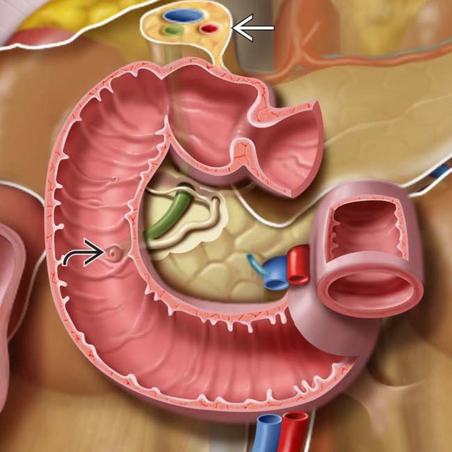
 lies along the medial wall of the 2nd duodenum. The hepatoduodenal ligament
  attaches the duodenum to the porta hepatic and contains the bile duct, portal vein, and hepatic artery.
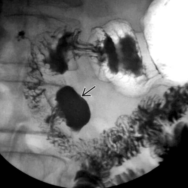
 extending from the medial border of the 2nd portion of the duodenum.
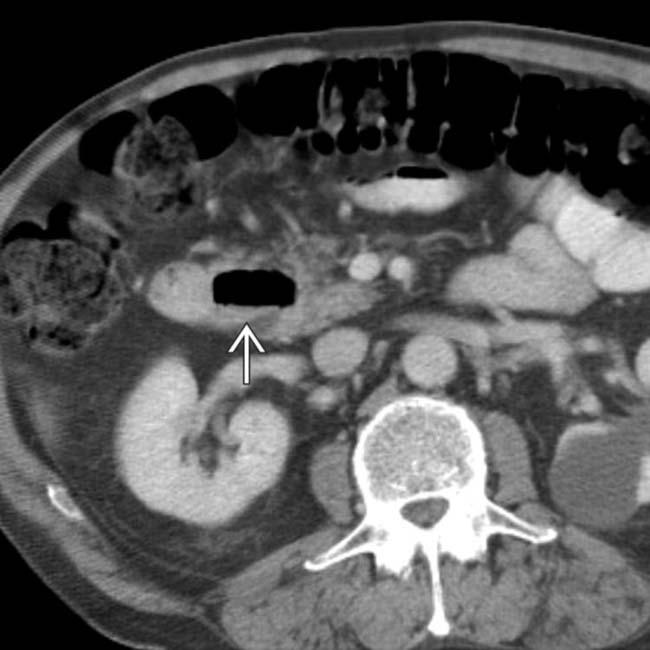
 . A completely fluid-filled diverticulum may mimic a cystic mass in the head of the pancreas.
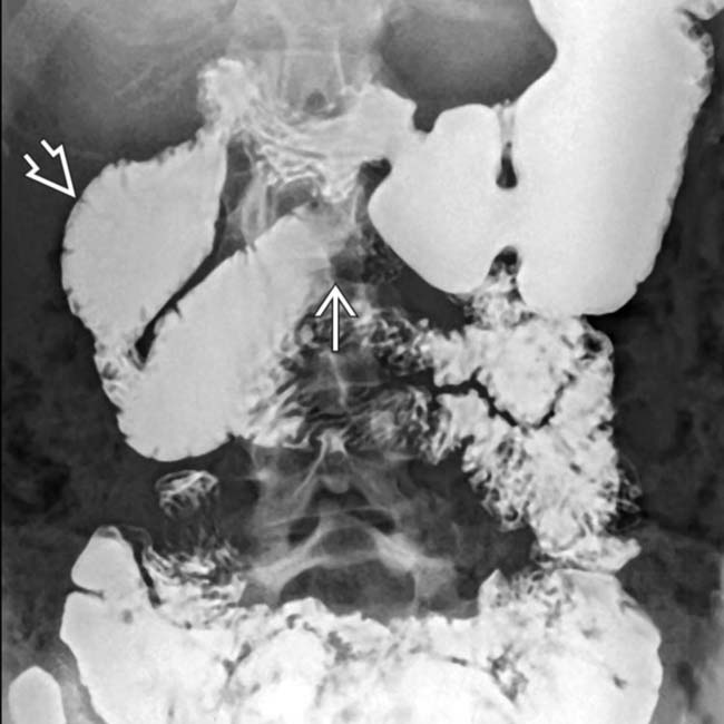
 of the midline duodenum, with dilation of the upstream duodenal lumen
  . The remainder of the small bowel is normal.
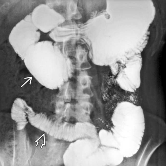
 . Dilation of the small bowel lumen and a “hidebound” appearance of the small bowel folds (thin and closely spaced)
  are typical features of this disease.
Related posts:
Stay updated, free articles. Join our Telegram channel

Full access? Get Clinical Tree


