(Left) Graphic shows the nasopharynx (A, purple, base of skull to palate), oropharynx (B, blue, palate to base of epiglottis), hypopharynx, (C, green, epiglottis to cricopharyngeus), and esophagus (D, below cricopharyngeus muscle). The cricopharyngeal muscle usually lies at the C5-6 level.
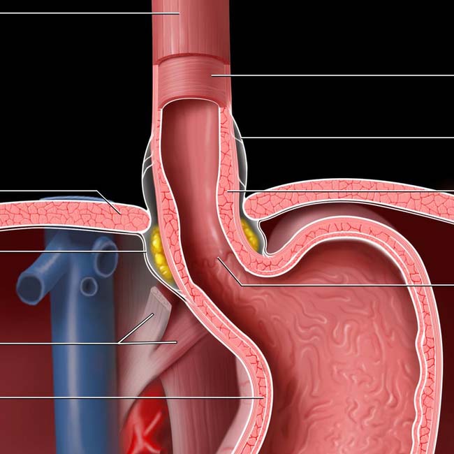
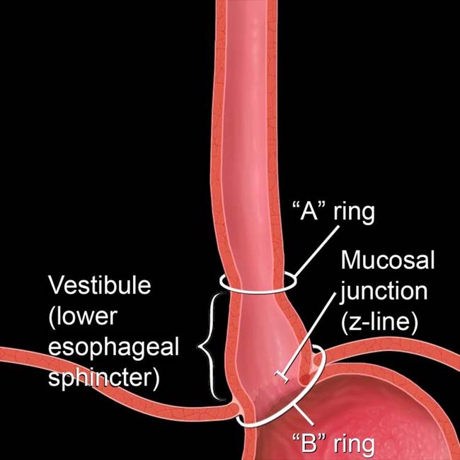
(Right) Graphic shows normal esophageal landmarks and anatomy. The lower esophageal sphincter extends from the “A” to the “B” ring and is sometimes referred to as the phrenic ampulla, or vestibule.
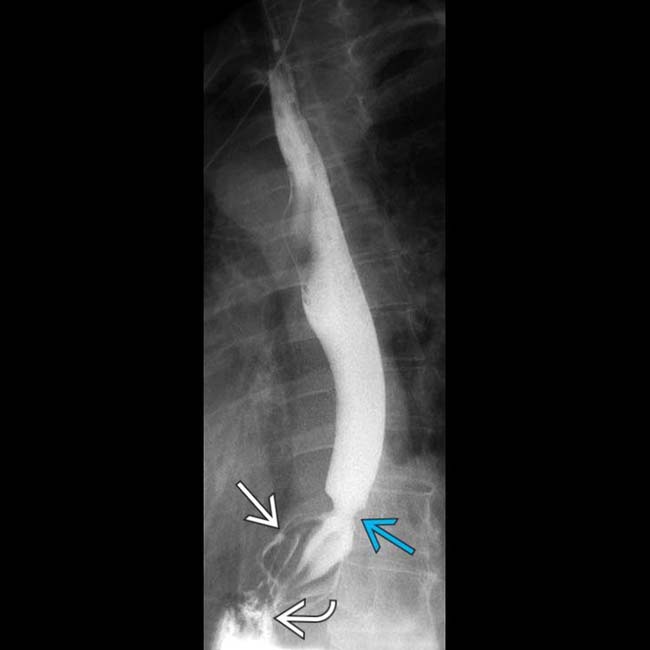
(Left) Spot film from an esophagram shows the lower esophageal sphincter, marked by the “A” ring proximally and the “B” ring distally. Just below the “B” ring is the herniated portion of the gastric cardia .
(Right) Spot film from an esophagram shows a type 1 hiatal hernia above the diaphragm . The esophagus is shortened, probably due to esophagitis and spasm of the longitudinal muscles. GE reflux was demonstrated. The “B” ring marks the GE junction .
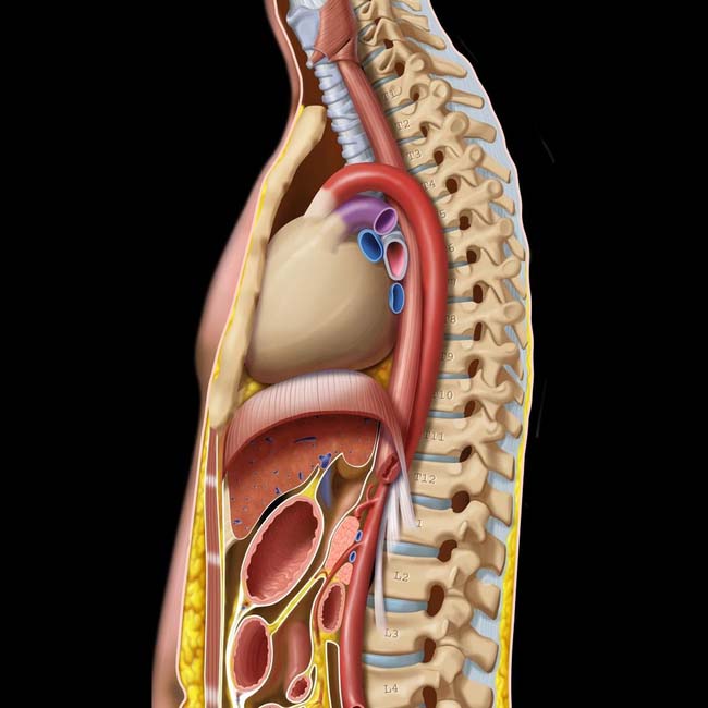
(Top) The esophagus is about 25 cm long and extends from the level of the cricopharyngeus muscle (at the C5-6 level) to the GE junction (at about the T10-11 level). Note the relationship between the esophagus and adjacent structures, including the heart, which may indent or displace the esophageal lumen. The mid esophagus is normally indented by the aortic arch and the left main bronchus. The esophageal hiatus is often at the level of the T10 vertebra.
Outer longitudinal muscle layer Diaphragm Phrenicoesophageal ligament (descending leaf) Right crus of diaphragm Circular muscle layer of stomach Inner circular muscle layer Phrenicoesophageal ligament (ascending leaf) Thickened muscle of LES “Z” line (Bottom) The esophageal wall musculature consists of an inner circular layer and an outer longitudinal layer. In the region of the lower esophageal sphincter (LES), the muscle layers are thickened. The “Z” line marks the junction of the esophageal and gastric mucosa.
Only gold members can continue reading. Log In or Register to continue
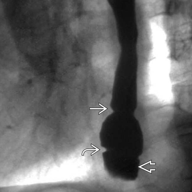
 proximally and the “B” ring
  distally. Just below the “B” ring is the herniated portion of the gastric cardia
  .
.
 above the diaphragm
  . The esophagus is shortened, probably due to esophagitis and spasm of the longitudinal muscles. GE reflux was demonstrated. The “B” ring marks the GE junction
  .
.