Longitudinal muscle
Circular muscle
Submucosa
Mucosa
(Top) There are 5 layers of the bowel wall. The innermost is the mucosa, the absorptive surface of the gut. The other layers are the submucosa, circular muscle, longitudinal muscle, and serosa.
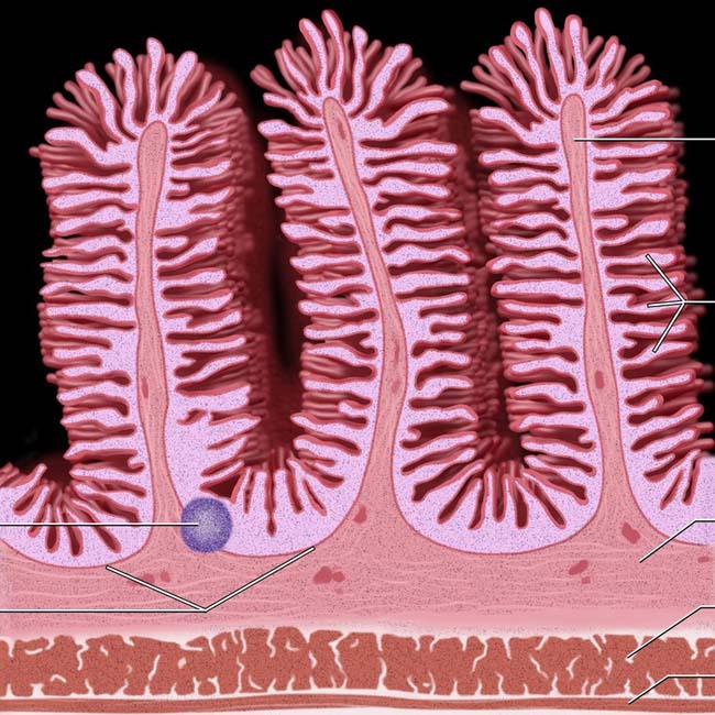
Muscularis mucosa
Circular fold & lamina propria
Villi
Submucosa
Circular muscle
Longitudinal muscle
(Bottom) The mucosal surface of the jejunum is increased by prominent villi, which are finger-like projections of mucosa. The submucosa has a network of capillaries, lymphatics, and a nerve plexus. The jejunum has few small, discrete lymphoid nodules. Although there is no sharp point of transition between the jejunum and ileum, the wall of the ileum becomes thinner and less vascular, with shorter and more widely spaced transverse folds, and more prominent lymphoid follicles.
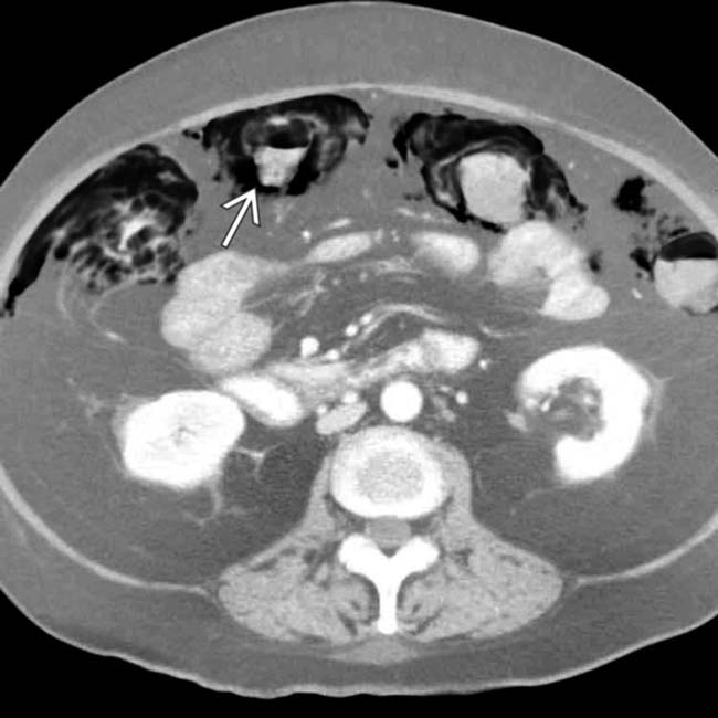
 , but no ascites or ileus. These findings, along with no clinical evidence for bowel ischemia, correctly suggested benign, medication-induced pneumatosis.
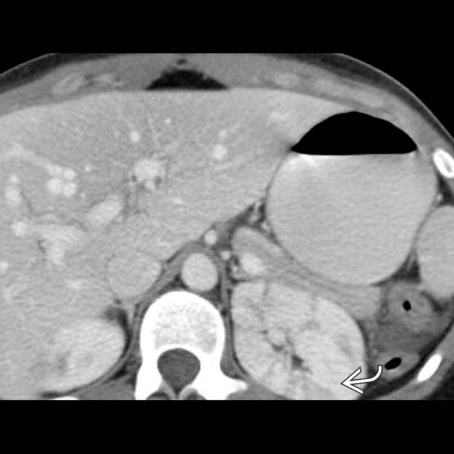
 within the kidneys.
Related posts:
Stay updated, free articles. Join our Telegram channel

Full access? Get Clinical Tree


