Naveen Kalra, Pankaj Gupta, Shuvro Roy Choudhury Image-guided ablation is an integral part of the management algorithm in patients with hepatocellular carcinoma (HCC) and hepatic metastases. Ablation is recommended for very early and early HCC as a potentially curative therapy as well as for downstaging tumours. Ablation is also effective for the treatment of limited small, liver only metastatic disease, especially in patients with colorectal metastases. It is also effectively utilized as an adjunct to transarterial therapies. Ablation is used in special situations for ablation of locally advanced pancreatic cancer, thyroid and parathyroid nodules when surgery is not feasible or is contemplated to be associated with increased risk of damage to adjacent structures. There are several ablative techniques currently available. Although, most widely used technique is radiofrequency ablation (RFA), microwave ablation (MWA), and cryoablation (CA), and irreversible electroporation are being increasingly utilized for ablation of HCC. Chemical ablation, RFA, laser ablation, and MWA are the ablation methods employed for thyroid and parathyroid lesions. In this chapter, we discuss the mechanism of action, hardware, indications, contraindications, and complications of the commonly utilized ablative techniques for malignant liver lesions and thyroid/parathyroid nodules. We also review the recent literature on the role of ablative therapies for these diseases. Ablation of lung lesions, renal lesions, and bone lesions has been discussed under different sections by other authors. The ablative methods are classified based on the mechanism of cellular action into chemical, thermal, and non-chemical non-thermal IRE. Alcohol and acetic acid induce coagulative necrosis by cellular dehydration and cytoplasmic protein denaturation. Thrombosis of tumour microvasculature promotes local ischemia. Compared to ethanol, acetic acid dissolves interstitial collagen allowing it to permeate through tumour septae and capsule. This causes a uniform distribution of acetic acid and greater degree of tumour necrosis at lower doses and fewer treatment sessions than ethanol. However, the completeness with chemical ablation is unpredictable, which is responsible for the higher rates of margin recurrence. Chemical ablation has a role primarily in HCCs as these tumours occur in “hard” cirrhotic livers. This along with the presence of a distinctive tumour capsule enables retention of the ablative agent within the tumour. RFA delivers alternating current at frequencies of 400 MHz, which causes ionic agitation and frictional heat. Following heat generation, regardless of the method of thermal ablation, the tissues respond in a similar fashion. With mild elevation of temperature (up to 40°C), cellular homeostasis is maintained. Between 42 and 45°C (hyperthermia), there is enhanced susceptibility of cells to cytotoxic agents. Irreversible cellular death occurs when tissues are exposed to 46°C for 60 min. Temperatures in the range of 50–52°C result in cell death in 4–6 min. Instantaneous cell death occurs at temperature between 60 and 100°C. Temperatures >105°C lead to vaporization and carbonization. The aim is to achieve a temperature between 50 and 100°C throughout the tumour volume. The presence of blood vessels near the zone of thermal ablation may take away the heat from the target and is known as heat sink effect. High-frequency microwaves generate an electromagnetic field (besides ionic polarization observed with RFA) resulting in rapid and homogeneous heating of tissues. This action is most potent in tissues with high water content and least in fat rich tissue. Temperatures between −20 and −50°C cause freezing of tissues. Ice formation within the extracellular space creates an osmotic gradient and causes cellular dehydration. The intracellular ice crystals cause cell membrane rupture and cell death. Additionally, there is vascular stasis and thrombosis. There is also an element of apoptosis. Slow thawing between cycles of freezing potentiates cellular damage. The connective tissue is relatively resistant to freezing. Hence, CA is safe for the tumours adjacent to the critical structures. Destruction of tissues by laser therapy involves conversion of the absorbed light (infrared) to heat. The optical penetration ranges from 12 to 15 mm, which is shown to be greater in tumour than normal tissue. The conduction of heat results in a lager ablation zone. IRE is a non-chemical, non-thermal ablative technique. It involves delivery of direct electric current at high voltage (up to 3 KV) and high intensity (up to 50 A) in short pulses. This results in pore formation in the lipid bilayer of cell membranes, the cells become leaky and irreversible cell death occurs by apoptosis. This mode of cell death is different from the coagulative necrosis which results from thermal ablation. IRE theoretically spares critical structures adjacent to the target lesion including bile ducts, blood vessels, and tissue stroma. Keeping with the non-thermal mechanism of action, heat sink effect is not observed with IRE. RF equipment comprises of a generator, electrodes (probes) and grounding pads. The RF generator produces alternating current. Electrodes deliver energy within the tumour. Monopolar electrodes require placement of grounding pads for completion of the electrical circuit, allowing the current to pass freely without causing significant heat generation. The grounding pads are placed on large body surface of the patients, typically, patient’s thighs or back. Bipolar RF electrodes do not require grounding pads. Based on the configuration, four types of RF electrodes are commercially available. These include two models of retractable-needle electrodes (model 70 and model 90 StarBurst XL needles, RITA Medical Systems, Mountain View, CA; LeVeen needle electrode, Boston Scientific, Boston, MA) that have multiple curved electrodes of varying length. Upon deployment, these assume the shape of a ‘Christmas tree’ or ‘umbrella’. The internally cooled electrode (Cool-Tip RF electrode; Medtronics, Minneapolis, MN, USA) is a 17-G insulated, hollow needle with a variable length of exposed tip. The shaft has two internal channels for chilled water perfusion. The clustered electrode (Octopus; STARmed, Goyang, Korea) is characterized by switching of energy between the pair of electrodes that creates relatively larger ablation zone in shorter time. Based on the mechanism of energy control, the RFA systems are classified into impedance-based, temperature-based, or both. The impedance-based systems include Cool-Tip (Medtronics), RF 3000 (Boston Scientific), CelonPower LAB (Celon), and VIVA RF (STARmed). The temperature-based systems include StarBurst (Angiodynamics). Those that utilize both the control mechanisms include M 3004 (RF Medical) and AMICA (HS Hospital Service Ltd.). Intraductal RFA (HabibTM PERF catheter, EMcision Ltd) is a bipolar device that can be placed over a 0.035-inch guide wire. The basic design is of an 8 F catheter (90-cm working length) that can be placed percutaneously. Proximal to the 5-mm leading tip at the distal end, there are two circumferential, 8-mm wide stainless-steel electrodes separated by 8 mm gap. This configuration allows the intraductal RF catheter to produce a 25 mm length cylindrical ablation. The terminal at the proximal end is connected to a power generator. MW generators have two frequency settings: 915 MHz and 2.45 GHz. MWA can be performed with single or multiple applicators, latter requires independent generators. MW generators produce different powers ranging from 45 to 100 W at 915 MHz and 2.45 GHz. Based on the size of the lesion and power settings, different protocols are recommended. The various MWA systems can be classified on the basis of the agent utilized for cooling. The non-cooled system is AveCure (MedWaves, Incorporated). Water cooled systems include Solero (Angiodynamics), Acculis Sulis Vp (Microsulis Medical), AMICA (HS Hospital Service Ltd.), ECO-100A1 (Nanjing ECO Medical Instruments), MicroThermX-100 (BSD Medical), Evident/Emprint (Covidien), and MimaPro Therm (Mima Pro). The only carbon dioxide cooled system is NeuWave Certus 140 (Ethicon, Johnson and Johnson). The nitrogen-based CA systems have now been replaced with argon-based unit. Joule-Thompson principle governs the freezing associated with CA systems. When argon gas is circulated through a thin probe, rapid expansion generates extremely low temperature and ice-ball formation around the tip of the probe. Cycles of passive slow thawing between freezing maximizes cell death. The probe removal at the end of procedure is facilitated by the helium gas circulated at the end of the thaw. To ablate larger tumours, multiple probes can be used simultaneously. The two commercially available argon-helium based systems are Cryocare (EndoCare) and ICEfx, Visual ICE (Boston Scientific). The only nitrogen-based system is Prosense (IceCure Medical Inc.). The most widely used LA technique is the Nd-YAG (neodymium: yttrium-aluminium-garnet) laser which has a wavelength of 1064 nm. Diode lasers are less expensive and have shorter wavelengths (800–980 nm) and are associated with a smaller volume of destruction. LA is performed using flexible quartz fibres of 300–600 μm diameter. The most commonly used are interstitial quartz fibres that have flat or cylindrical diffusing tips. These fibres are 10–40 mm long and provide an ablative diameter up to 50 mm. The use of beam splitting devices allows multiple fibres to be placed simultaneously with associated increase in ablative volume. The NanoKnife (AngioDynamics, New York) is the most commonly used commercial device. The IRE electrodes are monopolar 19 G electrodes with adjustable active tip length (5–40 mm). IRE procedures are performed under general anaesthesia and with muscle relaxation. The electrical pulses are delivered during the refractory phase of the myocardium (ST segment). The cardiac synchronization is achieved using a commercially available device (Accusync, Accusync Medical Research). TEACHING POINTS There are three methods of ablation. These are chemical, thermal and non-chemical non-thermal. RFA is the workhorse of ablation. MWA, CA and IRE are the other techniques.
2.17: Imaging guided ablation
Introduction
Ablative therapies: Classification
Mechanism of ablation
Chemical ablation
Thermal ablation
Radiofrequency ablation (RFA).
Microwave ablation (MWA).
Cryoablation (CA)
Laser ablation, LA (also known as laser interstitial tumour therapy, LITT)
Irreversible electroporation (IRE)
Hardware
RFA
Endobiliary (intraductal) RFA
MWA
Cryoablation
LA
IRE
Ablation of liver lesions
Indications
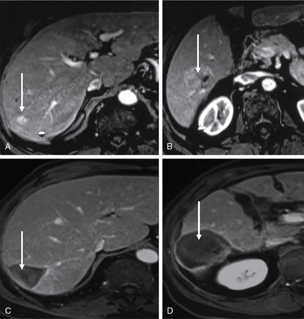
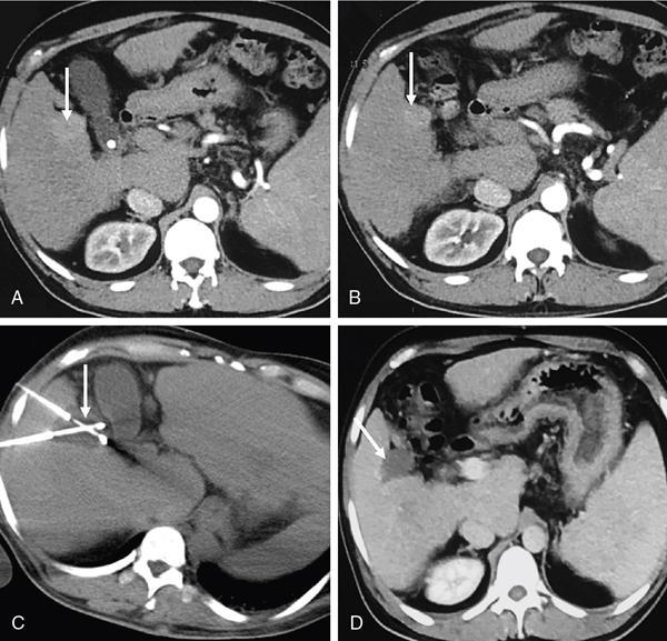
Contraindications of liver tumour ablation
Absolute
Stay updated, free articles. Join our Telegram channel

Full access? Get Clinical Tree


