Imaging Maxillofacial Trauma


Imaging Maxillofacial Trauma


Mark P. Bernstein

Alexander B. Baxter

John H. Harris Jr.


INTRODUCTION


Epidemiology

Facial fractures account for a large proportion of emergency room visits and 2% of all hospital admissions. Significant facial injuries are clinically occult in more than half of all intubated multitrauma patients. Mechanisms include motor vehicle collisions (MVCs), assault, falls, sports injuries, and civilian warfare. Together, MVCs and assault account for more than 80% of all injuries and commonly involve young adult males and alcohol use. A recent decline in MVC-related maxillofacial trauma appears to reflect improved automobile safety as a result of airbags, mandatory seatbelt laws, and improved road conditions.


Biomechanics and Associated Life-Threatening Injuries

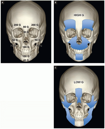
Direction and magnitude of an impacting force determines the pattern and severity of maxillofacial fractures. For example, the nose, mandibular body, and zygoma are typically injured in assault because of their prominent positions on the face and the relatively small amount of energy transferred in a strike or a punch. MVC, falls and other high-velocity injuries result in more complex, midfacial fractures. A collision of 30 miles per hour exceeds the tolerance of most facial bones (Fig. 4.1A).


Figure 4.1. Biomechanics of the facial skeleton. A: Maximum tolerable impact forces. (Modified from Swearingen JJ. Tolerances of the Human Face to Crash Impact. Oklahoma City, OK: Civil Aeromedical Research Institute; 1965.) B: High G-force facial injuries: minor injuries 32%, major injuries 51%, death 15%. C: Low G-force facial injuries: minor injuries 30%, major injuries 21%, death 3%. (Modified from Luce EA. Maxillofacial trauma. Curr Probl Surg. 1984;21(2):1-68.)


Luce et al. studied injuries associated with major facial fractures in 1,020 patients and grouped them into high and low G-force mechanisms. Life- threatening injuries included intra-abdominal injury requiring surgery, pneumothorax, chest trauma requiring ventilator support, and severe closed head injury. Twenty-one percent of patients with low G-force facial trauma had one or more of these associated injuries compared with 50% in patients with high G-force mechanisms (Figs. 4.1B, 4.1C). Mortality in the latter group was 12%.

Mulligan et al. investigated the relationship between facial fractures, cervical spine injuries, and head injuries in 1.3 million trauma patients between 2002 and 2006. Nine percent sustained one or more facial fractures. The 6.7% of facial fracture patients had concomitant cervical spine injury, and 61.8% had associated head injury. Almost 5% suffered injuries to all three areas.

Cole et al., in a study of 247 victims of facial gunshot wounds, found associated cervical spine injury in 8% and head injury in 17%.


IMAGING

Imaging in facial trauma aims to define the number and locations of facial fractures and to identify injuries that could compromise the airway, vision, mastication, lacrimal system, and sinus function. Individual fractures should be listed and associated soft tissue injuries described with attention to these areas. If possible, bony findings should be summarized in one of several typical fracture patterns.

Imaging in most emergency departments for significant facial trauma begins with computed tomography (CT) scanning. Helical CT and, more recently, multidetector CT (MDCT) have supplanted plain radiography and have revolutionized the imaging of the maxillofacial trauma. CT is more cost efficient and more rapidly performed than radiographs of the face and mandible. MDCT is now considered the optimal imaging modality, particularly in the polytrauma setting because it allows safe and rapid image data acquisition and multiplanar reconstruction without patient manipulation. MDCT accurately depicts both bony and soft tissue injury. Submillimeter slice thickness permits exquisite multiplanar reformations (MPRs) and three- dimensional (3D) reconstructions. Fracture fragment displacement and rotation are easily determined and fracture patterns may be readily classified and assessed for stability. Volume reformations from helical and MDCT datasets enhance diagnostic accuracy and allow the surgeon to better plan operative repair by depicting complex injuries in three dimensions.

Magnetic resonance imaging (MRI) can be a useful adjunct in patients with cranial nerve deficits not explained by CT, evaluation of incidentally discovered masses, and suspected vascular dissection. Its advantages include multiplanar imaging, excellent soft tissue contrast, and lack of ionizing radiation. The practical limitations of long scan times, limited patient access, poor evaluation of bone and contraindication in patients with pacemakers, some aneurysm clips, and ocular metallic foreign bodies prevent its primary application in the emergency setting.


Multidetector Computed Tomography Technique

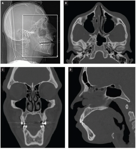
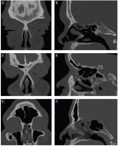
At Bellevue Hospital, patients with direct facial injury and suspected maxillofacial fractures are scanned from the hyoid through the top of the frontal sinuses. Acquisitions using 64-MDCT with 0.625-mm detector width and 0.4 mm overlapping sections allow high-quality MPRs to be generated and evaluated at the workstation. The 2 mm thick images in three planes oriented parallel and perpendicular to the hard palate provide symmetrical images for interpretation (
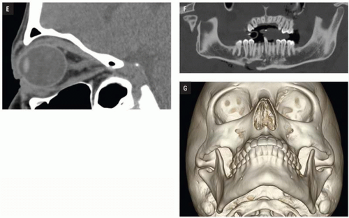
Figs. 4.2A-4.2D). Reconstructions in bone and soft tissue algorithm and specialized reformations may be generated depending on the presence and type of fractures. For example, oblique sagittal reformations along the plane of the optic nerve elegantly characterize orbital floor fractures with respect to depression, orbital depth, and relation to the inferior rectus muscle (Fig. 4.2E). Panoramic or oblique sagittal planes optimize evaluation of mandibular angle and ramus fractures (Fig. 4.2F). The 3D reconstructions can be oriented in any plane and are often acquired in patients with complex injuries as an aid to surgical planning (Fig. 4.2G).

The multitrauma patient requires a comprehensive examination to evaluate multiple body regions in a single visit to the CT suite. With current technology, scanning of the head, face, and cervical spine may be acquired as a single acquisition and no longer requires patient repositioning for direct coronal plane imaging.


Figure 4.2. CT acquisition and imaging planes. A: CT scout. B: Transaxial CT image at level of zygomatic arches. C: Coronal CT reformation through the mid-orbits and maxillary sinuses. D: Midsagittal CT reformation. (continued)


Figure 4.2. (continued) E: Oblique sagittal reformation along the plane of the optic nerve. F: Curved reformat panoramic CT simulates the Panorex radiograph. G: 3D CT reconstruction in Water’s projection.


FACIAL FRACTURES

Facial fracture complexes are classified by location and pattern: nasal, naso-orbito-ethmoid (NOE), frontal sinus, orbital, zygomatic, maxillary, and mandibular. Manson et al. have proposed further categorizing each area by the energy of the injury, namely low, moderate, and high energy. Low-energy injuries show little or no comminution or displacement. Moderate-energy injuries, the most common, demonstrate mild to marked displacement, whereas high energy is reserved for cases of severe fragmentation, displacement, and instability. Impact energy subclassifications dictate management from simple closed reduction to wide exposure open reduction and internal fixation.


NASAL FRACTURES


Anatomy

The upper third of the nose is supported by a bony skeleton consisting of the nasal bones proper, the frontal process of the maxilla, and the nasal process of the frontal bone. The middle and lower thirds are composed of the upper lateral and lower alar cartilages, respectively. The anterior nasal septum is cartilaginous. The posterior perpendicular plate of ethmoid, vomer, nasal crest of maxilla, and nasal crest of the palatine bone form the bony nasal septum (Fig. 4.3).


Injuries

Nasal bone fractures are common and account for half of all facial fractures. Most of these involve the distal third because this represents the most prominent projection of the facial skeleton. Peak incidence is in the second to third decades, with
injuries twice as common among men. Potential sequelae include nasal obstruction, cosmetic deformity, and cerebrospinal fluid (CSF) leak. Septal hematoma, if unrecognized, may result in later ischemic septal necrosis and “saddle nose” deformity.


Figure 4.3. Nasal bone anatomy.

Nasal injuries are classified by the energy and direction of the impact force. Lateral force from assault is the most common mechanism and causes contralateral displacement of the nasal bones and frontal processes of the maxilla. In low-velocity injuries, detachment of the nasal septal cartilage from the vomer may accompany the fracture. High- velocity injuries and frontal impacts result in central, comminuted, septal fractures. Inferior forces typically cause an isolated septal injury. The nasal bones are most resistant to frontal impact; once the force is great enough to fracture the upper nasal bones, the delicate ethmoid air cells behind them offer little resistance to further impaction and allow the nasal bones to telescope into the deep face. This fracture pattern usually also involves the medial orbital walls and is referred to as an NOE fracture.

Epistaxis is a serious complication of nasal fractures. Even minor trauma can result in hemorrhage from Kiesselbach’s plexus (Fig. 4.3), a robust vascular network that supplies the nose. Severe bleeding is usually caused by injuries to the anterior ethmoid artery branch of the ophthalmic artery (anterior bleeding) and to the sphenopalatine artery branches (posterior bleeding).


Imaging

CT analysis aids operative management of severe nasal bone fractures and identifies associated facial soft tissue and bony injuries. Fractures are described as unilateral or bilateral, simple or comminuted, displaced or undisplaced, impacted or non- impacted, and with or without nasal septal involvement. A proposed classification scheme is illustrated in Figure 4.4. Posterior packing with balloon tamponade may be seen as treatment for epistaxis (Fig. 4.5). Septal involvement can complicate nasal bone realignment and should be specifically addressed in the radiology report. Generally, nasal fractures with associated septal fracture, significant dislocation, or severe concomitant soft tissue injury require open repair, whereas most others can be treated with closed reduction.


Figure 4.4. Nasal bone fractures—classification. (N) Normal. (I) Simple, undisplaced. (IIA) Simple, displaced, unilateral without telescoping. (IIAs) Simple, displaced, unilateral with septal fracture (arrow). (IIB) Simple, displaced, bilateral. (IIBs) Simple, displaced, bilateral with septal fracture (arrow). (III) Comminuted with telescoping.


Figure 4.5. Posterior nasal packing with Foley balloon tamponade. Transaxial (A) CT reformation show bilateral transnasal Foley catheters with balloons inflated (greater on the right; arrows). Right parasagittal CT reformation (B) shows Foley and balloon posterior to the hard palate (arrows). 3D reconstruction (C) shows bilaterally placed nasal catheters (arrows).


NASO-ORBITO-ETHMOID FRACTURES


Anatomy

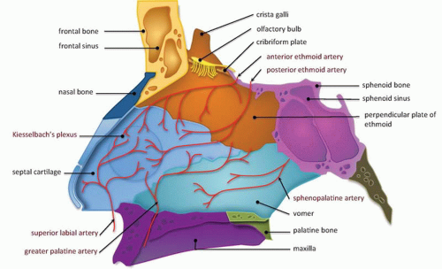
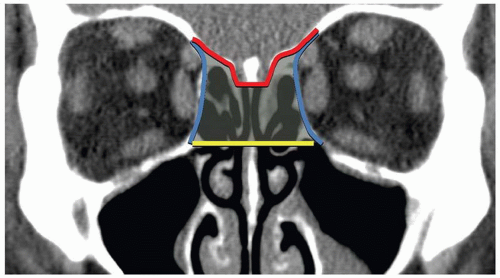
The NOE region refers to the space between the eyes or interorbital space. The interorbital space represents the confluence of the bony nose, orbit, maxilla, and cranium. It is bound laterally by the thin medial orbital walls and posteriorly by the sphenoid sinus. The cribriform plate and the medial floor of the anterior cranial fossa define its superior margin and separate the NOE region from the dura, CSF, and brain. Inferior margin is the lower border of the ethmoid air cells (Fig. 4.6). The frontal process of maxilla, nasal process of the frontal bone, and thick proximal nasal bones comprise the anterior border of the interorbital space. Together, these form the relatively strong central facial “pillar.” Important soft tissue structures of the interorbital space include the olfactory nerves, lacrimal sac, nasolacrimal duct, ethmoid vessels, and the medial canthal ligaments.


Injuries

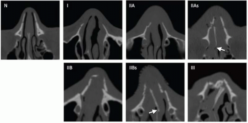
NOE injuries result from direct anterior impact to the upper nasal bridge and are characterized by fracture of the nasal bones, nasal septum, frontal process of the maxilla, ethmoid bones (lamina papyracea and cribriform plate), lacrimal bones, and frontal sinus (
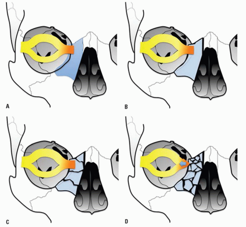
Fig. 4.7). They may be associated with other facial fractures and remote multisystem trauma. NOE fractures have been classified by Gruss who grouped them by extent of injury, displacement, orbital involvement, and associated facial fractures (Table 4.1).


Figure 4.6. Naso-orbito-ethmoid (NOE) regional anatomy. Coronal CT reformation shows the interorbital space (shaded green) defined by medial orbital walls (lateral margins; blue), cribriform plate (superior margin; red), and lower border of the ethmoid sinuses (inferior margin; yellow).


Figure 4.7. Naso-orbito-ethmoid (NOE) injury.

Type 1 fractures detach the frontal process of maxilla, displacing the fragments posteriorly and laterally without severe comminution. Type 2 fractures are more severely comminuted and impacted through the interorbital space, shattering the nasomaxillary buttress (discussed with maxillary fractures subsequently), and surround the piriform aperture. Subtypes a-c describe the integrity of the zygomaticomaxillary buttresses, from intact to unilateral to bilateral involvement, respectively. Type 3 fractures occur in conjunction with more extensive craniofacial injuries and reflect superolateral extension, including cribriform plate disruption with intracranial involvement and dural violation (superior extension), or LeFort II and III fractures (lateral extension). Type 4 injuries include varying degrees of orbital detachment and displacement; whereas type 5 injuries are associated with significant bone destruction or loss, potentially complicating reconstructive strategies.


TABLE 4.1 Classification of Naso-Orbital-Ethmoid Injuries


Type 1


Isolated bony NOE injury


Type 2


Bony NOE and central maxilla


2a


Central maxilla only


2b


Central and one lateral maxilla


2c


Central and bilateral lateral maxillae


Type 3


Extended NOE injury


3a


Superiorly—craniofacial injuries


3b


Laterally—with LeFort II and III fractures


Type 4


NOE injury with orbital displacement


4a


With oculo-orbital displacement


4b


With orbital dystopia


Type 5


NOE injury with bone loss


From Gruss JS. Naso-ethmoid-orbital fractures: classification and role of primary bone grafting. Plast Reconstr Surg. 1985; 75(3):303-317.


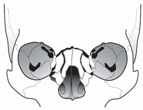
The medial and lateral canthal ligaments support the globe and keep the eyelid apposed to it. Fracture through the inferomedial orbital rim suggests injury to both the medial canthal ligament and lacrimal apparatus. Patients present with nasal and periorbital ecchymosis, depression of the nasal bridge, telecanthus, enophthalmos, and a shortened palpebral fissure.

The junction of the frontal process of maxilla and the inferomedial orbital rim make up the bony anchor of the medial canthal ligament. In the setting of NOE fracture, this bony anchor is referred to as the “central” fragment and may be either intact or comminuted or fractured through the medial canthal ligament insertion site. Markowitz et al. have devised a classification system to address its integrity and dictate optimal repair (Fig. 4.8) (Table 4.2).


Figure 4.8. A: Medial canthal ligament injury. Medial canthal ligament (orange) is shown anchored to the bony “central fragment.” The central fragment lies at the junction of the frontal process of maxilla and inferomedial orbital rim. B, C, and D: Medial canthal ligament injury. Manson classification.


TABLE 4.2 Classification of Central Fragment (the Bone Bearing the Medial Canthal Ligament Insertion) Injury, and Incidence


Unilateral


Bilateral


Type I


Single segment


50%


13%


Type II


Comminuted


44%


85%


Type III


Comminuted into canthal insertion


6%


2%


From Markowitz BL, Manson PN, Sargent L, et al. Management of the medial canthal tendon in nasoethmoid orbital fractures: the importance of the central fragment in classification and treatment. Plast Reconstr Surg. 1991;87(5):843-853.


Imaging

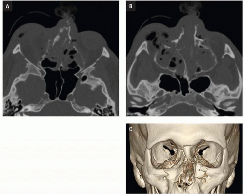
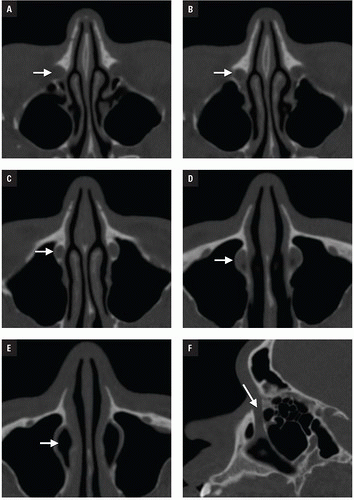
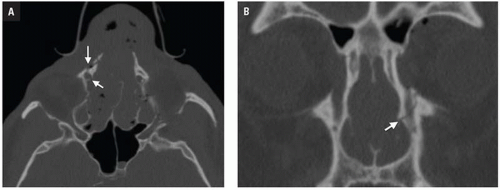
CT shows impaction of the intraorbital contents with posterior telescoping of ethmoid air cells, nasal septal buckling, and intrasinus hemorrhage. Inferomedial orbital rim fracture with displacement of the central fragment indicates medial canthal ligament involvement (Fig. 4.9). Injuries may be unilateral or bilateral and of variable comminution depending on impact energy.

Low-energy injuries are exclusively unilateral with a single displaced inferomedial orbital rim fracture fragment. Moderate-energy NOE fractures are more common and are characterized by several fractures of the inferomedial orbital rim without fragmentation of the bony medial canthal ligament insertion. High-energy injuries disrupt the medial canthal ligament anchor and require more complex surgical repair. NOE fractures are often associated with LeFort II and III injuries and close attention should be paid to the pterygoid plates.

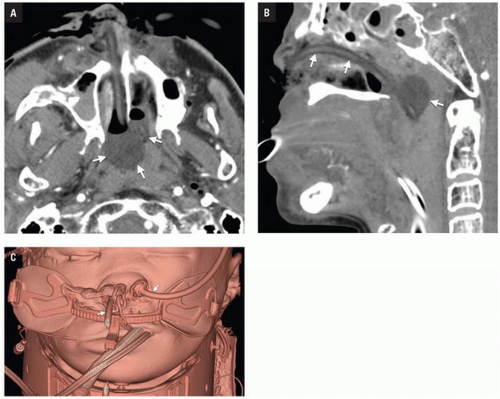
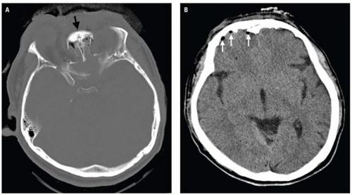
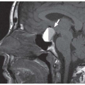
Clinical consequences include telecanthus, enophthalmos, ptosis, and lacrimal system obstruction. Associated cribriform plate fracture may result in anosmia, CSF leak, and pneumocephalus (
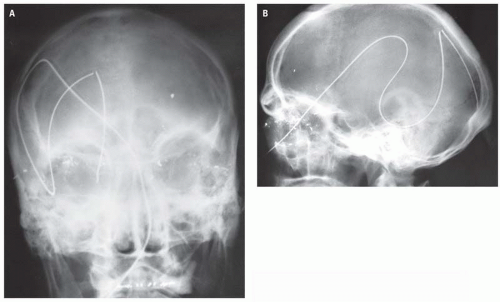
Fig. 4.10). The latter can evolve into tension pneumocephalus after cardiopulmonary resuscitation efforts. Disruption of the sinus roof predisposes to intracranial nasogastric tube and Foley catheter (for control of posterior nasal bleeding) malposition (Fig. 4.11).


Figure 4.9. A and B: NOE fracture. Two transaxial CT images in the same patient showing comminuted, impacted fractures centered on the nose and central maxilla. C: 3D CT reconstruction shows comminuted NOE midface fractures.


Figure 4.10. A and B: NOE fracture. A: Transaxial CT image shows comminuted and impacted NOE fractures at the level of the upper nasal bridge (black arrow). Pneumocephalus (white arrows in B) from fracture of the cribriform plate.


Figure 4.11. Intracranial nasogastric tube.


NASOLACRIMAL INJURIES


Anatomy

The nasolacrimal fossa and canal make up the bony lacrimal excretory system. The fossa originates in the medial orbital wall and is made up of the thick anterior lacrimal crest of the frontal process of the maxilla and the posterior lacrimal crest of the lacrimal bone. The nasolacrimal canal descends into the thinner nasal portion of the maxilla, terminating beneath the inferior turbinate (Fig. 4.12). The lacrimal drainage system, consisting of the nasolacrimal sac and duct, is protected within the bony fossa and canal, respectively.


Figure 4.12. Nasolacrimal fossa and canal anatomy. Nasolacrimal fossa originates in the medial orbital wall (arrow) (A) and lies behind the thick anterior lacrimal crest of the frontal process of maxilla (arrow) (B). The nasolacrimal canal descends into the thin nasal portion of maxilla (arrow) (C and D) and terminates below the inferior turbinate (arrow) (E). Sagittal CT reformation (F) shows the entire course (arrow).


Injuries and Imaging

Nasolacrimal injuries are anticipated with NOE fractures, but can occur in other injuries as well. Symptomatic lacrimal obstruction (epiphora and dacryocystitis) has been reported in 0.2% of nasal fractures, 4% of LeFort II and III fractures, and 21% of NOE fractures.

Unger studied the CT appearance of nasolacrimal injuries in 25 patients and found that all nasolacrimal fractures were associated with other facial fractures. Fractures limited to the stronger nasolacrimal fossa were less common than injuries combined with the fragile nasolacrimal canal. Canal fractures are mostly comminuted (Fig. 4.13) whereas fossa fractures are mostly avulsions in which the fossa remains intact but is separated from its normal attachments. The sac and duct normally contain either air or fluid and duct obstruction must be diagnosed clinically.


FRONTAL SINUS FRACTURES


Anatomy

Frontal sinus anatomy is variable—10% have a unilateral sinus, 5% a rudimentary sinus, and 4% have no sinus (Fig. 4.14). The anterior sinus wall is much thicker and stronger than the posterior wall and can tolerate up to 2,200 lb of force before fracturing. The dura and frontal lobes are immediately posterior to the sinus; the orbital roof is inferior and lateral. Inferomedially, the frontal sinus drains into the middle nasal meatus via the nasofrontal duct more commonly referred to as the nasofrontal outflow tract (NFOT) (Fig. 4.15).


Injuries

Frontal sinus fractures account for 5% to 15% of all craniomaxillofacial fractures and result from anterior upper facial impact. Frontal sinus fracture indicates high G-forces that propel the head and cervical spine into extension, often with severe associated intracranial injury and facial fractures. Management decisions depend on fracture type, neurologic status, CSF leak, posterior table fracture pattern, and NFOT injury.

NFOT integrity is the most critical determinant and a reliable sign of high energy transfer. Patients with frontal sinus fractures and NFOT injury have two to three times as many associated facial fractures, most commonly orbital roof and NOE fractures than patients with frontal sinus fracture alone. The incidence of cerebral injury with frontal sinus fracture rises from significant (31%) to striking (76%) when the NFOT is involved. Patients suffering frontal sinus fractures have a 25% overall mortality and frequently present in shock (52%) or coma (42%). Table 4.3 summarizes associated injuries based on integrity of the NFOT in 857 patients with frontal sinus fractures.


Figure 4.13. Nasolacrimal injuries. Transaxial CT image shows comminuted fractures across the right nasolacrimal fossa (arrows).


Figure 4.14. Frontal sinus variability. Coronal and sagittal CT reformations in patients with no frontal sinus (A and B), small unilateral frontal sinus (C and D), and larger bilateral frontal sinuses (E and F).


Figure 4.15. NFOT anatomy. A: Coronal CT reformation show the normal nasofrontal outflow tracts bilaterally (arrows). B: Sagittal CT reformation depicts the outflow tract draining the frontal sinus (FS) in a different patient (arrowheads).


Imaging

Frontal sinus fractures may involve the anterior table, the posterior table, or both (Figs. 4.16, 4.17). One-third are limited to the anterior table and half involve both anterior and posterior tables. Separation of fracture fragments by more than one table width constitutes displacement. NFOT injury occurs in 70% of cases (Table 4.4) and is indicated by (1) anatomic outflow tract obstruction, (2) frontal sinus floor fracture, and (3) medial anterior table fracture. A fracture fragment partially or entirely within the tract indicates outflow tract obstruction. Any of these features permits diagnosis of NFOT injury. MPRs aids in identification of the NFOT and are often necessary for comprehensive assessment (Fig. 4.18).


TABLE 4.3 Associated Injuries in Frontal Sinus Fractures


Injury


NFOT Intact


NFOT Injury


Intracranial


31%


76%


NOE


12%


31%


Orbital roof


13%


40%


Orbital wall


7%


13%


Orbital floor


2%


7%


Zygoma


8%


18%


LeFort


2%


17%


Mandible


3%


5%


Cervical spine


7%


14%


Upper extremity fracture


15%


25%


Lower extremity fracture


13%


23%


Pneumothorax


12%


24%


Abdominal


7%


13%


NFOT, nasofrontal outflow tract; NOE, naso-orbitoid-ethmoid. From Stanwix MG, Nam AJ, Manson PN, et al. Critical computed tomographic diagnostic criteria for frontal sinus fractures. J Oral Maxillofac Surg. 2010;68(11):2714-2722.


Isolated and undisplaced anterior table fractures require no operative fixation. Displaced posterior table fractures indicate that the dura has been breached and there is potential contiguity between the sinus and brain. Ninety-eight percent of displaced posterior table fractures are associated with NFOT injuries. Posterior table injuries require sinus obliteration or cranialization to prevent mucocele or mucopyocele formation. Cranialization is also necessary for persistent CSF leak and involves the stripping of mucosa, obliteration of the nasofrontal duct, and removal of posterior table fragments (Fig. 4.19). Complications of posterior table fractures can be life threatening and include meningitis, encephalitis, brain abscess, and cavernous sinus thrombosis.

Jun 20, 2016 | Posted by in GENERAL RADIOLOGY | Comments Off on Imaging Maxillofacial Trauma

Full access? Get Clinical Tree

Get Clinical Tree app for offline access