Fig. 12.1
Normal pregnancy. (a) Intradecidual sac sign: ultrasound demonstrating a 4-week gestational sac in thickened endometrium, without any identifiable yolk sac or embryo. (b) Double decidual sac sign: ultrasound demonstrating concentric hyperechoic rings produced by chorion and decidua (arrowheads), around the gestational sac. (c) Six weeks: ultrasound demonstrates a yolk sac (arrow) and tiny embryo within a 6-week-old pregnancy. (d) Nine weeks: ultrasound demonstrates the anechoic amniotic cavity separated from chorionic cavity by the amnion. The chorionic cavity has low-level echoes due to the presence of high protein concentration
The gestational sac is eccentric to the endometrial canal and measures 1 mm in diameter when a 4-week blastocyst cavity is present. It is possible to see the gestational sac along with the yolk sac (in chorionic cavity) on ultrasound at 4.5–5-week gestational age, when it measures up to 1 cm in diameter. The embryo and the amniotic cavity are very small at this gestational age. The gestational sac typically grows at the rate 1.1 mm/day and measures 1.6 cm by 6 weeks.
Double decidual sac sign refers to the presence of two concentric rings: the inner representing chorion and outer one representing the decidua (Fig. 12.1b). The presence of double decidual sign does not exclude the presence of ectopic pregnancy. It is seen in majority of the pregnancies and is best seen when the gestational sac is approximately 10 mm in diameter.
Yolk sac is the first element identified within the gestational sac, typically at 5 weeks of gestation. It is usually less than 5 mm in diameter and spherical in shape (Fig. 12.1c). A yolk sac which is too small or too large is considered to be abnormal (Table 12.1). The number of yolk sac is equal to the number of amniotic sac.
Table 12.1
Criteria for abnormal yolk sac
1. More than 5.6 mm (between 5 and 10 weeks) in diameter |
2. Less than 2 mm in diameter (between 8 and 12 weeks) |
3. Absence of yolk sac in a gestational sac more than 8 mm in diameter |
4. Absence of yolk sac in presence of embryo during early pregnancy |
The amnion is barely appreciable when the crown-rump length is 2 mm, at gestational age of 6 weeks. It is normal to see an amnion as a separate membrane between 2 and 4 months of gestation (Fig. 12.1d). The chorionic fluid is more echogenic than the amniotic fluid because of the presence of a high protein concentration. The amniotic cavity progressively increases in size and fills the subchorionic cavity by 14–16 weeks. Double bleb refers to the visualization of two concentric blebs due to the presence of a yolk sac and amniotic sac.
Although the embryonic cardiac activity begins at day 26 of menstrual age, they are first seen on sonography when the crown-rump length is 5 mm, at approximately 6.2 weeks. If the crown-rump length is less than 5 mm and no embryonic cardiac pulsations are demonstrated, then the ultrasound should be repeated. The embryonic cardiac activity of less than 80 beats per minute is associated with high risk of embryonic demise.
The reliable criteria to verify the presence of early intrauterine pregnancy include demonstration of yolk sac within the gestational sac and presence of an embryo with cardiac activity (Table 12.2).
Table 12.2
Morphologic findings of pregnancy and the corresponding gestational ages when they appear
Gestational sac | 4.5 weeks (in all by 5 weeks) |
Yolk sac | 5 weeks |
Embryo | 5.7–6.1 weeks |
Embryonic cardiac pulsations | 6.2 weeks |
Typically transabdominal ultrasound is positive for pregnancy when beta-hCG level is more than 3,600. Intrauterine pregnancies should be seen on transvaginal ultrasound in all when the beta-hCG level is more than 2,000.
Abnormal Gestational Sac
Anembryonic gestation refers to the presence of gestational sac without an embryo. It is due to embryonic death with continued development of the trophoblast and is often due to underlying chromosomal abnormality.
The most important criteria for detection of anembryonic pregnancy are the absence of embryo when the mean sac diameter on transvaginal ultrasound study is more than 18 mm and the absence of yolk sac when the mean sac diameter is more than 13 mm on transvaginal sonography (Fig. 12.2a) (Table 12.3). Since these criteria are not full proof, a follow-up ultrasound should be performed in 7–10 days’ time to rule out the possibility of normal pregnancy.
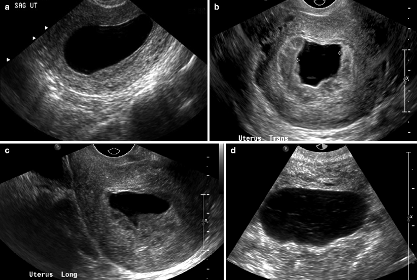
Fig. 12.2
Abnormal gestational sac: anembryonic pregnancy. (a) Ultrasound demonstrates anembryonic gestational sac in 8-week-old pregnancy. Lack of embryo in a gestational sac measuring more than 13 mm in diameter is considered abnormal on transvaginal ultrasound. (b, c) Ultrasound shows irregular outline of the anembryonic gestational sac on transvaginal ultrasound. (d) Ultrasound shows anembryonic 8-week gestational sac, containing low-level internal echoes
Table 12.3
Imaging findings of anembryonic pregnancy
1. Absent embryo when mean sac diameter is 18 mm |
2. Absent yolk sac when the mean sac diameter is 13 mm |
3. Low-lying gestational sac |
4. Irregular shape of the gestational sac (Fig. 12.2b, c) |
5. Thin decidual reaction of less than 2 mm thickness |
6. Absence of double decidual sac sign |
7. Presence of fluid-fluid level or debris within the gestational sac (Fig. 12.2d) |
When the fetal pole is more than 6 mm in diameter and no fetal cardiac pulsations are demonstrated, the findings are suggestive of silent miscarriage. If the fetal pole is less than 6 mm in diameter and no fetal cardiac pulsations are demonstrated, then the patient should be rescanned in a week’s time to distinguish live from failed pregnancy. If no fetal cardiac activity is seen on 1 week follow-up ultrasound study, then the findings indicate a silent miscarriage. Small sac size, defined by difference of less than 5 mm between mean sac diameter and crown-rump length, is associated with high miscarriage rates. Missed abortion refers to retention of pregnancy for more than or equal to 2 months.
Pregnancy Termination Failure
Termination of pregnancy could be performed within 72 h with more than 80 % success rate with the use of oral misoprostol and mifepristone. In case of failed termination, the sonographic features include presence of intrauterine mass and increased vascularity. The lack of blood flow however does not exclude the presence of retained products of conception. The absence of color Doppler flow favors that the products of conception are nonviable and therefore will pass out spontaneously without the use of dilatation and curettage (Fig. 12.3a, b). Retained products of conception typically produce irregular interface between endometrium and myometrium. If no endometrial mass or fluid is seen and the endometrial stripe thickness is less than 10 mm, retained products of conception are very unlikely. MR imaging may show intracavitary uterine mass with variable enhancement and junctional zone obliteration (Fig. 12.3c, d).
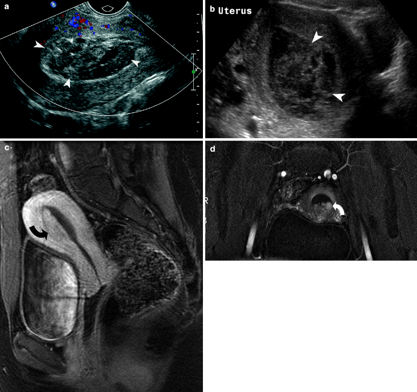
Fig. 12.3


Retained products of conception. Ultrasound demonstrates retained blood clots (a) (arrowheads) and products of conception (b) (arrowheads) present within the endometrial cavity. Gadolinium-enhanced MRI (c, d) demonstrates enhancement of the retained products of conception (curved arrows)
Related posts:


Stay updated, free articles. Join our Telegram channel
Full access? Get Clinical Tree
