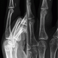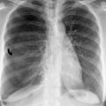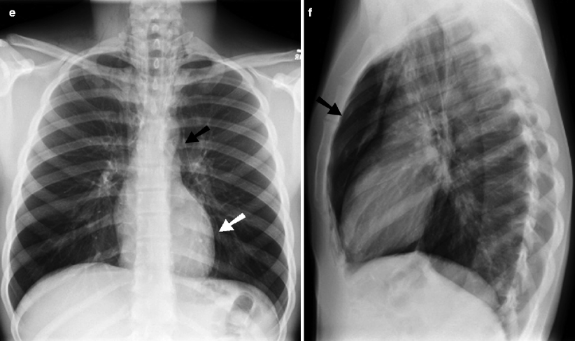
Fig. 24.1
(a) Pneumomediastinum and pneumothorax. Single frontal chest radiograph from a trauma patient after a gunshot wound reveals air between the epicardium and the pleura on the left (curved arrow). Pneumothorax on the right with collapsed lateral lung margin (arrowheads) and loss of peripheral lung markings. The ballistic fragment can be seen within the right mid lung. Extensive subcutaneous emphysema is present. (b) Tension pneumothorax. Frontal radiograph shows relative lucency and loss of lung markings within the right hemithorax with shift of mediastinum to the left. The medial margin of the right tension pneumothorax is seen crossing the midline (arrowheads). (c) Tension pneumothorax. In this pediatric patient, there is a large lucency in the right hemithorax with loss of lung markings. There is associated shift of the mediastinal structures, including the heart to the left. Medial margin of the enlarged pleural cavity crosses the midline (arrowheads). (d) Pneumomediastinum. Frontal radiograph shows air outlining the heart (arrows) as well as extensive subcutaneous emphysema. (e) Pneumomediastinum and pneumopericardium. Frontal radiograph reveals are outline structures in the upper mediastinum outlined by air (black arrow). Pneumopericardium is best seen outlining the left ventricle (white arrow). (f) Pneumomediastinum. Lateral radiograph shows the pneumomediastinum (arrow) as a large lucent area posterior to the sternum and surrounding the heart
The early diagnosis of a small pneumothorax can become clinically important especially if the patient is going to be mechanically ventilated as this can expand the pleural space leading to greater mass effect and mediastinal shift. This, in turn, can cause decreased blood return to the heart more likely from the loss of negative intrapleural pressure rather than the presence of positive intrapleural pressure [6]. Large, symptomatic pneumothoraces are usually treated with chest tube (24 Fr) or smaller caliber image-guided catheter (6–12 F). Smaller pneumothorax may be conservatively monitored with serial radiographs, provided the patient is not breathless. If the patient is breathless, a chest tube or catheter drainage should be performed irrespective of the size of pneumothorax.
Hemothorax
When blood fills the pleural space, a hemothorax results and occurs in approximately 50 % of major thoracic trauma victims [7]. Blood may originate from injury to the lung, chest wall, heart, great vessels, or abdominal organs. Although, bleeding from venous structures is often self-limiting secondary to the low pressure, hemorrhage from high-pressure arterial sources such as the internal mammary, intercostal, or subclavian arteries can cause rapid mass effect on the mediastinum.
CT can accurately identify a small hemothorax more readily than plain radiographs. On plain radiographs, the density of the pleural fluid is difficult to determine but with the use of CT, a density of 30–70 HU suggests blood with greater than 90 HU indicative of acute blood (Fig. 24.2) [6]. Of note, hemorrhage of extrapleural origin is usually associated with rib fractures.
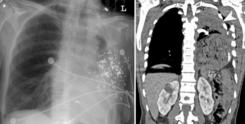

Fig. 24.2
(a) Hemothorax after gunshot injury. Frontal radiograph shows multiple shotgun pellets within the left hemithorax and a large hemorrhagic left pleural fluid collection. (b) Hemothorax and diaphragmatic rupture. Coronal CT reformation demonstrates left hemothorax (arrowhead) with left rib fractures and left diaphragmatic hernia (curved arrow)
Injuries to the Lung Parenchyma
Pulmonary Contusion
Pulmonary contusion is seen in 17–70 % of trauma patients and represents the most common injury to the lung parenchyma [8]. Contusions result from the direct transmission of energy through adjacent solid structures and thus are usually found along the ribs, sternum, and vertebral bodies. They may be unilateral or bilateral, focal, multifocal, or diffuse depending on the mechanism of injury. Pathologically, the opacification of the lung is caused by hemorrhage and edema filling the alveoli which originates from damage and resultant leak from the alveolar-capillary membrane [9]. Radiographically, this is seen as peripheral focal ground-glass opacities or patchy consolidative opacities that are distributed along a bronchopulmonary segmental anatomy (Fig. 24.3a) [6].
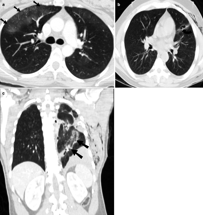

Fig. 24.3
(a) Pulmonary contusion. Axial CT shows non-segmental distribution of ground-glass opacification (arrows) in the right upper lobe, indicative of contusion. (b) Pneumatocele. Axial CT shows a pneumatocele (curved arrow) with adjacent hemorrhage in this patient who suffered a knife stab injury to the left anterior chest wall. The cavity is formed due to retraction of the edges of the lacerated lung. (c) Lung laceration. Coronal CT shows the linear pulmonary laceration (arrows) following the bullet path through the left lung as a central lucent area with adjacent hemorrhage. Subcutaneous emphysema is also present
Plain radiograph tends to underestimate the size of pulmonary contusion and lag behind the clinical presentation by up to 6 h. The appearance of pulmonary opacities greater than 24 h after initial injury should suggest other possible diagnoses like pneumonia or fat embolism [7]. Resolution of contusions is within 24–48 h and is complete between 3 and 10 days.
Pulmonary Laceration
Pulmonary lacerations are less common than contusions and result from disruption of the lung parenchyma, most commonly from penetrating trauma. The result is formation of a cavity within the lung as the surrounding parenchyma pulls back from the laceration itself (Fig. 24.3b, c). Because of this, they are more common in younger patients who have more optimal lung elasticity [10].
The formed cavity from the laceration may contain air, blood, or a combination of the two and the terms pneumatocele, hematocele, and hematopneumocele correlate to the contents of the cavity.
CT is more sensitive than radiograph in the evaluation of pulmonary laceration. Pulmonary lacerations are usually more difficult to diagnose on plain radiographs, often due to the presence of surrounding contusion. They become more apparent as the densities from contusion clears up. The pulmonary laceration classification by Wagner et al. is based on the mode of injury and the imaging indication (Table 24.1).
Table 24.1
Classification of pulmonary laceration
Type 1 laceration | They are the most common laceration and are seen within the deep lung parenchyma. They tend to be large and result from direct compressive force to the lung. |
Type 2 laceration | They occur in paraspinal region and are caused by a sudden blow to the lower thorax, resulting in shift of the lower lobes across the spine. |
Type 3 laceratio | They occur when the lung is torn by penetrating ribs and are thus usually seen in the lung periphery. |
Type 4 laceration | It occurs when the lung tears away from an adhesion. |
Traumatic Lung Herniation
A segment of the peripheral lung rarely can herniate outside the confines of the thoracic cavity either as the result of blunt or penetrating thoracic trauma. The anterior thorax is often the site of herniation because of a general lack of support (Fig. 24.4). Surgical reduction of intercostal lung hernia is recommended because of the potential for incarceration or strangulation. This potential risk is much greater in mechanically ventilated patients.
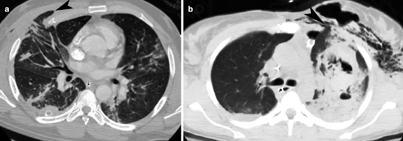

Fig. 24.4
(a) Lung herniation. Axial CT shows anterior herniation (arrowhead) of a portion of the right upper lobe, between consecutive ribs. An area of ground-glass density likely representing pulmonary contusion can be seen in the posterior left upper lobe. A chest tube is noted in the posterior right hemithorax. (b) Lung herniation. Axial CT shows herniation (arrowhead) of the medial portion of the left upper lobe into the adjacent anterior chest wall. Subcutaneous emphysema, pulmonary hemorrhage, and a pneumatocele are also present on the right side. There is a small left anterior pneumothorax present
Injuries to the Airways
Injury to the tracheobronchial tree is rarely seen in clinical practice, because up to 78 % of the patients die before arriving at the hospital [10]. The majority of these cases occur during deceleration when the airways may be compressed against the sternum or spine or when elevated thoracic pressure from compression occurs in the setting of a closed glottis. Anatomically, the thoracic trachea is more commonly affected than the cervical trachea and the right main stem bronchus is more commonly injured than the left. CT is an important tool for early diagnosis as up to 68 % of injuries are not accurately diagnosed [11].
Clinically, these patients may present with respiratory distress, cough, and hemoptysis among other symptoms. These symptoms are more common in the setting of bronchial injuries as opposed to tracheal injuries.
Stay updated, free articles. Join our Telegram channel

Full access? Get Clinical Tree


