Fig. 7.1
A 19-year-old Division I female basketball player with acute tear of the anterior cruciate ligament. Sagittal (a) proton density-weighted and (b) STIR MRI through the affected knee demonstrate disruption of ACL fibers (arrow). The remaining ACL fibers are amorphous and wavy with areas of discontinuity. Other sequences demonstrated the associated bone contusions in the lateral femoral condyle and lateral tibial plateau (With permission from Newman and Newberg (2010))
A number of theories have been posited to explain the higher incidence of sports-related ACL tears among women as compared with men. These include decreased intercondylar notch width in women as compared with men, a predisposition to knee abduction on landing in female athletes, and an elevated risk in the preovulatory phase of the menstrual cycle (Renstrom et al. 2008). Many ACL injuries in female basketball players occur during the sidestep cutting maneuver, particularly during the stop phase. This results in an increase in landing knee valgus angle, possibly predisposing to ACL injury (Xie et al. 2013; Munro et al. 2012). Other investigations do not support that width of the intercondylar notch is a predisposing factor in ACL injury (Lombardo et al. 2005).
Given the prevalence of ACL injury in basketball, many players will undergo ACL reconstruction, subsequent rehabilitation, and attempt to return to competitive play. In a 9-year review of athletes entering the Women’s National Basketball Association (WNBA) combine, 14.4 % reported previous ACL reconstruction (McCarthy et al. 2013). Among male and female professional basketball players who undergo ACL reconstruction, the majority return to competitive play (Busfield et al. 2009; Namdari et al. 2011).
Basketball-related injuries to the posterior cruciate and medial collateral ligament (Fig. 7.2) as well as multi-ligament injuries are much less common than ACL injuries. Isolated PCL tears may be treated nonoperatively (Moyer and Marchetto 1993) (Fig. 7.3). In a recently published investigation of concomitant injuries identified in athletes undergoing ACL reconstruction, basketball players were considerably more likely to have concomitant meniscal and cartilage injuries than multi-ligament injuries (Granan et al. 2013).
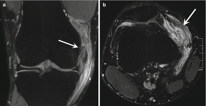
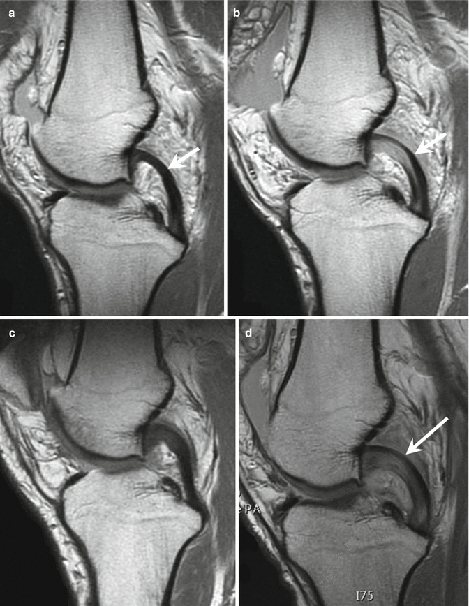

Fig. 7.2
A 25-year-old professional basketball player with medial collateral ligament tear and medial patellofemoral ligament tear with retinacular injury. (a) Coronal fat-suppressed proton density-weighted MRI demonstrates proximal medial collateral ligament tear (arrow). (b) Axial fat-suppressed T2-weighted MRI demonstrates medial patellofemoral ligament tear and medial retinacular injury (arrow)

Fig. 7.3
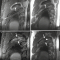
A 32-year-old professional basketball player with recurrent posterior cruciate ligament strain. (a) Sagittal proton density-weighted MRI in February 2009 shows normal posterior cruciate ligament (PCL) (arrow). (b) Sagittal proton density-weighted MRI in March 2009 after injury. Thickened PCL with increased signal intensity within the ligament consistent with strain related to a hyperextension injury (arrow). (c) In September 2009, the PCL strain is healing with some residual proximal thickening and slight increase in signal intensity. (d) In December 2009, the PCL strain has recurred (arrow) (With permission from Newman and Newberg (2010))
7.2.1.2 Menisci and Articular Cartilage
While meniscal injuries are associated with a variety of athletic activities, they are less frequently encountered in basketball players than are other knee injuries, including ACL tears or patellar tendinopathy. Despite this, meniscal injuries are not uncommon in basketball players. Nearly 10 % of elite female basketball players at the WNBA combine reported a history of prior meniscal surgery (McCarthy et al. 2013). In contrast, other investigators have reported no meniscal tears in a study where knee MRI was performed on asymptomatic collegiate basketball players (Major and Helms 2002). Many of these athletes manifest abnormal signal intensity in the menisci, without surfacing tears, chiefly on the medial side (Kaplan et al. 2005).
Meniscal tears may accompany other knee injuries including ligament tears. Isolated meniscal tears are reported far less commonly (Fig. 7.4). In a review of NBA athletes over 21 seasons, only 129 isolated meniscal tears were reported with a modest predilection for the lateral meniscus over the medial meniscus. Of those, 19.4 % did not return to play. A body mass index greater than 25 was associated with an increased risk of meniscal tears (Yeh et al. 2012).
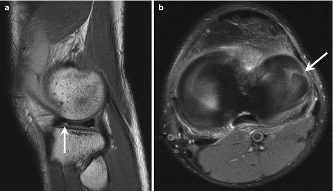

Fig. 7.4
A 24-year-old professional basketball player with lateral meniscal tear. (a) Sagittal and (b) axial fat-suppressed proton density-weighted MRI demonstrates a parrot beak-type tear of the lateral meniscus (arrow). The tear can be well seen on the axial sequence, extending to the periphery (arrow)
Injuries to articular cartilage may be seen in basketball players, especially at the patellofemoral joint. A subset of cartilage injuries are likely asymptomatic (Major and Helms 2002; Kaplan et al. 2005). The predilection for patellofemoral injuries in basketball, particularly to the extensor mechanism will be detailed below. On MRI, manifestations of chondromalacia may range from altered cartilage signal to fissuring, fibrillation, delamination, and full-thickness cartilage defects (Newman and Newberg 2010). In addition to careful evaluation of the patellar cartilage, the femoral sulcus cartilage should be examined in both sagittal and axial planes and the femorotibial cartilage in both sagittal and coronal planes. In a study of asymptomatic basketball players signed by the NBA, patellar cartilage lesions were identified in 35 %, femoral trochlear lesions in 25 %, medial femoral condyle in 10 %, and lateral tibial plateau in 5 % of cases (Kaplan et al. 2005). In all cases of cartilage injury, particularly when discrete cartilage defects are identified, careful inspection of the entire knee joint is recommended in order to detect and localize displaced cartilaginous bodies (Newman and Newberg 2010).
7.2.1.3 Extensor Mechanism
Overuse injuries at the knee in basketball players predominate at the extensor apparatus. While the entity “jumper’s knee” typically refers to proximal patellar tendinopathy, some investigators apply the term to reflect pathology occurring from the quadriceps tendon to the tibial tubercle (Newman and Newberg 2010; Molnar and Fox 1993) (Fig. 7.5). In fact, anterior knee pain, patellofemoral dysfunction, and extensor tendon abnormalities are common in basketball players across all age groups (Drakos et al. 2010; Barber Foss et al. 2012).
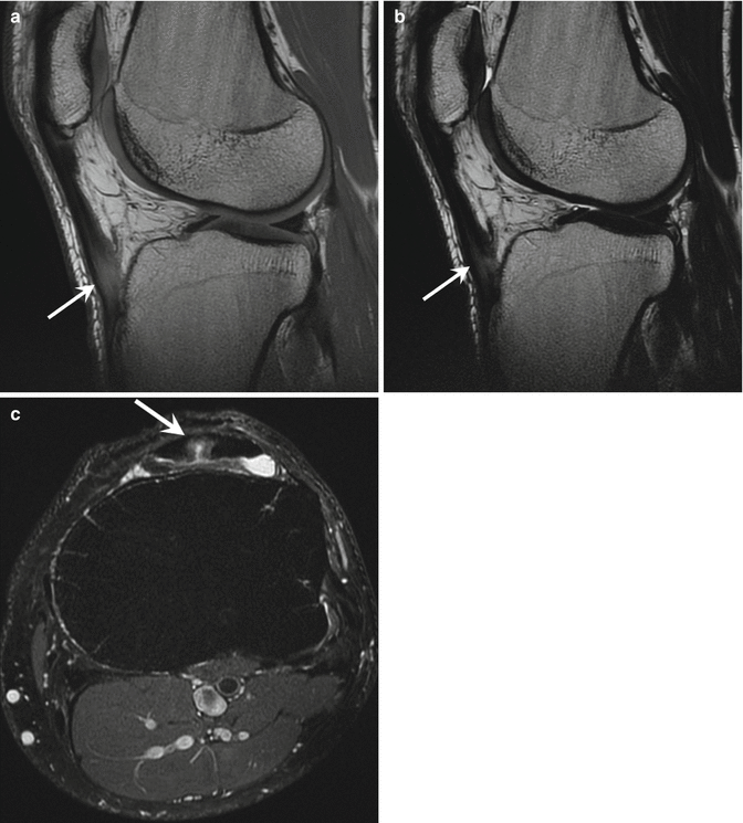

Fig. 7.5
Distal patellar tendinopathy. Exacerbation of distal patellar tendinopathy in professional center play-off competition. (a) Sagittal proton density-, (b) T2-, and (c) axial fat-suppressed proton density-weighted MRI demonstrates prominent distal patellar tendinopathy (arrows) with partial tear (c)
Extensor mechanism injuries predominate in sports requiring high demand for the leg extensors. In basketball, sudden decelerations and jumping result in eccentric muscle contraction, predisposing to tendon tear, particularly in the proximal patellar tendon. Excessive force may be generated across the patellofemoral articulation, exacerbated in individuals with underlying joint malalignment. In rare cases, extensive, repetitive stresses can result in rupture of the extensor tendons or even patellar stress fractures (Molnar and Fox 1993; Lian et al. 2005).
On MRI, patellar tendinopathy manifests as focal thickening of the proximal patellar tendon. Instead of a homogeneous, low signal intensity, intratendon signal intensity is typically elevated (Fig. 7.6). Tendinopathy may have a similar appearance in the distal quadriceps with thickening and elevated, globular appearing signal, instead of the linear, longitudinally oriented striations of higher signal that are present in the normal tendon (O’Keefe et al. 2009; Sonin et al. 1995; Dupuis et al. 2009).
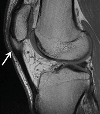

Fig. 7.6
A 23-year-old professional basketball player with patellar tendinopathy (jumper’s knee). Sagittal proton density-weighted MRI demonstrates tendinopathy and partial tear (arrow) of the proximal patellar tendon
Sonography is also well suited for evaluation of the extensor mechanism at the knee. In addition to changes in tendon thickness, tendinopathy manifests as abnormal tendon hypoechogenicity (Fig. 7.7). Ultrasound exhibits high sensitivities for the detection of patellar tendinopathy (Warden et al. 2007). It must be emphasized, however, that findings of patellar tendinopathy on sonography may be detected in a substantial portion of basketball players who are asymptomatic (Khan et al. 1997; Cook et al. 2000). In a study of Australian elite athletes participating in several sports, including basketball, hypoechoic areas on ultrasound were present in 14 % of patellar tendons of participants with no history of anterior knee pain, but were present in only 4 % of nonathlete controls (Cook et al. 1998). The use of power Doppler ultrasound has also been investigated in jumper’s knee, though the finding of elevated flow does not always correlate with the presence of symptoms (Tersley et al. 2001).
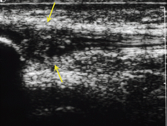

Fig. 7.7
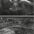
A professional basketball player with patellar tendinopathy (jumper’s knee). Sagittal ultrasound image of the central proximal patellar tendon demonstrates abnormal tendon hypoechogenicity (arrows) consistent with tendinopathy
Patellar dislocation is associated with a number of athletic activities, including basketball. Peak incidence is in the late teens. Approximately half of all reported dislocations in the USA occurred during athletic activity. By a significant margin, basketball accounted for a higher percentage of dislocations than any other sport (Waterman et al. 2012).
7.2.2 Ankle and Foot
7.2.2.1 Ankle Sprains
The ankle is the most commonly injured joint in basketball with ankle sprains the most common acute injury (Drakos et al. 2010; Dick et al. 2007; Starkey 2000). In the NBA, ankle injuries accounted for 14.7 % of all injuries reported and 17.9 % of game-related injuries. Lateral ankle sprain, in particular, accounted for 17.0 % of game-related injuries (Drakos et al. 2010). Ankle injuries sustained in basketball in the pediatric population who presented to hospital emergency departments exceeded 70,500 per year during the period from 2000 to 2006 (Pappas et al. 2011). In a 2-year prospective study of Greek female professional basketball players, there were 1.12 sprains per 1000 exposure hours. Most injuries occurred within the three-point line (Kofotolis and Kellis 2007). The very high incidence of ankle sprains in basketball must be considered in light of the overall incidence across all activities. Between 2002 and 2006, in excess of 3,000,000 ankle sprains presented to emergency departments in the USA, nearly half occurred with athletic activity. Of these, 41.1 % occurred with basketball, far exceeding all other athletic-related sprains (Waterman et al. 2010).
As noted above, lateral ankle sprains account for the vast majority of ankle sprains sustained in basketball (Drakos et al. 2010; Sickles and Lombardo 1993) (Fig. 7.8). Lateral ankle sprains typically occur in the setting of excessive inversion, often accompanied by a varying degree of plantar flexion. In basketball, these conditions may occur when a player steps on the foot of another player or when a player lands, cuts, turns, or pushes off awkwardly. Injuries to the anterior talofibular ligament predominate, followed by those involving the calcaneofibular ligament (Sickles and Lombardo 1993; Sonzogni and Gross 1993; Morrison and Kaminski 2007).
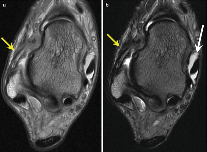

Fig. 7.8
A 24-year-old professional basketball player with anterior talofibular ligament (ATFL) injury following acute ankle sprain. Axial (a) proton density- and (b) T2-weighted MRI demonstrate ATFL tear with distal rupture. The ligament is thick, and distal fibers are indistinct (yellow arrows). Posterior tibialis tenosynovial fluid (white arrow) is noted incidentally (With permission from Newman and Newberg (2010))
Eversion injuries, resulting in medial or deltoid ligament sprain, are encountered far less frequently in basketball than are lateral injuries. High ankle sprains with syndesmotic injury are also less common injuries than lateral sprains, both within the general population and among basketball players. In a study of military cadets, syndesmotic sprain accounted for 6.7 % of ankle sprains and medial sprains 5.1 %. Among specific athletic activities, basketball was only the fourth most common cause of syndesmotic sprains. Syndesmotic injuries typically result from external rotation and hyperdorsiflexion. The biomechanics of high ankle sprain may result in a spectrum of findings, including injuries to the tibiofibular ligaments, syndesmotic membrane, and, in extreme cases, fibular or even medial malleolar fractures (Drakos et al. 2010; Sickles and Lombardo 1993; Sonzogni and Gross 1993; Norkus and Floyd 2001).
Many ankle sprains in basketball players are not imaged and are managed conservatively. Some injuries warrant ankle radiographs to exclude fracture or joint malalignment (Johnson and Teasdell 1993). Additional osseous injuries may be detected, including fifth metatarsal fractures (Fernandez Fairen et al. 1999). While most ankle sprains do not require evaluation with cross-sectional imaging, MRI may be employed in select cases. These include serious sprains with recalcitrant symptoms. In addition to the evaluation of ligament integrity (Perrich et al. 2009), MRI of the ankle may aid in the detection of chondral or osteochondral injuries of the talar dome (Figs. 7.9 and 7.10) (O’Loughlin et al. 2010), osseous contusions (3), anterolateral process calcaneal fractures, or peroneal tendon subluxation (McDermott 1993).
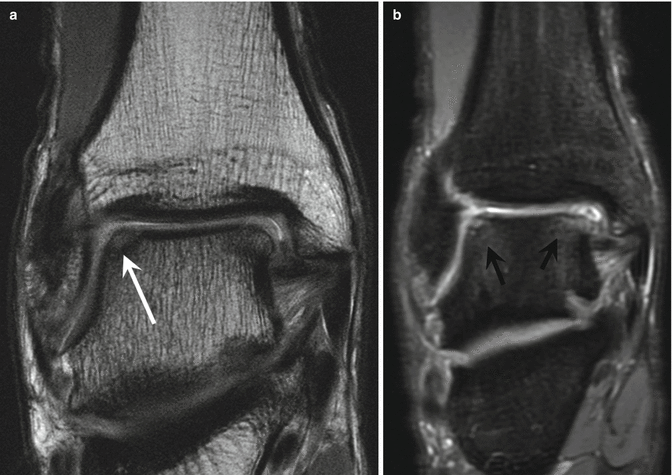
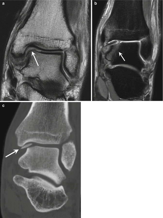

Fig. 7.9
A 32-year-old professional basketball player with osteochondral talar dome injury. Coronal (a) high-resolution and (b) fat-suppressed proton density-weighted MRI show cartilage defects (white arrows) with mild underlying bone marrow edema in both the medial and lateral talar dome (black arrows) (With permission from Newman and Newberg (2010))

Fig. 7.10
A 22-year-old professional basketball player with talar dome chondro-osseous shear injury. (a) Coronal proton density-weighed high-resolution MRI demonstrates a large full-thickness cartilage defect at the medial talar dome (arrow). (b) Coronal fat-suppressed proton density-weighted MRI demonstrates the displaced chondro-osseous fragment and underlying osseous edema (arrow). (c) Coronal reformatted CT image demonstrates the displaced chondro-osseous fragment
7.2.2.2 Fractures at the Ankle and Foot
Basketball players are at risk for both acute osseous injuries and overuse injuries in the lower appendicular skeleton. Avulsion fractures of the base of the fifth metatarsal and navicular avulsions may present with symptoms of ankle sprain. Avulsion fractures at the base of the fifth metatarsal may occur when a rebounding player lands on the foot of another player. This results in an inversion injury to the rebounding player, contraction of the peroneus brevis muscle, and avulsion of the peroneus brevis attachment on the fifth ray metatarsal (McDermott 1993).
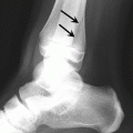
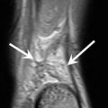
Fractures of the anterolateral process of the calcaneus may also present with symptoms of lateral ankle sprain. These fractures occur with foot inversion/abduction and plantar flexion resulting in tension on the bifurcate ligament or as a compressive injury due to forced dorsiflexion (McDermott 1993; Judd and Kim 2002). Some anterolateral process calcaneal fractures may be difficult to visualize on lateral foot radiographs, but can be readily detected on MRI (Fig. 7.11). As these fractures may be slow to heal (Berkowitz and Kim 2005), serial imaging may be necessary. Computed tomography (CT) with multiplanar imaging reformation is recommended to assess for fracture healing (Newman and Newberg 2010).
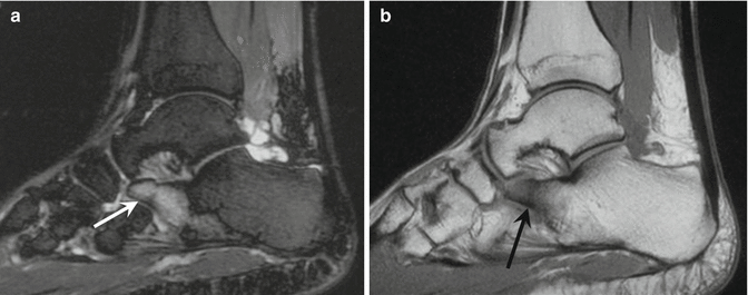

Fig. 7.11
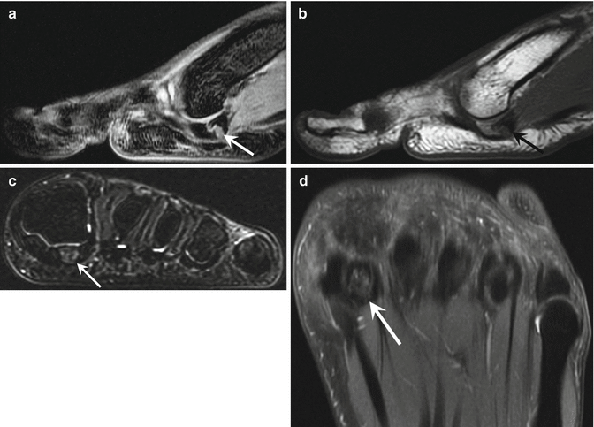
A19-year-old Division I female college basketball player with anterior lateral calcaneal process fracture. (a) Sagittal STIR MRI of the right ankle demonstrates intense osseous edema of the anterior lateral process of the calcaneus (arrow). (b) Sagittal T1-weighted MRI demonstrates low signal intensity of the anterior lateral process with a fracture line seen as a low signal intensity line (arrow) (With permission from Newman and Newberg (2010))
Stress fractures in basketball players occurring below the knee may involve the tibia or distal fibula or may occur in the navicular, calcaneus, metatarsals, or cuneiforms (Meyer et al. 1993). MRI has supplanted bone scintigraphy for the early diagnosis of stress injuries by virtue of its superior contrast resolution, resulting in its ability to detect bone marrow and soft tissue changes early in the injury course. It must be emphasized that the appearance of osseous stress injuries on imaging studies varies by bone structure and composition. Fractures in trabecular bone, such as the calcaneus, demonstrate no extraosseous callus with healing. In contrast, metatarsal stress fractures typically heal with exuberant callus (Newman and Newberg 2010).
In the earliest phases of stress injury, subtle bone marrow edema may be evident on MRI; with progression, a low signal intensity fracture line will be observed (Figs. 7.12 and 7.13). In fractures involving metatarsals or the tibia, periosseous soft tissue edema may accompany intramedullary and/or cortical signal changes (Fig. 7.14). These soft tissue changes may be absent in trabecular bone stress injuries.
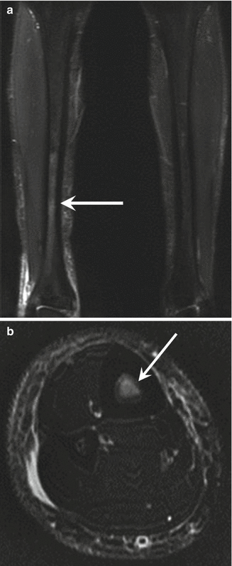
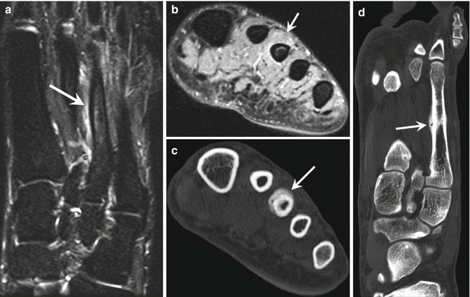
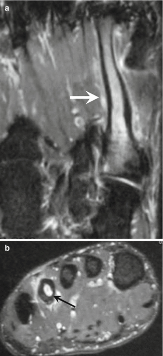

Fig. 7.12
A 21-year-old basketball player with stress reaction of the tibia. (a) Coronal STIR and (b) axial fat-suppressed T2-weighted MRI demonstrate osseous edema in the medullary cavity of the right tibia (arrows) in keeping with osseous stress injury (With permission from Newman and Newberg (2010))

Fig. 7.13
A 30-year-old professional basketball player with stress fracture of the third metatarsal. (a) Axial and (b) coronal STIR MRI of the forefoot demonstrate minimal periosteal high signal intensity along the shaft of the third metatarsal (arrow). A fracture was not demonstrated at this time. (c) Axial and (d) coronal reformatted CT images 6 weeks later demonstrate a healing stress fracture with periosteal new bone formation (arrows) (With permission from Newman and Newberg (2010))

Fig. 7.14
A 26-year-old professional basketball player with stress fracture of the fourth metatarsal and a history of Jones fracture with surgical fixation. (a) Axial and (b) coronal STIR MRI demonstrate fourth metatarsal osseous edema and periosteal and periosseous soft tissue edema (arrows) (With permission from Newman and Newberg (2010))
The ability of MRI to detect early stress injuries may allow for intervention in the competitive athlete, preventing progression to an actual fracture. This is particularly true of metatarsal injuries. In fact, one investigator has demonstrated the benefit of preseason MRI examinations of the foot in preventing metatarsal stress fractures in National Collegiate Athletic Association (NCAA) male basketball players (Major 2006).
Jones fractures occur at the junction of the metaphysis and diaphysis of the fifth ray metatarsal (Fig. 7.15) and can be a problematic foot injury in basketball players. Symptoms may predate radiographic changes for a significant period, precluding early diagnosis (Meyer et al. 1993). These injuries may represent acute fractures or stress injuries. Moreover, Jones fractures may be slow to heal, often requiring operative fixation, particularly in this athletic population (Fernandez et al. 1999). While MRI may facilitate early diagnosis, evaluation of healing requires radiographic or CT follow-up; both techniques allow detection of healing even in the presence of metallic hardware.
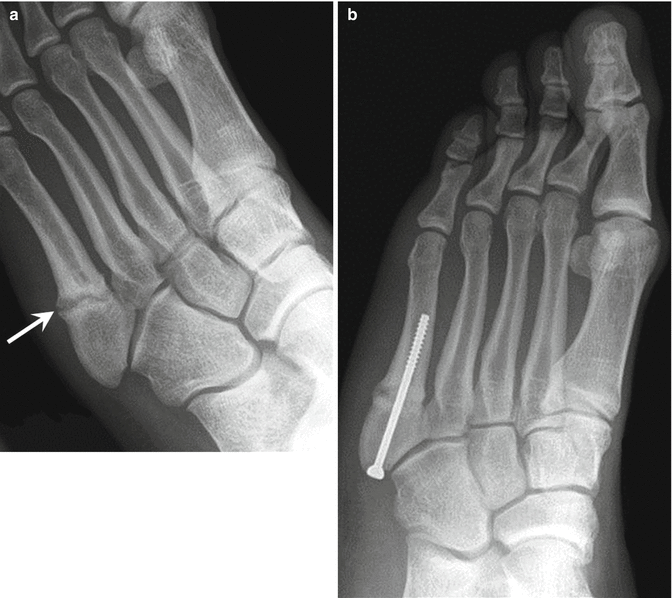

Fig. 7.15
A 28-year-old professional basketball player with Jones fracture. (a) Internal oblique radiograph of the left foot demonstrates a fracture of the proximal fifth metatarsal shaft (arrow). The fracture line is well seen with peripheral callus and sclerosis along the fracture margins. (b) Postoperative anteroposterior radiograph demonstrating screw placement within the fifth metatarsal (With permission from Newman and Newberg (2010))
Navicular stress fractures represent another problematic injury which may be seen in basketball players. These fractures are very difficult to diagnose on radiographs as they are oriented in the sagittal plane and manifest no significant callus. These fractures typically propagate from proximal to distal and dorsal to plantar aspects of the navicular bone (Kiss et al. 1993) (Fig. 7.16). While both CT and MRI are useful in diagnosing this injury, CT is required to assess fracture healing (Newman and Newberg 2010) (Fig. 7.17).
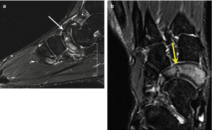
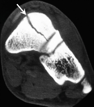

Fig. 7.16
A 19-year-old Division I female basketball player with tarsal navicular stress fracture. (a) Sagittal fat-suppressed T2-weighted MRI demonstrates hyperintensity in the navicular bone (arrow). (b) Axial (long axis) STIR MRI demonstrates a fracture line in the middle third of the tarsal navicular (yellow arrow) consistent with a stress fracture (With permission from Newman and Newberg (2010))

Fig. 7.17
A 24-year-old professional basketball player with tarsal navicular stress fracture. Coronal reformatted CT image demonstrates the oblique fracture (arrow) through the navicular. The fracture healed with nonoperative treatment (With permission from Newman and Newberg (2010))
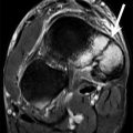
The sesamoids of the first ray or hallux are prone to injury due to the significant shear and loading forces placed on these small structures during ambulation. It is estimated that the sesamoid complex transmits 50 % of the body weight, increasing to greater than 300 % during push off (Boike et al. 2011; Richardson 1999). In basketball, running as well as jumping would be expected to further increase the risk of sesamoid injury. Sesamoid injury may be acute, resulting in fracture or contusion or chronic (sesamoiditis). On imaging, fracture may be difficult to distinguish from a bipartite sesamoid, a common normal variant. In our experience, superimposed acute or repetitive injuries on a bipartite sesamoid are commonly observed (Fig. 7.18).

