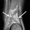
Fig. 4.1
A 25-year-old man with clinical concern for a rotator cuff tear and bicipital tendinosis. Oblique coronal inversion-recovery (a) and intermediate-weighted (b) MRI of the shoulder demonstrate a subperiosteal osteoid osteoma (arrows) within the proximal humerus, eliciting an intense, surrounding bone marrow edema pattern. Coronal CT-reformatted image (c) demonstrates the nidus of the osteoid osteoma (arrow), with faint central sclerosis, successfully treated with radiofrequency ablation under CT guidance (d)
4.2.3 Contrast Use
MR arthrography, with the injection of intra-articular gadolinium, is used by many institutions to better evaluate the dynamic and static stabilizers of joints, most commonly the shoulder and hip. MR arthrography of the shoulder is frequently advocated to differentiate tendinosis from partial or full-thickness rotator cuff tears (Stetson et al. 2005) as well as labral pathology of the shoulder (Major et al. 2011) and hip (Sutter et al. 2014). It has been our experience that noncontrast MR techniques, with optimized spatial resolution and fluid contrast, can reliably and accurately demonstrate both partial thickness rotator cuff tears and labral tears of the shoulder (Connell et al. 1999; Gusmer et al. 1996) and hip (Mintz et al. 2005). This eliminates the potential, albeit low, risk of an adverse reaction to gadolinium and added cost while maintaining the throughput of each magnet (Hash and Potter 2012).
Perfusion imaging, on the other hand, requires intravenous administration of gadolinium. Dynamic contrast-enhanced MRI techniques can be used to confirm the suspicion of avascular necrosis of the scaphoid in the setting of nonunion (Ng et al. 2013) or of the femoral head following traumatic posterior hip subluxation or dislocation.
4.2.4 Imaging Planes
Depending on the type of injury in question and body part, specific modifications to routine protocols are made at the radiologist’s discretion. For example, when the clinical question is the presence and/or stability of an osteochondral lesion or chondral shear injury of the talar dome, a straight coronal, cartilage-sensitive sequence is performed in addition to the routine oblique coronal sequence (Fig. 4.2).
Fig. 4.2
A 37-year-old man with an inversion injury to the ankle. Coronal intermediate-weighted MRI of the ankle demonstrate a full-thickness chondral shear from the lateral talar dome (arrow, a), with the displaced fragment, complete with the tidemark as denoted by the hypointense line (arrows, b), in the anterior aspect of the lateral gutter. Coronal CT-reformatted image (c) confirms the presence of subchondral bone attached to the chondral defect (circle)
When evaluating injuries to the chest and abdominal wall and underlying structures, e.g., the external oblique, pectoralis major, or latissimus dorsi, imaging planes orthogonal to the anatomic structure of clinical concern are obtained (Fig. 4.3). Orthogonal planes are also used when structures are in nonanatomic alignment; e.g., when imaging a patient with fixed flexion following an elbow dislocation, 2 FSE moderate TE axial sequences, 1 perpendicular to the axis of the humerus, and 1 perpendicular to the axis of the ulna are prescribed. In addition, the coronal sequence should be parallel to the axis of the ulna to evaluate the collateral ligaments (Fig. 4.4).
Fig. 4.3
A 42-year-old male skier with thoracic injury. Coronal inversion-recovery (a) and intermediate-weighted (b) MRI oblique to the axial plane of the anterior chest wall (scout view, lower right-hand corner of images) demonstrate interstitial edema within the sternal head of the pectoralis major (arrows, a), with perimuscular edema, related to distal avulsion of the tendon, which is retracted to the deltopectoral groove (arrow, b)
Fig. 4.4
A 34-year-old woman status post elbow dislocation. Sagittal inversion-recovery MRI of the elbow (a) demonstrates flexion deformity at the elbow and marked posterior subcutaneous edema. Axial intermediate-weighted MRI (b) is prescribed from the sagittal images perpendicular to the distal humerus (b) and proximal ulna (d), with enlarged, corresponding scout views (c, e). Coronal intermediate-weighted MRI prescribed parallel to the long axis of the proximal ulna (f) demonstrates an intra-articular radial head fracture (arrow), also seen in d (arrow), as well as complete disruption of the lateral ulnar collateral ligament (f, arrowhead) and medial collateral ligament (f, black arrows) from their humeral origin
4.2.5 Field of View and Coil Type
MRI provides the necessary high spatial resolution and soft tissue contrast to evaluate small joints with complex anatomy, such as the wrist. Morphological changes seen on MRI correlating to a patient’s clinical presentation can indicate injuries to the interosseous intercarpal ligaments and triangular fibrocartilage complex.
Another advantage of MRI lies in its ability to provide a large field of view of the area of concern. As such, clinically unsuspected entities based on patient history and physical examination, which are subjective and sometimes nonspecific, can be diagnosed (Fig. 4.5). For example, when imaging the hip, it is important to routinely obtain large field-of-view coronal STIR and axial intermediate-weighted FSE sequences, which afford visualization of the entire osseous pelvis, including the sacrum, as well as intrapelvic structures, such as the ovaries and inguinal region, pathologic conditions of which may refer pain to the hip and/or groin area.
Fig. 4.5
A 38-year-old female dancer with hip injury. Sagittal intermediate-weighted MRI (a) of the hip demonstrates a fluid cleft in the anterior labrum confirming the presence of clinically suspected tear (arrow). Coronal inversion-recovery MRI of the pelvis (b) and coronal intermediate-weighted MRI of the left hip (c) demonstrate erosive changes of the left sacroiliac joint (arrows), in this patient with circumferential rectal wall thickening (arrowheads, b, d) and unsuspected inflammatory bowel disease
4.2.6 Cartilage Imaging
Due to its inherent high soft tissue contrast capabilities, MRI is particularly sensitive for evaluating cartilage injury and wear. For example, MRI can detect surface and basilar delamination in the setting of femoroacetabular impingement, not infrequently diagnosed in the elite athlete with hip and groin pain (Gold et al. 2012). In the setting of an acute injury, it can identify whether a chondral shear has occurred and, if so, the integrity and location of the displaced chondral fragment. MRI aids in preoperative planning for cartilage repair, in which lesion size and 3D joint geometry are important determinants of the type of repair to be performed and of the potential osteochondral donor site, particularly in the case of autologous osteochondral transplantation. MRI also functions as a noninvasive, objective means of long-term monitoring following cartilage repair surgery by assessing percent fill, morphology and signal intensity of the repair tissue, adverse synovial reaction, and the integrity of the underlying subchondral bone (Hayter and Potter 2011). Quantitative MRI techniques, such as T2 mapping to assess collagen orientation (Xia et al. 2001), and delayed gadolinium-enhanced MRI of cartilage (dGEMRIC) (Williams et al. 2004) and T1rho (Wheaton et al. 2005), to assess proteoglycan content depletion, provide more objective measurements of repair cartilage ultrastructure and integrity.
4.2.7 Physeal Imaging
In adolescent athletes, a 3D, fat-suppressed T1-weighted gradient recalled echo physeal-sensitive sequence is obtained to evaluate the degree of skeletal maturity. This is particularly helpful in the setting of a complete anterior cruciate ligament (ACL) tear. Orthopedic surgeons will often perform a physeal-sparing ACL reconstruction to prevent growth arrest and use either allograft or the iliotibial band, rather than autologous hamstring or patellar tendon, to restore anterior-posterior stability and rotational control in prepubescent adolescents (Fabricant et al. 2013) (Fig. 4.6).
Fig. 4.6
A 14-year-old boy with knee injury. Coronal (a) and sagittal (b) intermediate-weighted MRI demonstrates an all-epiphyseal anterior cruciate ligament reconstruction with hamstring autograft (arrows). Sagittal physeal-sensitive MRI (c) demonstrates open distal femoral and proximal tibial physes
A physeal-sensitive sequence is also valuable when evaluating little league shoulder, i.e., a chronic type I Salter-Harris fracture of the proximal humeral physis most commonly seen in adolescent baseball throwers (Fig. 4.7). MRI will demonstrate irregularity, sclerosis, and widening of the lateral physeal margin, preferentially affecting the metaphyseal side. Physeal-sensitive MR sequences, which are amenable to semiautomated segmentation and 3D modeling, can also be obtained to accurately quantify the volume of physeal interruption and illustrate its geometry following fracture involving the physis (Lurie et al. 2014) (Fig. 4.8).
Fig. 4.7
A 12-year-old little league baseball pitcher status post shoulder injury. Oblique coronal intermediate-weighted (a) and physeal-sensitive (b) and sagittal intermediate-weighted (c) MRI demonstrate a chronic stress reaction, manifest as sclerosis at the lateral metaphyseal margin of the proximal humerus physis (arrows)
Fig. 4.8
A 12-year-old boy status post removal of fixation hardware for a Salter-Harris Type III fracture of the distal tibia. Sagittal physeal-sensitive (a) and intermediate-weighted (b) MRI of the ankle demonstrate a physeal bar as a linear transphyseal connection between the epiphysis and metaphysis (arrows). A 3D physeal segmentation map superimposed on the source image (c) demonstrates the bar (blue) and the uninjured, open physis (red), with corresponding areas of both and percentage involvement by the bar (rectangle)
4.2.8 Nerve Imaging
While the focus of most sports imaging is on soft tissue structures, peripheral nerves are often involved, particularly with high-velocity injuries. In the setting of a high-velocity mechanism, such as a posterior knee dislocation resulting in a posterolateral corner injury, the common peroneal nerve is frequently affected, with palsy of the nerve seen in 25 % of all dislocation cases in 1 series (Niall Et al. 2005) (Fig. 4.9).
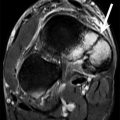
Fig. 4.9


A 20 year-old man after a recent posterior knee dislocation with severe posterolateral corner injury and common peroneal nerve injury. Coronal intermediate-weighted MRI of the knee (a) demonstrates complete tear of the fibular collateral ligament and biceps femoris with a degenerative stump near the fibular head (black arrowhead) as well as complete detachment of the popliteus tendon at the femur (a, white arrow). The axial image (b) shows the fascicles of the common peroneal nerve as diffusely thickened and hyperintense (white circle). Compare with the normal signal intensity and morphology of the tibial nerve (black circle)
Related posts:

Stay updated, free articles. Join our Telegram channel
Full access? Get Clinical Tree
