Grade
Pain
Imaging findings
Description
I
No
Imaging evidence of stress fracture, no fracture line
Asymptomatic stress fracture
II
Yes
Imaging evidence of stress fracture, no fracture line
Symptomatic stress fracture
III
Yes
Non-displaced fracture line
Non-displaced fracture
IV
Yes
Displaced fracture (≥2 mm)
Displaced fracture
V
Yes
Nonunion
Nonunion
Imaging modalities suitable for the evaluation of stress injuries are conventional radiography, bone scintigraphy, computed tomography, and magnetic resonance imaging.
Conventional radiography is only reliable in the later stages of stress injury, because it cannot depict early features of stress injury. In cortical bones, such as the metatarsals and the tibia, radiographs may eventually demonstrate periosteal reaction and endosteal thickening with or without a fracture line. In cancellous bones, stress fractures may be seen as a linear sclerotic and lucent area perpendicular to the trabeculae. The calcaneus is a common site of cancellous stress fractures (Berger et al. 2007; Dixon et al. 2011). Although radiographic examination is not recommended as a diagnostic method for stress fractures, it is performed to rule out other pathologies like tumor or infection.
Bone scintigraphy is highly sensitive in detecting stress fractures and is therefore an effective diagnostic technique. However, it lacks specificity as it reflects any condition associated with osteoblastic activity. Furthermore, the high radiation exposure is undesirable in pediatric patients.
CT is less sensitive than MRI or bone scintigraphy in the detection of early stress fractures (Spitz and Newberg 2002). However, it can be valuable in addition to MRI to confirm the diagnosis or if the MRI findings are inconclusive. CT often provides excellent imaging of bony structures. Additionally, CT offers more accurate diagnosis of stress injuries in specific anatomical sites such as the navicular bone (Fig. 20.1) (Mann and Pedowitz 2009) or longitudinal stress fractures of the tibia (Spitz and Newberg 2002). The added value of CT is the ability to detect a discrete lucent or sclerotic fracture line in the affected bone (Spitz and Newberg 2002).
Fig. 20.1
A 17-year-old female gymnast with stress fracture of the navicular bone. Axial fat-suppressed T2-weighted MRI (a) depicts high signal intensity of the navicular bone. Axial CT image (b) clearly delineates the fracture line (arrow)
Several studies have stated that ultrasound may be useful to detect early stress fractures in the metatarsals and the lower leg as it is an easy and rapid technique with low costs and good sensitivity and specificity. Ultrasound may show periosteal reactions or a cortical fracture line (Banal et al. 2009; Dixon et al. 2011). However, ultrasound is not able to rule out stress fractures. Since no large trials have been carried out, ultrasound is not the preferred method of choice to diagnose bone stress injuries.
The gold standard in the diagnostic evaluation and follow-up of stress fractures is MRI using STIR, T1-weighted and T2-weighted sequences (Berger et al. 2007). MRI findings vary in relation to the severity of the bone stress injury. Despite the development of a universal scoring system that is applicable for any imaging modality, an MRI-specific scoring system for stress fractures exists as well (Kaeding and Miller 2013; Berger et al. 2007). Early injury (grade 1) is only visible on STIR sequences, showing endosteal edema. Grade 2 injuries show periosteal and endosteal edema on STIR images and marrow edema on T2-weighted images. In grade 3 stress fractures, bone marrow edema is also visible on T1-weighted images. Grade 4 stress fractures are seen as a clear low signal fracture line on T1-weighted and T2-weighted images (Berger et al. 2007).
20.2.2 Ankle and Foot
20.2.2.1 Calcaneal Apophysitis
Calcaneal (traction) apophysitis or Sever disease is a common overuse injury of the calcaneal tuberosity in the immature skeleton. The calcaneal apophysis is the attachment site of the Achilles tendon. The apophyseal physis is unfused in children and adolescents and therefore forms a weak link to the bone. Repetitive and chronic forces placed on the apophysis by the tendon result in an accumulation of microscopic avulsions along the apophyseal physis with subsequent inflammation at the attachment site (Davis 2010a).
Calcaneal traction apophysitis is mostly seen in the 9–12 year age group (Chang et al. 2013; Hoang and Mortazavi 2012). Calcaneal apophysitis is usually a clinical diagnosis. Conventional radiography might be useful to rule out other causes of heel pain or to show fragmentation and sclerosis of the apophysis, which is indicative of Sever disease. However, neither conventional radiography nor MRI are helpful in the initial evaluation of suspected apophysitis of the calcaneus, as imaging abnormalities are also often observed in asymptomatic children. MRI is only valuable in the follow-up of this injury in patients not responding to conservative therapy, to exclude a calcaneal stress fracture (Chang et al. 2013).
20.2.2.2 Osteochondritis Dissecans of the Talus
Osteochondritis dissecans (OCD) of the talar dome, also referred to as osteochondral lesions of the talus, is a condition of the subchondral bone and overlying articular cartilage. OCD can result from a single trauma or repetitive microtrauma and is also associated with inversion injuries (Davis 2010a). These lesions usually occur at the medial or lateral aspects of the talar articular surface. The medial lesions typically affect the posterior third of the talar dome, causing a deep cup- or crater-like defect. Lateral lesions are more shallow and mostly occur at the middle third of the talar dome (Iyer and Thapa 2013).
Conventional radiographs may demonstrate a focal radiolucent area subjacent to the articular surface, which may contain a central fragment of bone (Chang et al. 2013). Although OCD of the talus can be diagnosed using conventional radiography, this is not helpful in the evaluation of stability of the lesion. Assessing the stability of the osteochondral fragment is essential as it influences management decisions.
MRI and CT are equally suitable for the detection or exclusion of OCD lesions (Verhagen et al. 2005). Both techniques have their own advantages. CT is valuable for preoperative planning, as it adequately depicts the true extent of the OCD lesion. Moreover this technique is widely available and has a short acquisition time. MRI has its value in assessing the integrity of the talar articular cartilage and to evaluate the stability of the osteochondral defect (Narvaez et al. 2003). The most reliable sign of an unstable OCD fragment is a high-signal line on T2-weighted images interposed between the talus and the fragment. Furthermore, defects to the cartilage greater than 5 mm, cysts underlying the lesion, and loose bodies can be detected (Iyer and Thapa 2013; Narvaez et al. 2003).
20.2.2.3 Ligament Sprain
The most common injury to the lower extremity among gymnastic athletes is the ankle sprain (Caine and Nassar 2005). Inversion trauma generally occurs during dismounts and landing from great heights, resulting in lateral ligamentous injury. The ligaments often tear in a predictable sequence, affecting first the anterior talofibular ligament, then the calcaneofibular ligament, and finally the posterior talofibular ligament (Narvaez et al. 2003).
The majority of ankle ligament injuries are diagnosed clinically and no further examination is indicated. When confirmation of the diagnosis is essential or when osseous lesions such as osteochondral fractures are suspected, MRI is considered the modality of choice to evaluate the ankle ligaments (Narvaez et al. 2003). MRI protocols for assessment of the ankle structures include axial and coronal T1-weighted, proton density-weighted, and T2-weighted images. Morphologic changes and increased intraligamentous signal intensity within and around the ligaments on T2-weighted images are indicative of ligament tears. Additionally, secondary signs of acute ligament injury, such as bone bruises or extravasation of joint fluid into the adjacent soft tissues, can be detected (Narvaez et al. 2003).
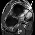
Injuries of the medial ligament complex are less common and are often secondary to an inversion injury of the ankle. Posteromedial impingement is a cause of residual medial ankle pain subsequent to an inversion injury. During an inversion injury, the deep fibers of the deltoid ligament may become compressed between the medial wall of the talus and the medial malleolus (Giannini et al. 2013). This lesion can resolve without any symptoms. However, hypertrophic and fibrotic scar tissue may persist and can impinge between the medial talus and the malleolus (Cerezal et al. 2003). MRI demonstrates the lesion and hypertrophic soft tissues (Fig. 20.2). Additionally it may show evidence of subchondral bone bruising of both the medial talus and medial malleolus (Cerezal et al. 2003).
Fig. 20.2
A 17-year-old female gymnast with posteromedial impingement. Axial CT image of the ankles (a) shows soft tissue swelling and obliteration of the fat of the left side (arrow) compared to the contralateral side. Axial proton density-weighted MRI of the ankles (b) shows normal fiber appearance of the right deltoid ligament (white arrow). The left deltoid ligament cannot be recognized and soft tissue edema is present posterior to the medial malleolus and around the flexor digitorum longus (black arrow)
20.2.3 Knee
20.2.3.1 Traction Apophysitis
Traction apophysitis of the knee or Osgood-Schlatter disease is an overuse injury resulting from repetitive microtrauma to the insertion of the patellar tendon on the tibial tuberosity.
The apophyseal physis on the tibia is exposed to traction forces from the attached muscle. Repetitive and chronic submaximal forces at the site of attachment results in microscopic avulsion injury to the apophysis that exceeds the body’s capacity for healing. This overuse injury is referred to as traction apophysitis of the knee or Osgood-Schlatter disease.
It is most often seen in children 10–15 years of age who participate in sports involving jumping, running, and squatting (Chang et al. 2013; Stevens et al. 1999).
Conventional radiographs may reveal thickening and irregularity of the patellar tendon. Furthermore, fragmentation or irregularity of the tibial tubercle ossification center and soft tissue swelling can be seen (Chang et al. 2013). Ultrasound can be used to support clinical findings of traction apophysitis of the tibial tuberosity. The patellar tendon is often thickened and intratendinous calcifications can be detected. MRI features of Osgood-Schlatter disease include soft tissue swelling anterior to the tibial tuberosity, loss of the sharp inferior angle of the infrapatellar fat pad and surrounding soft tissues, thickening and edema of the inferior patellar tendon, and infrapatellar bursitis (Stevens et al. 1999).
Sinding-Larsen-Johansson disease is another traction apophysitis affecting the inferior pole of the patella, the proximal attachment site of the patellar tendon. As in Osgood-Schlatter disease, this injury results from chronic traction forces of the patellar tendon. Conventional radiography or MRI of Sinding-Larsen-Johansson disease demonstrates identical findings to Osgood-Schlatter disease, although they are seen at the lower pole of the patella (Davis 2010a).
20.2.4 Pelvis/Hip and Upper Leg
20.2.4.1 Apophyseal Injuries
Apophyseal avulsion fractures of the pelvis in adolescent competitive athletes occur most frequently in soccer and gymnastics. The apophyseal physis is the weakest site of the bone, especially in the pelvis as it tends to fuse later than many other epiphyseal centers (Vandervliet et al. 2007). The ischial tuberosity, the insertion site of the hamstring muscle group, is the most common site of injury in gymnasts. Injuries to other apophyseal locations such as the anterior superior iliac spine and the anterior inferior iliac spine are less common (Rossi and Dragoni 2001). The underlying biomechanics of avulsion injuries of the pelvis can again be divided into chronic and acute. Repetitive forces placed by the tendon on the unfused apophysis may result in an overuse injury referred to as traction apophysitis (Davis 2010a). Acute injuries to the ischial tuberosity in gymnasts are associated with sudden and excessive lengthening of the hamstring muscles during floor exercises (Rossi and Dragoni 2001).
Conventional radiography is the modality of choice in the imaging workup of suspected apophyseal injury. Clinical history and radiographic appearance may provide a definitive diagnosis. Acute avulsion injuries appear as a sharply marginated displaced fragment adjacent to its origin. However, the displacement of fragments can be subtle and requires attention to detail (Davis 2010a).
Traction apophysitis can demonstrate both irregularities or smooth margins of the bone or prominent bone formation. Additionally, areas of lysis and sclerosis can be seen secondary to healing of the repetitive microtrauma. These findings may be confused with more aggressive processes like neoplasm or infection (Stevens et al. 1999). Therefore understanding the underlying biomechanics of pelvic apophyseal injuries is essential and can help differentiate between these conditions. Additionally, CT or MRI may be helpful in the diagnostic process.
CT is also valuable in the follow-up of avulsion injuries as it is often the best imaging modality for bony structures (Chang et al. 2013). MRI characteristics of traction apophysitis in adolescent athletes include increased signal intensity on fluid-sensitive sequences (Fig. 20.3) and mild widening of the physis, often with adjacent bone marrow edema (Hodnett et al. 2009). Furthermore, avulsion injuries may demonstrate irregularity and bony protuberance at the site of avulsion with visibly displaced bone fragments. Non-displaced avulsion injuries are seen as sharply marginated pieces of bone adjacent to their donor site (Hodnett et al. 2009).
Fig. 20.3
A 12-year-old female gymnast with traction apophysitis of the ischial bone. Anteroposterior radiography of the pelvis (a) shows an irregular aspect of the ischial apophysis. Additional imaging is necessary to rule out, for example, neoplasm in this aggressive lesion. Coronal fat-suppressed T2-weighted MRI of the pelvis (b) shows high signal intensity in the hamstring muscles (arrow) and in the iliac bone representing edema. Coronal T1-weighted MRI (c) shows a broadening of the apophyseal area (arrow)
20.3 Upper Extremity
The movements in gymnasts causing injuries to the upper extremity include repeated weight-bearing impact loads on the elbow and the wrist. As these joints are not adapted to bear these high-impact loads, performing gymnastics causes repetitive microtrauma to the upper extremity joints (Davis 2010b). With respect to the wrist, this includes axial loading to the dorsiflexed wrist or wrist loading varying from ulnar to radial deviation.
“The rings” is a discipline in gymnastics requiring a lot of extreme traction-force movements to the shoulder. As this discipline is only performed by male gymnasts, it is logical that shoulder injuries are the most frequent upper extremity injuries in male gymnasts, followed by the wrist. The most vulnerable sites of the upper extremity in female gymnasts are the wrist, followed by the elbow (Caine and Nassar 2005). The most typical injuries occurring in the upper extremities of gymnasts include the gymnast wrist, osteochondritis dissecans of the elbow, and SLAP tears in the shoulder.
20.3.1 Wrist
Gymnastic wrist injuries can be categorized into traumatic and overuse injuries. Acute traumatic wrist injuries commonly arise from a fall on the outstretched hand and include fractures of the radius, ulna, and carpal bones. This section will focus on gymnastic wrist injuries caused by repetitive stress, which is the most common mechanism of wrist injury in gymnasts and can lead to chronic wrist pain. The prevalence of chronic wrist pain ranges from 46 to 79 % (Mandelbaum et al. 1989; Caine et al. 1992). Chronic wrist pain in gymnasts can be caused by various disorders. These gymnastic overuse injuries of the wrist have been classified by Dobyns and Gabel into osseous and soft tissue injuries (Table 20.2) (Dobyns and Gabel 1990).
Table 20.2
Causes of chronic wrist pain in gymnasts
Causes of chronic wrist pain | |
|---|---|
Osseous | Soft tissue |
Stress reaction of the distal radial epiphysis | Dorsal impingement syndrome |
Scaphoid impaction syndrome | TFCC tears |
Ulnar impaction syndrome | Ganglia |
Scaphoid stress fracture | Carpal instability |
Avascular necrosis of the capitate | Distal radial ulnar joint instability |
Distal radial physeal arrest | Wrist splints |
Triquetrohamate impingement | |
Acquired Madelung deformity | |
Carpal chondromalacia | |
20.3.1.1 Radial Epiphysitis
The term “gymnast wrist” commonly refers to radial epiphysitis but is sometimes used to describe the total spectrum of overuse wrist injuries in gymnasts or wrist pain in general. In radial epiphysitis, the repetitive high forces on the wrist produce a stress injury of the distal radial growth plate. This injury is sometimes described as a non-displaced Salter Harris type I stress fracture of the growth plate (Davis 2010b). The pathogenesis is analogous to other growth plate injuries such as Little Leaguer’s shoulder and Little Leaguer’s elbow (Davis 2010b). Immature gymnasts are susceptible to this disorder because their radial growth plates have not fused yet. Additionally, the ulna has been found to be relatively shorter than the radius in growing children, causing the radius to carry up to 96 % of the load applied to the wrist (Palmer and Werner 1984; Hafner et al. 1989).
Patients with radial epiphysitis commonly present with pain on the dorsal side of the wrist, increasing with wrist dorsiflexion and weight bearing and sometimes with concomitant wrist stiffness.
On conventional radiographs, signs of epiphyseal damage may be evident and include asymmetric widening or breaking of the growth plate, cystic changes or sclerosis, and haziness and irregularities within the usually radiolucent area of the epiphysis (Zetaruk 2000; Roy et al. 1985). In certain cases, however, gymnasts presenting with chronic wrist pain may display no radiological changes at all (Davis 2010b). On the other hand, asymptomatic gymnasts have been found to sometimes show changes in MRI of the wrist joint (Fredericson et al. 2009). This presents difficulty in the interpretation of imaging abnormalities in gymnasts.
On MRI, widening of the radial growth plate is often evident (Fig. 20.4) (Ecklund 2002). Additionally, diffuse bone marrow edema can be seen on the metaphyseal and epiphyseal sides of the growth plate (Dwek et al. 2009). On gradient echo (GRE) sequences, an area of increased signal intensity can indicate the presence of unossified cartilage extending into the metaphysis, due to arrested growth of the physis (Zetaruk 2000).
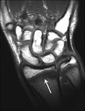
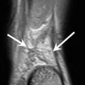
Fig. 20.4
A 14-year-old male gymnast with radial epiphysitis or “gymnast wrist.” Coronal T1-weighted MRI of the wrist visualizes widening of the growth plate (arrow)
20.3.1.2 Triangular Fibrocartilage Complex Tear
The triangular fibrocartilage complex (TFCC) is a complex structure consisting of a triangular fibrocartilage disk with a function analogous to the meniscus in the knee and a collection of ligaments connecting the osseous structures within the wrist joint. In gymnastics, the TFCC can be injured by acute trauma such as a fall on the outstretched hand, but repetitive stresses as seen in bar or ring exercises can cause TFCC tears as well. Damage to the TFCC can also be a consequence of distal radial epiphyseal injury leading to relative shortening of the radius (Mandelbaum et al. 1989). Unlike radial epiphysitis, TFCC injuries are more common in skeletally mature gymnasts (DiFiori 2006). Palmer has classified TFCC lesions into traumatic and degenerative lesions, the latter ranging from simple TFCC wear to perforation of the TFCC with lunate and ulnar chondromalacia and lunotriquetral ligament perforation (Palmer 1989).
Patients with a tear of the TFCC often present with ulnar-side wrist pain (Kocher et al. 2000). Pain increases with weight bearing or rotation of the wrist (Jacobson and Plancher 1996). The ulnar side of the wrist can be tender on palpation (Gerbino 1998).
Little evidence is available on the use of imaging compared to arthroscopy in the diagnosis of TFCC injuries. MRI can be of use but requires the application of a magnetic field of 3 Tesla or higher and a wrist coil in order to obtain high-quality images. Tears of the TFCC can be visible on MRIs as high-signal abnormalities, sometimes with distal radioulnar joint effusion (Lomasney et al. 2013). Partial-thickness TFCC tears can be seen on coronal T2-weighted images, where they present as an abnormal fluid signal traversing the TFCC (Lisle et al. 2009). Bone marrow edema on the dorsal side of the ulna can also be indicative of TFCC injury (Lisle et al. 2009). Dorsal ganglion cysts of the wrist are common in gymnasts and often seen on imaging when wrist pain is present. They can be symptomatic and can also be a sign of underlying pathology such as TFCC injury (Lisle et al. 2009). MRI or ultrasound can both be used to depict dorsal wrist ganglion cysts (Davis 2010b).
20.3.1.3 Carpal Stress Fracture
In gymnasts, stress fracture of the carpal bones can arise due to repetitive loading of the wrist in dorsiflexion. Of the carpal bones, the scaphoid is the most vulnerable for this type of injury. Not many cases have been described, but most commonly the scaphoid waist is involved (Manzione and Pizzutillo 1981; Hanks et al. 1989; Nakamoto et al. 2011). Despite its low incidence, the long-term risk of avascular necrosis of the scaphoid as a complication of fracture renders it a diagnosis not to be missed.
Presentation of patients with a stress fracture of the scaphoid is similar to that of patients with an acute scaphoid fracture, particularly pain at the radial side of the wrist, tenderness on palpation of the anatomical snuff box and the scaphoid tubercle, and impaired range of motion (Nakamoto et al. 2011). Pain can worsen with hyperextension and radial deviation (Webb and Rettig 2008).
Scaphoid stress fractures can be visible on conventional radiography as sclerosis at the scaphoid waist in an early stage or later as a clear fracture line (Webb and Rettig 2008). However, not all scaphoid stress fractures are visible on conventional radiographs. In suspect cases with negative radiographs, MRI can be of use (Davis 2010b). On MRI, early stress fracture of the scaphoid presents as bone marrow edema and periosteal fluid (Lisle et al. 2009). These are the signs of an early stress reaction. When a full-blown stress fracture is present, a low-signal line is seen with disruption of the bone cortex (Lisle et al. 2009). Bone scans have a high sensitivity for the diagnosis of stress fractures, but specificity is low, leading to a large number of false-positives (Fredericson et al. 2006). Additionally, the high radiation burden of bone scan makes it a less desirable imaging technique, especially in the relatively young population of gymnasts.
20.3.1.4 Scaphoid Impaction Syndrome
Scaphoid impaction syndrome is thought to be caused by impaction of the dorsal rim of the scaphoid against the dorsal part of the radius when the wrist is in hyperextension (Webb and Rettig 2008). This is believed to cause inflammation of the joint capsule (Jacobson and Plancher 1996).
Related posts:

Stay updated, free articles. Join our Telegram channel

Full access? Get Clinical Tree


