Fig. 26.1
Bull riding event. The cowboy’s riding arm is positioned in supination, with the elbow in extension. The free arm should not touch the bull, the rope, or the cowboy during the ride (Image courtesy of John Tilley)
In the Chute
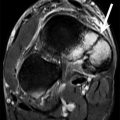
The rider can be injured in the chute, before the chute is opened. The most common injury in the chute is a broken lower leg. Tibial plateau fractures can occur from the lower leg being crushed between the bull and the bars of the chute. If the rider’s foot slips between the bars in the chute and the bull jumps forward, the ankle can invert, resulting in lateral malleolus fracture (Fig. 26.2). Lateral ankle ligament injuries, most often the anterior talofibular ligament and calcaneofibular ligament, can occur when the bull jumps forward in the chute, the spur catches on the bull, and the ankle twists and inverts. The cowboy’s head can also hit the bars at the front of the chute when the bull jumps forward. In some rodeo venues, the bars are covered with padding to help prevent concussion from occurring in the chute. There are also reports of riders losing their balance in the chute and falling between the bull and wall, with the risk of being trampled.
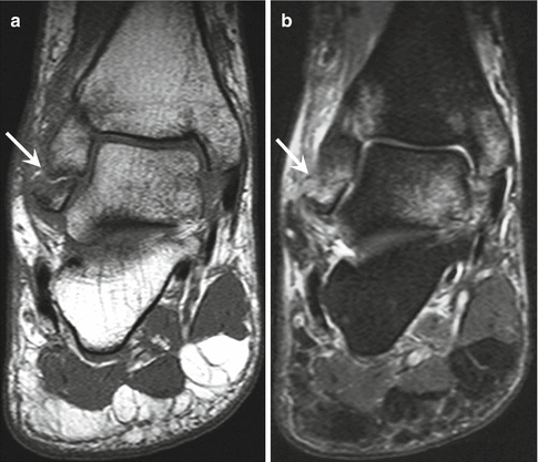
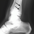
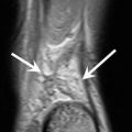
Fig. 26.2
Coronal T1- (a) and fat-suppressed T2-weighted (b) MRI of the ankle demonstrates an infrasyndesmotic fracture of the distal fibula (arrow). This is an isolated type A Weber fracture. There were osseous contusions of the medial malleolus and medial talus
During the Ride
Concussion is one of the more common injuries during the ride. Some authors report an increase in incidence of concussion over recent years due to the higher-quality bucking bulls that are bred for competition, with overall fewer “easy draws” (the Justin Sportsmedicine Team 2006). Concussion can occur due to the whiplash effect of impact from the rider’s head hitting against the bull’s head (Downey 2007). While there is increased awareness of concussion, not all riders wear protective helmets. Livingston et al. found only 33 % of riders wore helmets. Some authors hypothesized that lack of compliance relates to potential limitation of performance, such as decreased ability to chin tuck. Some authors suggest that helmets are avoided as they are not part of the traditional “cowboy image.” There are specialized helmets, for example, the Bull Tough helmet which purports to allow for the chin-tuck position. Many bull riders find the extra weight from the helmet makes it more difficult to control their head position and hold a chin tuck, increasing the risk of whiplash. Other protective gear includes chaps, mouth guard, and cowboy boots.
Multiple upper extremity injuries can occur during the ride due to the extreme force through the riding arm. The Justin team found the shoulder was the most commonly injured body part; shoulder fractures and shoulder dislocations are common.
Complete distal biceps ruptures can occur during the ride (Butterwick 2003). Biceps tendon rupture mechanism of injury commonly involves biceps muscle contraction when the elbow is rapidly straightened under a load, during eccentric contraction (Fig. 26.3). This is likely the mechanism of injury in bareback bronc riding as well.
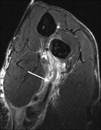
Fig. 26.3
Coronal fat-suppressed FABS (flexed elbow with shoulder abducted and forearm in supination) view T2-weighted MRI of the elbow demonstrates rupture of the distal biceps tendon with retraction of the tendon (arrow)
Injury to the elbow with elbow ligament injuries and elbow dislocations in the riding hand is thought to occur from the supinated fixed position of the elbow during the ride. Sprains of the medial ulnar collateral ligament are common (Fig. 26.4a). Meyers et al. reported radiographic findings of elbow osteoarthritis, olecranon fractures, elbow joint laxity, and medial collateral ligament traction spurs in rough stock athletes (Fig. 26.4b).
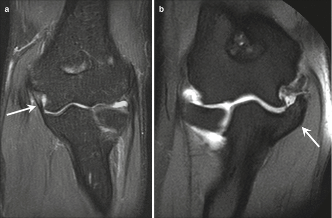
Fig. 26.4
Coronal fat-suppressed T2-weighted (a) MRI of the elbow demonstrates a grade 1 sprain of the medial ulnar collateral ligament (arrow). Coronal fat-suppressed T1-weighted MR arthrogram (b) of the elbow shows a traction spur (arrow) at the ulnar insertion of the medial collateral ligament
Not surprisingly, wrist and hand injuries are common during the ride. In collegiate athletes participating in bull riding or bareback bronc (age 19–23 years old), hand and wrist injuries included healed and nonhealed fractures of the carpal and metacarpals, injuries to the triangular fibrocartilage complex, and osteoarthritis (Meyers et al. 2003) (Fig. 26.5). Scaphoid fractures are common in bull riders; the proposed mechanism of injury is jamming an extended thumb during the ride. The thumb can be jammed by the motion of the ride with the forces extending along the first digit into the scaphoid, although riders are supposed to keep the thumb curled when gripping the braided resin rope to avoid this injury. Forearm injuries included radial stress fractures (Meyers et al. 2003). A common chronic injury reported in bull riders is carpal tunnel syndrome.
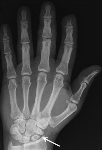
Fig. 26.5
Anteroposterior radiograph of the left hand depicts nonunion of a transverse fracture through the waist of the scaphoid (arrow) with cystic changes in the proximal scaphoid fragment and amputation of the distal second phalanx
Groin injuries can occur during the ride and accounted for 6 % of all injuries in professional bull riding with first- or second-degree strains to the iliopsoas or hip adductors (Butterwick 2003). The Justin team reported thigh and groin injuries as formerly the most common injury in bull riding; the decrease is possibly due to improved pre-event preparation. A search of our database revealed multiple cases of injury to the rectus abdominis/adductor longus aponeurosis in bull riders (Fig. 26.6).
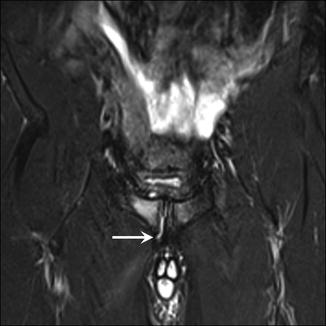
Fig. 26.6
Coronal fat-suppressed T2-weighted MRI of the pelvis shows tearing of the rectus abdominis/adductor longus aponeurosis. The tear extends from the midline into the right aponeurosis and right adductor tendon origin (arrow). There is edema in the adjacent right pubic tubercle and a grade 1 strain of the right adductor longus musculature
There is controversy about whether to “take an extra wrap” or “suicide wrap” in which the hand is further secured by cinching an extra section of the rope around the hand a second time. This strengthens the grip but increases the risk of getting hung up. Getting “hung up” is one of the most dangerous situations for a bull rider. This is when the rider is unable to free the riding hand from the rope, or the spurs get caught in the rope, the bell strap or the flank strap and the rider cannot dismount from the bull. The rider can get dragged along and stepped on by the bull, or the bull can spin and spear the rider with its horns. Knee injuries occur if the bull rope wraps around the ankle while the rider is dismounting, causing a whipping effect as the rider is dragged behind the bucking bull. Many bull riders tie their boots on with rope around the ankle to hold the boot on. If the rider is hung up by his boot, the rodeo clown may have to cut him free. Upper extremity fracture, acromioclavicular joint separation (Fig. 26.7), or elbow ligament injuries can occur as well when the rider gets hung up (Downey 2007).
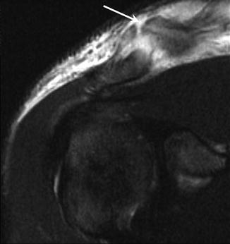
Fig. 26.7
Coronal fat-suppressed T2-weighted MRI of the right shoulder depicts a grade 3 acromioclavicular joint separation. This image shows complete disruption of the ligaments at the acromioclavicular joint and elevation of the distal clavicle (arrow)
Dismount
Areas injured during the dismount (planned or thrown from the bull) include soft tissue, chest, abdomen, and limb. The ideal dismount involves using the free hand to jerk the riding hand free from the rope. At the same time, the rider leans his body back and turns toward the riding hand, while lifting the opposite leg over the bull to the same side as the riding hand. The rider then tries to land on hands and knees, low to the ground to avoid the kicking back legs of the bull.
The knee was the second most commonly injured body part in bull riders according to the Justin team. Knee injuries usually occur when the rider hits the ground. Planting and twisting lead to injuries of the anterior cruciate ligament (Fig. 26.8) and medial collateral ligament, with complete rupture in some cases, as well as meniscal injuries. Hyperextension or a fall on a bent knee can result in posterior cruciate ligament injuries. Rarely complete dislocation can occur with associate neurovascular injuries (Fig. 26.9). Tibial and fibular fractures are also common injuries (Butterwick and Meeuwisse 2003).
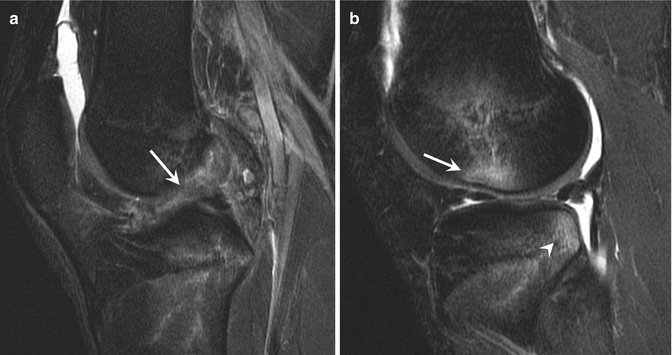
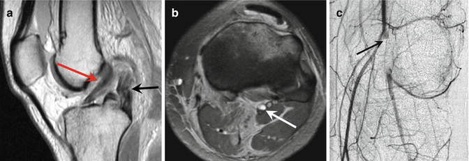
Fig. 26.8
Pivot shift mechanism of injury. Sagittal fat-suppressed T2-weighted MRI of the knee demonstrates complete rupture of the anterior cruciate ligament (a, arrow) and osseous contusions of the lateral femoral condyle (arrow) and posterolateral lateral tibial plateau (b, arrowhead)
Fig. 26.9
Sagittal fat-suppressed proton density-weighted MRI of the knee (a) depicts complete tear of the posterior cruciate ligament (black arrow) and high-grade partial tearing of the anterior cruciate ligament (red arrow). Axial T1-weighted MRI of the knee (b) shows T1 hyperintense signal in the popliteal artery related to thrombosis (arrow); there was laceration of the popliteal artery during the dislocation. Delayed arterial digital subtraction image of the knee (c) demonstrates abrupt cutoff of the popliteal artery (arrow) with reconstitution via the geniculate arteries more distally (Image courtesy of Richard Walker, MD, FRCPC, University of Calgary, Alberta, Canada)
Involuntary dismount from a moving bull can result in large forces with deceleration and blunt force to the rider when they hit the ground with resultant life-threatening injuries. Tripp and Robiseck report aortic transection in the case of a 27-year-old man who was thrown from a bull. Yetman et al. reported ventricular septal rupture in a 15-year-old bull rider who was thrown from the bull and kicked in the chest.
Interestingly, shoulder and elbow dislocations occur both in the free arm and riding arm during the ride (Butterwick 2003). Some riders have instability of the shoulder from previous dislocations; these riders are especially vulnerable to repeat dislocation during the ride. Injuries of the free arm can occur during dismount as the rider uses his free arm to protect himself and his head during the fall or as he escapes from the bull. The free arm also can be injured when the rider is hung up.
Other upper extremity injuries can occur as well. Hyperextension of the elbow during the dismount is a common injury. Other common upper extremity injuries from a fall include clavicle fracture, shoulder dislocation, humerus fracture, acromioclavicular joint separation, or rotator cuff injury. Butterwick and Meeuwisse also report brachial plexus neuropraxia as a possible injury.
On the Ground and Escaping from the Bull
When on the ground, the rider is at risk of being stomped on by the bucking bull. Thoracic compression from this mechanism can be fatal, even when the rider is wearing a vest. Some studies report decreased rib fractures and penetrating chest wounds with protective vests. However, other studies have found that the vest does not prevent more serious injury due to the large forces at play; Livingston et al. found that all of the injured riders were wearing safety vests.
Slawski and West (1997) reported on a series of syndesmotic ankle injuries in rodeo bull riders. Mechanism of injury involved the bull stepping on the lateral aspect of the riders’ ankle as they were escaping after dismount. This resulted in external rotation of the ankle which led to syndesmotic ligament tears with or without fibular fractures.
In some cases, the bull steps on the rider’s hand while the rider is crawling away to escape, fracturing the phalanges.
26.1.2.2 Bareback Bronc
Bareback bronc is essentially riding a horse that attempts to buck off the rider. The event originated in horse breaking skills that would have been necessary for working cowboys. Like bull riding, the event begins in a chute where the rider prepares for the ride. After a surcingle style rigging is placed on the horse at its withers, the rider grips a simple handle with one hand in the semipronated position. A bucking strap is applied around the flank of the horse to encourage bucking. The rider indicates when he is ready and the chute is opened, at which point the horse bursts out and the rider attempts to ride the bucking bronc for 8 s. On the first jump out of the chute, the rider “marks the horse out” with his heels in contact with the horse above the point of the animal’s shoulders before the horse’s front legs hit the ground. The rider leans back against the bucking horse in the supinated position and spurs in rhythm with the motion of the horse. The ride is one handed, and the cowboy cannot touch himself or the horse with his free hand. The ride is scored out of 100: rider is scored 0–50 and the horse is scored 0–50.
Bareback riding is not quite as dangerous as bull riding but has the second largest number of injuries (the Justin Sportsmedicine Team 2006). Butterwick et al. reported injury incidence as 25/1000 competitor exposures. Bareback riding involves higher acceleration forces than bull riding. A pilot study in 2009 tested acceleration in rough stock riders using an ear-mounted triaxial accelerometer. The bare back rider experienced up to 46 G, higher than bull riding where the rider experienced 26 G (Mathers 2009).
In the Chute
The rider can be injured in the chute in both bareback bronc and bull riding. Ankle fractures are common in bronc riders as well. Other injuries can occur too, such as lower thoracic fractures from bucking in the chute with spine hyperflexion (Fig. 26.10). Boham and O’Connell (2014) report a unique mechanism of a T12 chance fracture in an 18-year-old man; the horse bucked in the chute forcing the rider into thoracolumbar hyperflexion.
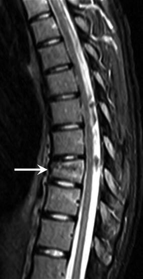
Fig. 26.10
Sagittal fat-suppressed T2-weighted MRI of the thoracic spine shows compression fracture of the T9 vertebral body (arrow). In this case there is minimal buckling of the posterior cortex with no compromise of the central canal. There is no extension into the posterior elements
During the Ride
Many of the injuries that occur during the ride in bareback bronc are similar to bull riding. Concussions can occur during the ride from whiplash effect or impact; in the case of bronc riding, the impact is with the horse’s hip (rather than the bull’s head) due to the supinated position of the rider (Downey 2007).
Patterns of injuries are similar to bull riding with some notable differences. The supinated position of the rider and the semipronated position of the grip result in increased acceleration and eccentric forces through the upper extremity. This results in greater risk of elbow hyperextension, degenerative joint disease, strains of the forearm musculature, and carpal fractures (Meyers et al. 2003). Similar to bull riding, shoulder and elbow sprains or dislocations can occur in both the riding arm and the free arm that is used to keep the balance; likely the majority of free arm injuries occur during the dismount (Fig. 26.11). Injured joints in bareback bronc (from most to least common) include shoulders, hands, and elbows (the Justin Sportsmedicine Team 2006). Sixty percent of radiographic changes to the upper extremity involve medial epicondyle or olecranon in riding and free arms. Ulnar hypertrophy is unique to professional bareback riders; Claussen (1982) describes the finding of chronic circumferential periostitis from the contact of the ulna with the pelvis and thigh during the ride. Rotator cuff tendinosis is another chronic injury seen in bareback bronc riders.
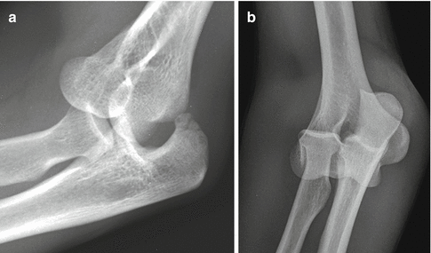
Fig. 26.11
Lateral radiograph of the elbow (a) depicts fracture dislocation of the elbow joint with comminuted minimally displaced fracture of the olecranon. Anteroposterior radiograph of the elbow (b) shows the overlap of the distal humerus with the proximal radius and ulna and extensive soft tissue swelling along the lateral and medial aspects of the elbow
Groin and hamstring injuries can occur during the ride. Pubic diastasis can occur from the wedge effect on the pelvis during the ride (Downey 2007).
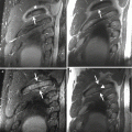
Dismount
Concussion can occur in an involuntary dismount or if the rider is kicked by the bronc while escaping after the ride (Downey 2007). Chest and rib injuries can occur if the rider is kicked or trampled (Downey 2007). Similar to bull riding, knee and ankle injuries can occur during the planned dismount, in a fall, or running from the animal after dismount on the uneven terrain (Figs. 26.12 and 26.13).
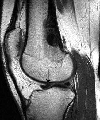
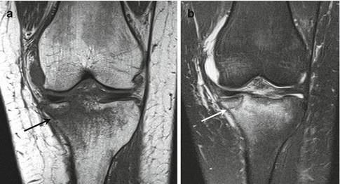
Fig. 26.12
Sagittal proton density-weighted MRI of the knee depicts a bucket handle tear of the lateral meniscus with flipped fragment (arrow) in the intercondylar notch. Incidental note was made of a nonossifying fibroma in the posterior aspect of the distal femur
Fig. 26.13
A 16-year-old rider who fell from a horse. Coronal T1- (a) and coronal fat-suppressed T2-weighted MRI (b) of the knee demonstrates medial tibial plateau fracture (arrow). There is mild depression of the medial fragment. This rider also had a sprain of the anterior cruciate ligament (not shown)
26.1.2.3 Saddle Bronc
Saddle bronc is similar to bareback bronc but has a saddle with free swinging stirrups and no horn. Using one hand, the rider grips a braided rein attached to a leather halter worn by the horse. The rider lifts on the rein and attempts to find a rhythm with the animal by spurring forward and backward with his feet (Fig. 26.14). The mark out rule is the same in saddle bronc as in bareback bronc. Saddle bronc riders have an added risk of becoming hung up in the stirrups, and to mitigate this risk, saddle bronc riders have been known to put baby powder in their boots so their foot will slide out if they become hung up in the stirrup. Lace up boots increase the risk of getting hung up in saddle bronc riders with the rider unable to slide out of the boot to free himself.
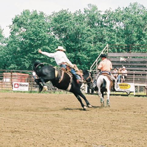
Fig. 26.14
Cowboy riding saddle bronc (black horse). The other rider (orange shirt) can assist the cowboy with his dismount from the bronc and loosen the flank strap on the bronc when the 8 s is over
Injuries can occur in the chute in this sport as well. If the horse flips backward in the chute, the rider can be pinned between the horse and the bars at the back of the chute. In this case the saddle pins the rider’s chest and traps the rider’s pelvis against the bars. This mechanism of injury can result in rib fractures posteriorly at the costovertebral junction.
Many of the injures in saddle bronc are similar to bareback bronc, although in this event the knee is the most commonly injured joint followed by the shoulder. The knee can be injured during the ride in the following way, if the door of the chute is close to the rider at the beginning of the ride, the rider is at risk of hitting the bar on the door with their knee while marking out the horse. PCL injuries can occur when the tibia hits the bar, similar mechanism to a dashboard injury.
Distal biceps injuries are common. Complete tears can occur in some cases during the ride when the horse’s head pulls forward to the ground and the force is transmitted through the rein to the rider’s upper extremity. Rotator cuff tears can occur during the ride as well.
Some riders have early osteoarthritis in the second metacarpal phalangeal joint from the increased strain through this joint during the ride. If the shank is threaded through the index and fifth finger, the second MCP takes the brunt of the forces.
Related posts:

Stay updated, free articles. Join our Telegram channel

Full access? Get Clinical Tree


