Fig. 1.1
A 20-year-old male volleyball player with injury to the rectus femoris. Transverse ultrasound image shows “bull’s-eye lesion” (arrows), indicating an injury to the indirect tendon of the rectus femoris. In clinical practice this is often confused with “muscle stiffness”
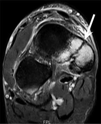
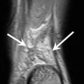
Fig. 1.2
A 17-year-old female track and field athlete with foot pain. Coronal T2-weighted fat-suppressed MRI shows stress fracture of the navicular bone (arrow). This is a common stress fracture in athletes usually with delayed presentation
References
Chan VO, Moran DE, Shine S et al (2013) Medial joint line bone bruising at MRI complicating acute ankle inversion injury: what is its clinical significance? Clin Radiol 68:e510–e523CrossRef
Cook JL, Purdam CR (2009) Is tendon pathology a continuum? A pathology model to explain the clinical presentation of load-induced tendinopathy. Br J Sports Med 43:409–416CrossRefPubMed
Related posts:

Stay updated, free articles. Join our Telegram channel

Full access? Get Clinical Tree


