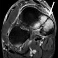
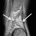
Fig. 2.1
A 23-year-old male professional football player with right knee contact trauma during a game. Sagittal (a, b) and coronal (c) fat-suppressed intermediate-weighted MRI shows fluid within the ACL (white arrow) and a degenerative tear of the anterior horn of the lateral meniscus (blue arrow). Symptoms were minor and the patient returned to play after 2 weeks. Eight weeks later, the patient had another trauma of the right knee without any contact during a game. Sagittal (d, e) and coronal (f) fat-suppressed intermediate-weighted MRI shows the ACL is torn (arrow) (d) with typical lateral femoral osteochondral lesion (yellow arrow) (e, f). The lateral meniscus tear (blue arrow) is unchanged (e). During the ACL surgical reconstruction, the torn ACL is visible (g), as well as degenerative meniscal tear (h) and the osteochondral lesion (i)
Fig. 2.2
(a, b, c) A 30-year-old male professional football player with an ACL graft. Multi-planar reconstruction of an isotropic fat-suppressed 3-D proton density-weighted SPACE MRI of a knee shows the orientation of the ACL in 3-D, and possible postoperative problems or a transplant elongation can be considered
Fig. 2.3
A 28-year-old male football player with an isolated, symptomatic cartilage defect of the medial femoral condyle. High-resolution sagittal (a) and coronal (b) proton density-weighted MRI of the knee demonstrates medial central femoral cartilage defect (arrow). Sagittal (c) and coronal (d) fat-suppressed proton density-weighted MRI shows the same chondral defect (arrow) with no subchondral bone marrow edema. Arthroscopic images show the debrided defect (e) and the microfracture therapy (f)
2.7 Conclusion
In this chapter only a portion of the various joint injuries and the impact of imaging to the sports medicine surgeon can be reviewed. Comparable data, for example, is available for the shoulder or other joints (Dwyer et al. 2015). However the role of diagnostic imaging for an exact diagnosis and following surgical therapy is similar in most of the anatomic regions. For recreational athletes, the importance of imaging joint injuries can be compared to other patients, but for professional athletes the sports medicine surgeon often needs even more safety in diagnosis, which might lead to the often discussed overuse of MRI in these patients. Nevertheless when it comes to predicting return to play at a professional level, the more information on the injury, the better for the surgeon and the patient. Also, especially for more severe injuries or more complex cases, diagnostic imaging often reduces the time of the diagnostic algorithm.
References
DiFiori JP, Benjamin HJ, Brenner J et al (2014) Overuse injuries and burnout in youth sports: a position statement from the American Medical Society for Sports Medicine. Clin J Sport Med 24:3–20CrossRefPubMed
Related posts:

Stay updated, free articles. Join our Telegram channel

Full access? Get Clinical Tree


