Fig. 5.1
Radiographic findings of small bowel obstruction. (a and b) Upright plain radiograph of the abdomen demonstrates dilated small bowel loops with differential air-fluid levels in a patient with high-grade obstruction. (c) Small bowel follow-through demonstrates dilated small bowel loops with abrupt transition in the mid-jejunum (arrow)
CT scan has higher sensitivity (90 %) than plain radiograph in making the diagnosis of small bowel obstruction and is considered the most appropriate imaging modality in diagnosing high-grade small bowel obstruction. The CT should be performed with intravenous contrast, but without oral contrast. CT enteroclysis and MR enteroclysis are both equally appropriate, first-line imaging modalities in diagnosing low-grade or intermittent small bowel obstruction.
CT study in patients with small bowel obstruction is characterized by small bowel loops more than 2.5 cm in caliber, transition point, and decompressed bowel loops beyond the transition zone. Although the adhesion band itself is rarely visible, the presence of a smooth transition zone often points to adhesions as the cause of the bowel obstruction.
Small bowel feces sign is characterized by the presence of fecal material along with air bubbles in dilated small bowel loops and is believed to be due to bacterial overgrowth and water resorption from incompletely digested food [3, 4] (Fig. 5.2). It is an indicator of the site of mechanical small bowel obstruction and points to the low-grade or subacute small bowel obstruction. It is present in longer segment of small bowel in patients with higher-grade obstruction and is useful in recognizing the transition point due to its close proximity. This sign has been reported in 7–82 % cases of small bowel obstruction and should not be mistaken for fecal material which can be normally present in terminal ileum in patients with incompetent ileocecal valve [5]. Fecal material can also be in small bowel in patients with cystic fibrosis, with bezoars, and with infectious and metabolic bowel disease.
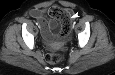
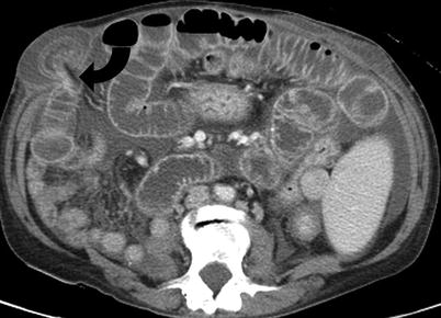
Fig. 5.2
Small bowel obstruction on CT. Contrast-enhanced CT study in a patient with small bowel obstruction demonstrates dilated small bowel loops with small bowel feces sign (arrowhead)
Fig. 5.3
Incarcerated hernia. Contrast-enhanced CT demonstrates small bowel obstruction secondary to bowel incarceration in hernia sac. The transition site (curved arrow) is located at the neck of the spigelian hernia sac
The other findings of small bowel obstruction include mesenteric edema, ascites, mesenteric venous congestion, decreased bowel wall enhancement, and free extraluminal air (Table 5.1).
Table 5.1
Causes of small bowel obstruction
1.Adhesions (most common, 75 %) |
2.Crohn’s disease |
3.Hernia (Fig. 5.3) |
4.Neoplasia |
5.Rare causes: radiation, internal hernia, intussusception, volvulus, gallstone ileus, tuberculosis, hematoma, bezoars |
Internal Hernia
Internal hernia is characterized by herniation of small bowel through a breech in the mesentery into another compartment of the abdomen. Although paraduodenal hernia has traditionally been the most common type of internal hernia, the frequency of transmesentric internal hernia has increased with increasing popularity of gastric bypass surgeries.
The internal hernia can be paraduodenal (most common), prececal, foramen of Winslow, transmesenteric, transmesocolic, intersigmoid, and retromesenteric. The right paraduodenal hernia extends in to the Waldeyer’s fossa, which is located posterior to the superior mesenteric artery and transverse colon. The left paraduodenal hernia extends in to the Landzert’s fossa, situated to the left of the fourth part of the duodenum.
The CT findings include saclike cluster of small bowel obstruction, anterior displacement of stomach, and crowding of mesenteric vessels [6] (Fig. 5.4). The CT findings of transmesocolic hernia, seen after gastric bypass surgery, include clustered small bowel loops cephalic to transverse mesocolon, high position of distal jejunal anastamosis, and clustering of mesenteric vessels [7]. The right paraduodenal hernia is usually larger than left paraduodenal hernia. Transmesenteric internal hernia may also cause central displacement of colon.
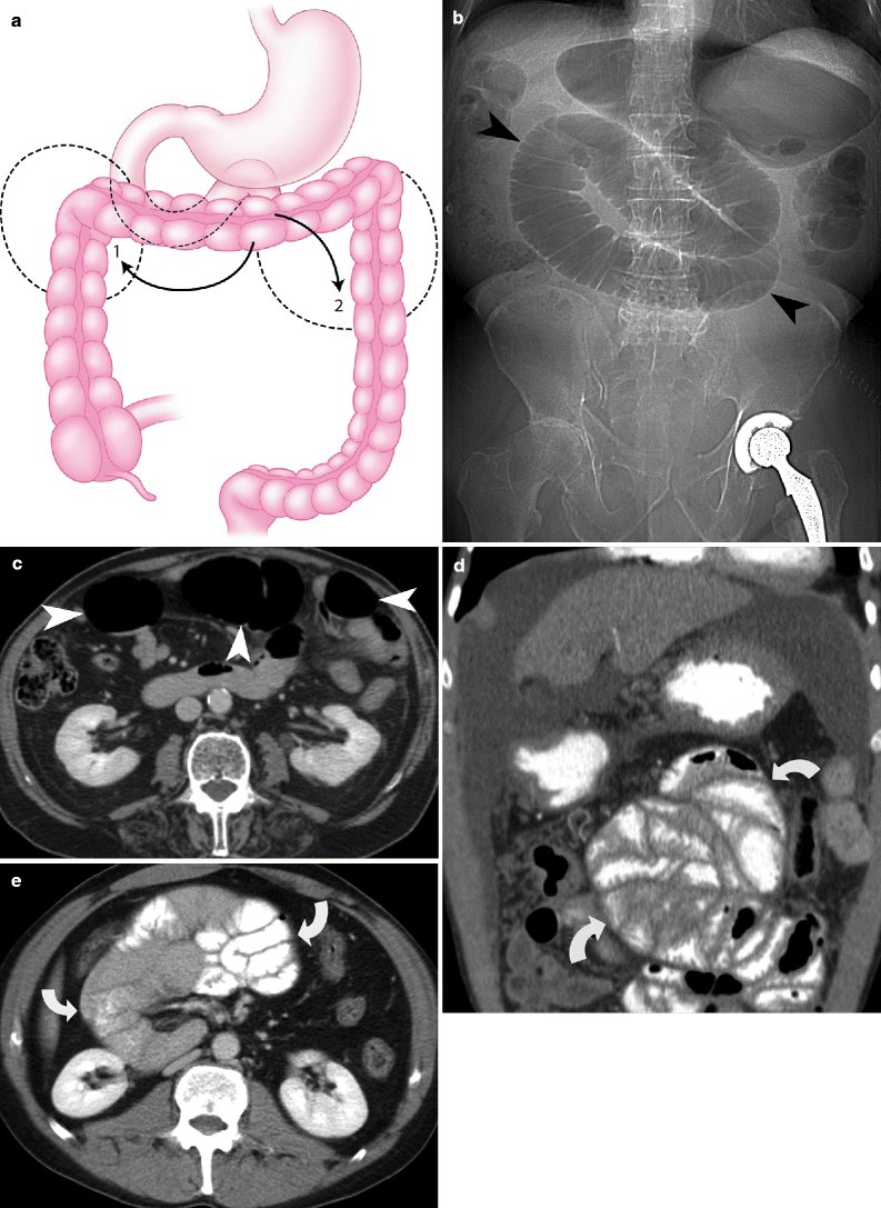
Fig. 5.4


Internal hernia. (a) Schematic representation of the right paraduodenal fossa (1) and left paraduodenal fossa (2). (b) CT scout film demonstrates dilated proximal small bowel (arrowheads) in the paraduodenal hernia. (c) Axial contrast-enhanced CT demonstrates dilated proximal small bowel loops (arrowheads) in paraduodenal hernia, without omental fat anterior to it. (d) and (e) Contrast-enhanced CT demonstrates clustering of small bowel loops in two different patients with paraduodenal hernia (curved arrows)
Related posts:


Stay updated, free articles. Join our Telegram channel
Full access? Get Clinical Tree
