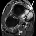
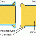
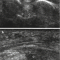
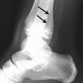
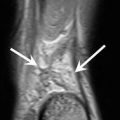
Fig. 8.1
The kinetic chain demonstrating the stages of the bowling action including the run-up, back foot contact, energy transfer to front foot, front foot contact, and release (see text for explanation of stages)
8.2.1.1 The Run-Up
The bowler experiences a series of impacts with the playing surface (grass) during the run-up. These forces are transmitted through the bones, cartilage, tendons, ligaments, and muscles of the foot, leg, thigh, and pelvis to the intervertebral discs and the facet joints of the vertebrae (Burnett et al. 1998). For spin bowlers, this involves a few paces at low speed, but for fast bowlers, this is usually a sprint over approximately 20 m. These forces are usually absorbed by the body, but when combined with a bowling technique that changes the axis of stress to the pars area, these forces can cause bony abnormalities within the spine (Portus et al. 2004).
8.2.1.2 Back Foot Contact
Just prior to ball delivery, the back leg (same side as the bowling arm) generates and transmits the linear velocity from the run-up to the lower limb, pelvis, spine, and trunk. The back leg performs triple extension at the hip, knee, and ankle making it rigid, and the trunk goes into hyperextension. The back leg gradually builds up forces of approximately 0.35 times the bowler’s body weight until the front foot impacts (MacWilliams et al. 1998).
8.2.1.3 Front Foot Contact
This is of vital importance as it is this movement that captures the energy generated by the back foot and transfers it to the throwing action via the lower limb to the pelvis, spine, and trunk. The lead foot applies a breaking force to halt forward momentum and begins to transfer the kinetic energy back up the kinetic chain. When the arm is in maximal external rotation, the front leg applies approximately 1.5 times body weight as well as a braking at force of approximately 0.75 times body weight (MacWilliams et al. 1998). The stride leg stabilizes the hip and knee joints to maintain standing posture for the trunk. The pelvis then tilts which increases movement of the upper torso forward. The humerus has to catch up with the trunk which it does at speed, i.e., forward and vertical momentums are transferred into rotational components. This is the point of greatest risk for injury in fast bowlers as the trunk experiences high degrees of lateral flexion, hyperextension, and rotation when ground reaction forces are at their highest (Foster et al. 1989) which leads to high contralateral facet joint stress (Worthington et al. 2013).
8.2.1.4 The Release
The final stage of delivery is the release phase. This occurs after the front foot impacts. The stride leg stabilizes the hip and knee joints, the arm extends, and the ball is delivered (Fleisig et al. 1999). A pelvis, trunk, and arm rotational follow-through movement then occurs which, along with knee flexion, dissipates the forces generated throughout the previous stages of the action.
Fast bowling at the elite level is a very complex dynamic sporting activity that requires athletes to repeatedly deliver the ball. In the case of the game’s longest version, Test cricket, this may occur up to approximately 180 times during a day’s play. Such repetitive highly complex mechanisms make fast bowlers the most prone to injury, at risk of sustaining a variety of injuries including muscle strain, joint impingement syndromes, and spinal injuries.
8.2.2 Spinal Injury
Fast bowlers, in particular, are prone to lumbar vertebral stress injuries. These are seen within the general population and other athletes, but the prevalence is notably higher in cricketing athletes (Hardcastle et al. 1992; Engstrom and Walker 2007; Millson et al. 2004; Johnson et al. 2012). The bowling mechanism of lumbar spine axial loading, hyperextension and rotation through delivery, is the primary factor for injury, but other contributing factors include the bowling workload and variation in bowling technique (Kountaris et al. 2012).
Three fast bowling actions have been described including front-on (Fig. 8.2), side-on (Fig. 8.3), and a mixed type, all relating to shoulder and hip alignment at ball release. The mixed action has been found to have the highest incidence of lumbar spine stress injuries (Johnson et al. 2012).
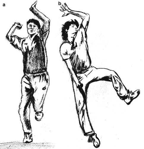
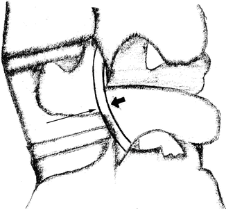

Fig. 8.2
(a) Front-on bowling action. The back foot points straight down the wicket causing the hips to align parallel with the bowling crease. The non-bowling arm is at the side of the head resulting in alignment of the hips and shoulders facing the batsman. (b) Side-on bowling action. The back foot is parallel to the bowling crease causing the hips to be side-on. The non-bowling arm is placed in front of the head causing the shoulders to align with the hips

Fig. 8.3
Diagrammatic representation of pars interarticularis. The short block arrow represents the point of maximum compressive load and the long thin arrow the site of maximal tensile stress on the pars interarticularis
The mechanism of injury is thought to relate to repeated lumbar hyperextension and axial loading on the non-bowling arm side (Gregory et al. 2004). The resulting tensile longitudinal stress across the pars interarticularis causes repeated osseous stress and eventual fracturing of the caudo-ventral aspect (Fig. 8.4 and 8.5).
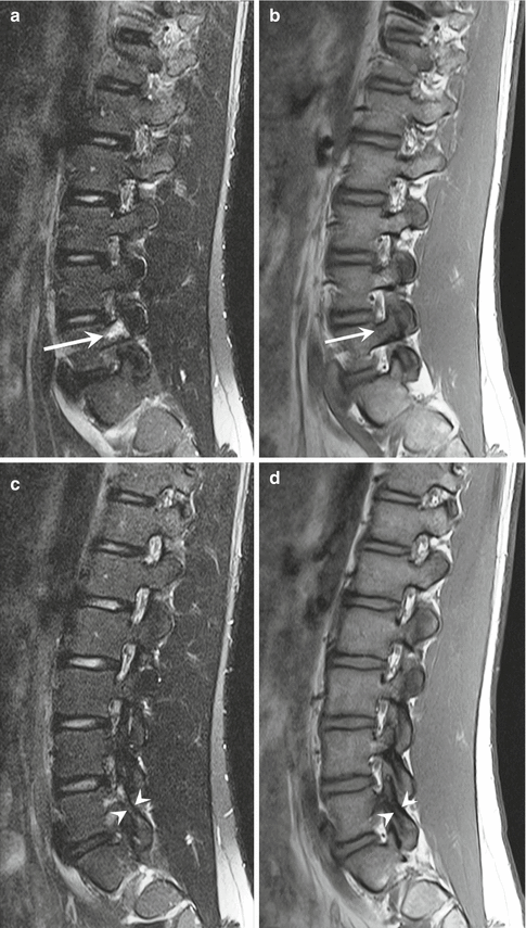

Fig. 8.4
Fast bowler with low back pain. (a) Sagittal fat-suppressed T2-weighted MRI and (b) corresponding sagittal T1-weighted MRI show marked bone marrow edema (arrow) with no definite fracture. (c) Sagittal fat-suppressed T2-weighted MRI and (d) corresponding sagittal T1-weighted MRI just medial to (a) and (b) confirms intact pars region (arrowheads)
If stress continues in this area, the fracture extends craniodorsally until becoming circumferential often within 2 months (Terai et al. 2010). These fractures are most commonly seen at L4 but can occur at any level from L3 to L5 depending on the degree of variation in the bowling action (Engstrom and Walker 2007; Gregory et al. 2004) and can be unilateral or bilateral with the latter more common at L5. Skeletally immature fast bowlers are most at risk of pars interarticularis stress fracturing as peak bone mass is not achieved before the age of 25 (Kountaris et al. 2012). If diagnosed early, these injuries can be treated conservatively and result in less match time lost. Imaging can play a vital role in confirming the diagnosis, its severity, and monitoring subsequent healing.
Players often present with insidious onset of low back pain on the opposite side to the bowling arm reproduced by maneuvers involving lumbar extension (Ranson et al. 2010). Historically plain films and CT have been used for assessment of lumbar spine injury, but MRI is now the investigation of choice. This modality can detect and differentiate early changes such as bone marrow edema, periostitis, and fracture (Figs. 8.4 and 8.5).
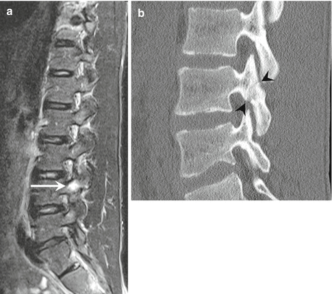

Fig. 8.5
Fast bowler with low back pain. (a) Sagittal fat-suppressed T2-weighted MRI shows pars edema (arrow) but no definite fracture. (b) Corresponding sagittal reformatted CT image confirms underlying acute fracture (arrowheads)
A classification system was suggested by Ranson et al. in 2005 (Ranson et al. 2005), grading stress injuries in the spine as seen in Table 8.1.
Table 8.1
Ranson et al. classification of spine stress injuries
Grade | MRI appearance of pars interarticularis |
|---|---|
0 Normal | No signal abnormality, intact |
0a Chronic stress reaction
Stay updated, free articles. Join our Telegram channel
Full access? Get Clinical Tree
 Get Clinical Tree app for offline access

|

