IMAGING OF THE HIP AND PELVIS

Chapter 11 IMAGING OF THE HIP AND PELVIS


Imaging of the hip is a rapidly growing area of interest to orthopedic surgeons, especially with regard to dysplasia, impingement, labral tear, and groin injuries. This chapter focuses on newly emerging areas, issues in which controversy exists, and management conundrums that commonly affect the practicing radiologist.


MODALITIES


Radiography


Radiography remains the initial tool for evaluation of the majority of musculoskeletal conditions involving the hip and pelvis. These include acute and chronic injury, infection, avascular necrosis, arthritis, metabolic disease, tumor, and dysplasia/impingement. Radiography is the mainstay for diagnosis of fractures, both acute and stress-related. In the acute trauma setting, views such as oblique Judet views and inlet/outlet views (with craniocaudal angulation) can be helpful. Anteroposterior (AP; actually AP with internal rotation) and “frog lateral” (AP with external rotation) views are excellent for detection of femoral neck fracture; the surgical lateral view is used to detect AP fracture angulation. The frog lateral view is especially useful for detection of avascular necrosis of the femoral head, as well as slipped capital femoral epiphysis. Evaluation of sacroiliitis is best performed with a modified Ferguson view (cranially angulated AP), which elongates the joint. Some radiologists prefer oblique views to image along the joint. The AP pelvis is useful as a screening examination for a variety of pathologic conditions, but care should be taken to align the pubic symphysis with the coccyx to ensure that the image is directly anteroposterior. On a properly positioned AP pelvis, the hips can be evaluated for symmetry as well as for dysplasia and femoral-acetabular impingement. A specialized view, the faux profile, has been described for evaluation of hip pathology, especially arthritis, dysplasia, and impingement. When evaluating a hip prosthesis, radiographs are especially useful for detection of loosening; using fluoroscopy, the joint can be aspirated for fluid analysis and culture. Confirmation of loosening can be obtained by injecting iodinated contrast and visualizing its extension around the bone/cement interface. The surgical lateral view is especially useful for evaluation of retroversion of the acetabular cup, which can lead to dislocation. The flamingo view (AP view acquired while standing on one foot, then the other) can be used to evaluate pelvic instability in patients with chronic pain and osteitis pubis.


Magnetic Resonance Imaging


For patients with acute trauma, nondisplaced fractures may not be visible radiographically or even on CT. This is especially true in elderly patients with osteoporosis; the combination of low bone density, thin cortices, volume averaging, and osteoarthritis/enthesopathy can make detection of fracture lines and cortical step-off very difficult. In this situation, MRI is the best test for detection of fracture as well as for the associated soft tissue injury. MRI is also excellent for diagnosis of stress-related injuries, which may not be apparent radiographically. In its early stages, AVN of the femoral head is also not visualized on radiographs. MRI is highly sensitive and can guide rapid intervention such as core decompression. It is also the test of choice for infection because it can give an overall picture of soft tissue disease and spread as well as underlying osseous involvement. MR arthrography is the test of choice for detection of acetabular labral tear; an additional advantage of this invasive procedure is that although the needle is in the joint, anesthetic can be injected to give additional information regarding the site of pain generation.


The approach to hip pain requires a flexible approach to imaging protocols. For example, hip MRI should not be approached from the perspective that one protocol can answer all questions. Most musculoskeletal radiologists have a number of protocols aimed at diagnosing different conditions at the hip and pelvis. (See also the protocols in the accompanying CD.)


AVASCULAR NECROSIS OF THE HIP (Figs. 11-1 through 11-6)


A number of predisposing conditions can result in AVN (the most common are listed in Box 11-1). Often, however, no cause is found (idiopathic is common). Patients present with groin pain, which becomes worse with weight-bearing.


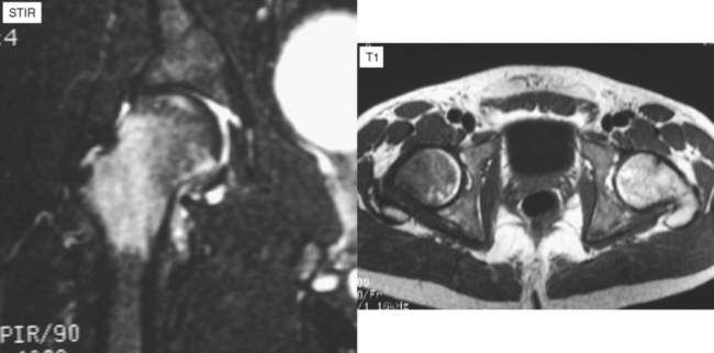
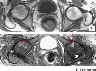
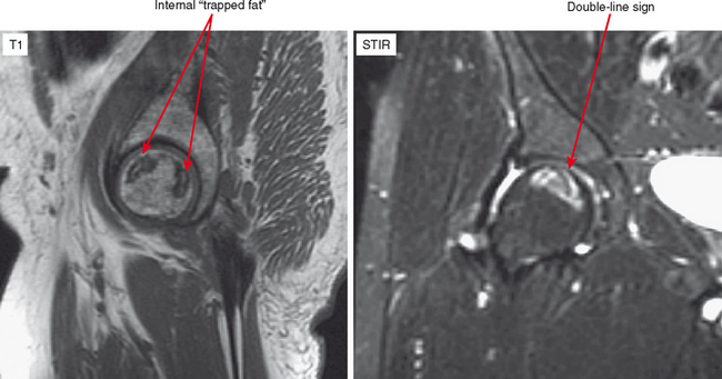
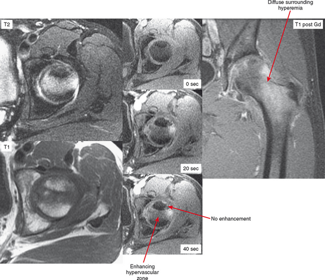
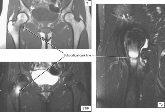
In the early stages of AVN, radiographs are negative. MRI will show diffuse marrow edema of the femoral head, possibly with some subchondral crescentic signal. At this stage, the differential diagnosis includes subchondral stress fracture or transient osteoporosis of the hip (TOPH). AVN does not show bone resorption because there is no blood flow. Therefore, to differentiate these entities the radiologist can perform a dynamic contrast-enhanced MRI looking for lack of enhancement (AVN) versus hyperemia (stress fracture, TOPH). Alternatively, noncontrast CT may show relative lucency of the femoral head in TOPH but not in the other conditions. However, care should be taken when comparing with the opposite side in patients who may have AVN bilaterally.


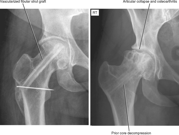
The Ficat staging system is a radiographic staging system, but it is still the most widely used. It has since been modified by the Subcommittee of Nomenclature of the International Association on Bone Circulation and Bone Necrosis; Table 11-1 summarizes the stages. The key is that appearance and progression are similar no matter what bone is affected. In other words, the disease progresses from radiographically occult to an increase in density, to collapse and fragmentation, to osteoarthritis. Treatment is based on the modified Ficat stage. However, in radiologic reports it always better to be descriptive rather than to list the stage because some surgeons may refer to the original Ficat staging, which is numbered differently.


In later stages, it is easier to document the finding as AVN. In stage 2 in which increased radiographic density is seen, MRI demonstrates a focal subchondral geographic signal abnormality, usually centered anterosuperiorly. The central signal is variable, often high on T1 (trapped/mummified fat) but occasionally bright on T2 or dark on all sequences (fibrotic). The margins demonstrate the classic “double line sign” of Mitchell, representing the interface between living and dead bone with a rim of granulation tissue. Typically, the surrounding marrow edema has resolved. At this point, patients may be asymptomatic.


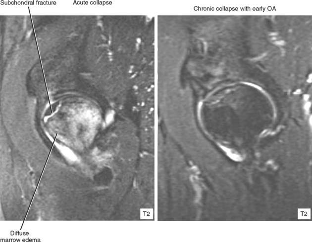
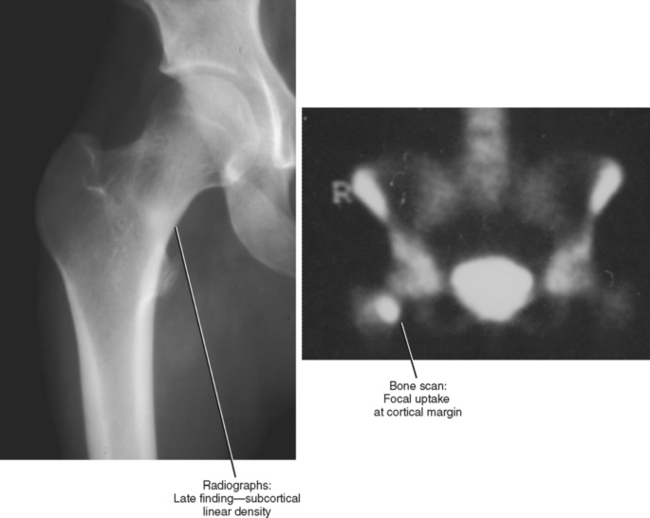
Patients may go on for many years at the prior stage. When a patient with established AVN presents with acute pain, the suspicion and imaging should be directed toward finding articular collapse, which is often very subtle. On radiographs, a subchondral lucent line may be seen, representing a fracture. Flattening of the normally spherical femoral head indicates collapse. If no radiographic signs of collapse are seen, CT with sagittal and coronal reformats is very useful, especially when using multidetector CT with thin cuts. MRI can also be used, although CT has higher resolution. The advantage of MRI is visualization of the diffuse marrow edema that results from collapse, edema that extends to the intertrochanteric region simulating early AVN. If established AVN is seen in addition to this finding, certainly acute-on-chronic AVN is a possibility, but subtle articular surface collapse should be sought. Like AVN, the collapse is usually anterosuperior. TOPH is in the differential for diffuse femoral head edema, but underlying established AVN (or AVN on the other side) helps exclude this possibility.


Osteoarthritis is the last stage of AVN, but distinction should be made between patients with AVN, collapse and subsequent osteoarthritis and those with incidental AVN, no collapse, and osteoarthritis from some other cause. These patients may be treated differently.


TRANSIENT OSTEOPOROSIS OF THE HIP


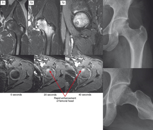
Transient osteoporosis of the hip is a painful condition that was first described in patients in the third trimester of pregnancy (Figs. 11-7 through 11-9). Radiographs and CT show asymmetric osteopenia of the femoral head. MRI shows diffuse edema of the femoral head extending to the intertrochanteric region, an appearance that mimics early AVN. However, unlike AVN, TOPH resolves spontaneously. One theory is that TOPH is ischemia that does not go on to frank necrosis; another theory is that TOPH represents a subchondral stress fracture, much like spontaneous osteonecrosis of the knee. Appearance in late pregnancy supports a stress-related etiology. Also supporting a stress origin is the presence of a subchondral crescentic low-signal region in many cases. CT (showing osteopenia) or dynamic contrast-enhanced MRI (showing early contrast enhancement) should be able to differentiate TOPH from AVN.


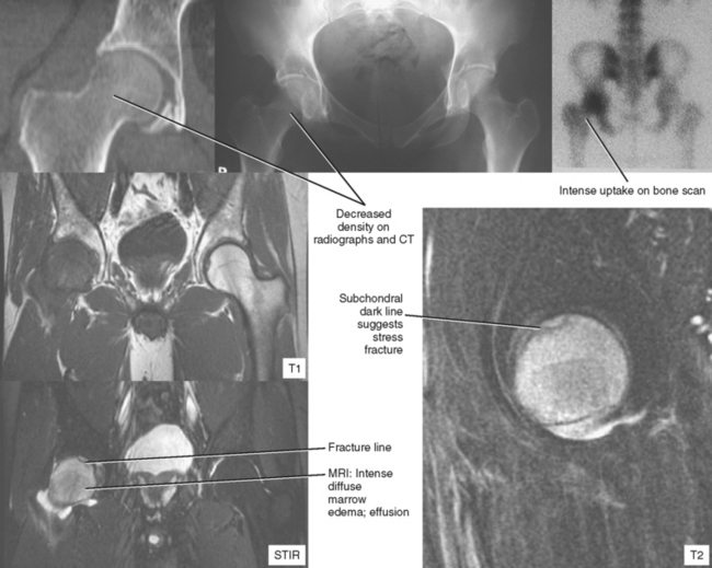
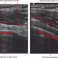
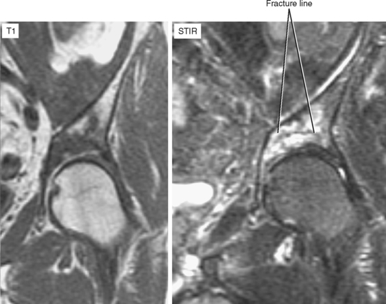
STRESS FRACTURE (Figs. 11-10 through 11-13)


Stress fracture is divided into two categories: fatigue (normal bone undergoing abnormal stress) and insufficiency (abnormal bone undergoing normal stresses). Stress fracture is a relatively common source of hip pain in certain populations, such as teens or young adults undergoing new physical activity (fatigue) and older patients with radiation therapy to the pelvis or with underlying osteoporosis and alteration of activity (e.g., recent total knee arthroplasty with shifting of weight-bearing to the other side) (insufficiency).


If MRI is ordered for a stress fracture, which is suspected either based on the clinical history or hip pain in a susceptible population, it is recommended to use a large field of view because these injuries often are multiple and bilateral in expected locations around the pelvic ring.


In young patients, common sites for stress fracture around the pelvis include the superior and inferior pubic ramus and the femoral neck. In older patients, the sacral alae and supra-acetabular region are more common.


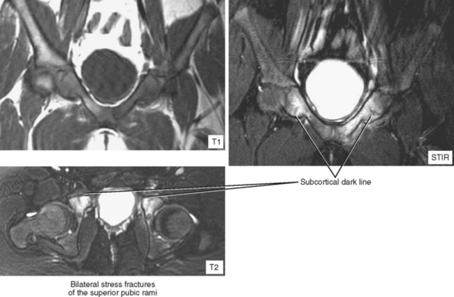
Stress fractures at the femoral neck tend to be solitary and unilateral. They tend to occur most frequently by far at the base of the medial femoral neck (the compressive aspect, where each step pushes the fracture sides together). Since bony apposition is essential for healing, stress fractures in this location have a good prognosis for healing. If the fracture occurs laterally, it is more unstable (this is the tensile aspect; weight-bearing tends to pull the fracture apart). The latter may be prophylactically treated with pin fixation.
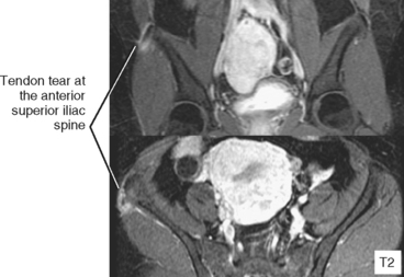
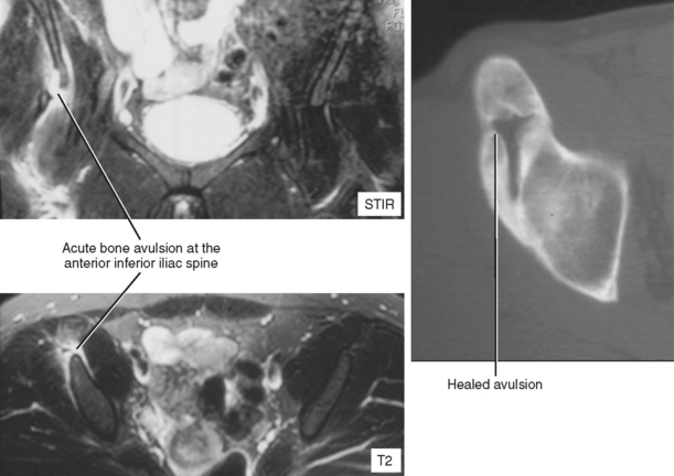

Stress fracture has a characteristic appearance on most modalities and should not be confused with tumor or another pathology. Despite this, sacral stress fracture is often referred for biopsy or more imaging, based on concern over the patient’s age and the presence of a sclerotic lesion.


Radiographs show a sclerotic region, which is linear and extends from the cortex. Sacral fractures are vertically oriented at the ala and often cross the midline, leading to the classic H sign on bone scan. Sacral stress fractures can be nonspecific on radiographs and present with ill-defined sclerosis. CT or MRI is the next test to confirm the diagnosis. Bone scan is useful if the injury is bilateral or if unilateral uptake in one sacral ala is nonspecific. CT shows sclerosis, which, if inspected closely or with reformatted images, shows a linear pattern. In the sacrum, inspection of the foraminal cortices often shows very subtle cortical step-off that can help confirm the diagnosis. MRI shows a subcortical low signal line on T1- and T2-weighted images, representing microcallus, surrounded by marrow edema.


TRAUMA


Occult Fracture (Figs. 11-14 through 11-17)


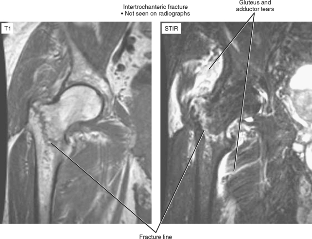
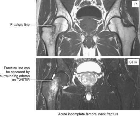
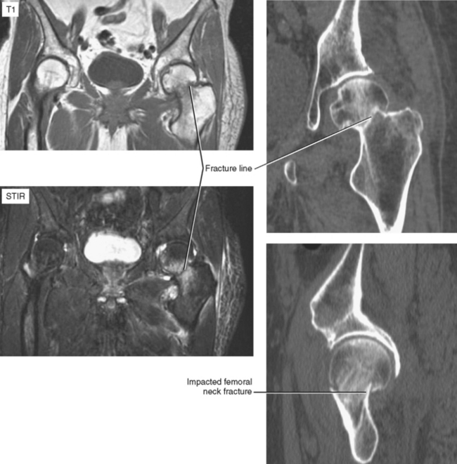
Hip and/or pelvic fractures may be undetectable (“occult”) on radiographs, even when they are reviewed after learning of the precise location. This situation is especially common in elderly patients who suffer a nondisplaced fracture through osteoporotic bone during a fall. Usually, a more severe injury is suspected clinically because the patient often cannot bear weight on the affected side, and additional imaging is requested. The best modality for definitive diagnosis of occult fracture is MRI. In fact, this exam is considered one of the few “musculoskeletal emergencies” (outside of spine imaging) that would prompt calling in the night technologist. During the day, an abbreviated hip survey exam (refer to protocols) can be performed in approximately 10 minutes, sandwiched between routine exams.


The survey exam is acquired using a large field of view to detect associated soft tissue injury and contralateral injury to the rigid pelvic ring. A coronal STIR sequence is used to provide maximal fluid conspicuity while limiting artifact from field heterogeneity. Bone marrow edema is easily seen, as are muscle tears and traumatic bursitis. Muscles that commonly tear include the adductors, glutei, and obturator externus and internus. In the acute stage, wherever they occur, fractures can be hard to distinguish from bone bruises on fluid-sensitive sequences (STIR or fat-suppressed T2-weighted imaging) because the fracture line is bright and so is the surrounding marrow. Although this is less of an issue in the pelvis after acute trauma, acquisition of a coronal T1-weighted sequence facilitates visualization of the fracture line itself. Location, whether complete or partial, and associated soft tissue injury should be reported. Just as on trauma radiographs, when one fracture is found, others should be sought.


CT can be used as an alternate modality, but this is less sensitive than MRI, especially for nondisplaced fractures that may have only slight cortical indentation or step-off or impacted femoral neck fractures that can be confused for a healed fracture or ring of osteophytes. Comparison from side to side is useful, as is close inspection of commonly fractured sites (femoral neck, acetabulum, superior/inferior pubic rami, sacral alae) and close inspection of bone adjacent to traumatic soft tissue infiltration.


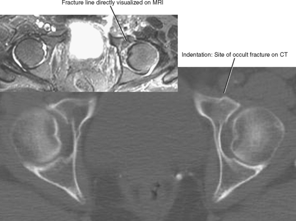
Nuclear medicine bone scan is too insensitive in the acute phase of occult fracture. A traditional low-cost alternative to advanced imaging, that being conservative therapy with follow-up radiographs, is not an option in this setting because the fracture can displace and become more complicated in addition to pulmonary embolism and other dreaded complications resulting from prolonged immobilization of the elderly.


Avulsion Fracture (Figs. 11-18 through 11-21)


Avulsion fractures are seen with sudden, forceful muscle contraction; adolescents are susceptible to avulsion of the sartorius from the anterior superior iliac spine (ASIS), and the rectus femoris from the anterior inferior iliac spine (AIIS). Hip pain in a young patient should always draw attention to these attachment sites, regardless of the modality. MRI of the hip with a small field of view can miss the ASIS, and if included, near field brightening or artifact can simulate pathology. Again, a large field-of-view STIR sequence is very useful as a survey for this and other pathology. In high-performance adolescent athletes, hamstring avulsion can be seen. Avulsions acquired during youth may heal back to the underlying bone leaving a bony prominence that can be mistaken for an osteochondroma. Often the avulsion remains separate from the native bone, becoming rounded and proliferative over time.


Jan 17, 2016 | Posted by in MUSCULOSKELETAL IMAGING | Comments Off on IMAGING OF THE HIP AND PELVIS

Full access? Get Clinical Tree

Get Clinical Tree app for offline access