Immunologic Diseases of the Lung


Immunologic Diseases of the Lung


Immunologic diseases of the lung may present in the bronchial system, lung parenchyma, and the pulmonary vessels. The classification system used in this book with breakdown into allergic, eosinophilic, and vascular pulmonary diseases is—just like other attempts to categorize these—arbitrary and inevitably results in overlapping. For example, allergic bronchopulmonary aspergillosis can be assigned to both the allergic and eosinophilic pulmonary disease groups. These overlaps will be pointed out in the introductory text to the respective sections.


7.1 Allergic Pulmonary Diseases

Allergic reactions essentially involve four different immune mechanisms. These are summarized in ▶Table 7.1. These different types of allergic reaction account for varying degrees for the diseases presented here.


7.1.1 Asthma

Asthma is characterized by major changes in intrapulmonary respiratory tract resistance within a very short period of time. This is caused by reversible bronchoconstriction, increased mucus secretion, and edema of the bronchial mucosa, giving rise to reversible hyperinflation of the airspaces. In two-thirds of asthma patients, this involves allergic bronchial asthma. The bronchial changes described above are caused by an allergic type I reaction. The remaining third of patients have nonallergic, intrinsic asthma.

The main clinical manifestations are cough and attack-like shortness of breath, in particular, at night and in the morning, wheezing, and transparent, viscous sputum. With regard to lung function, asthma is associated with increased lung volume and higher functional residual volume. Concomitant reduced vital capacity attests to the presence of peripheral obstruction and resultant air trapping. Severe complications are very rare. Pneumothorax or mediastinal emphysema is seen especially in children. Increased endobronchial mucus viscosity may cause hyperinflation due to an endobronchial valve mechanism. The resultant clinical picture is similar to that of tension pneumothorax.


Table 7.1 Types of allergic reactions and associated allergic pulmonary diseases


Allergy type


Description


Pulmonary diseases


Type I


Classic immediate-type allergy:


  • Immunoglobulin-E-mediated:


    – Mast cell activation


    – Histamine release


  • Reaction in seconds to minutes


  • Allergic bronchopulmonary aspergillosis


  • Asthma


Type II


Cytotoxicity: antibody production → opsonization of autologous structures (antibody marking)


  • Type IIa


Destruction by immune system


Goodpasture syndrome


  • Type IIb


Activation


Type III


Formation of immune complexes: antibodies against soluble antigens:


  • Multivalent complex formation


  • Deposition in capillaries or complement activation


  • Hypersensitivity pneumonitis


  • Allergic bronchopulmonary aspergillosis


Type IV


Delayed-type: T cell activation by soluble antigens → macrophage activation


Hypersensitivity pneumonitis


The use of chest radiography is controversial in different guidelines.1,2 The following conditions may constitute an indication for chest radiography:


  • Patients to be admitted to the hospital because of asthma.


  • Suspected pneumonia or pneumothorax.


  • Before mechanical ventilation.


  • In cases of poor response to treatment.


  • If methotrexate treatment is intended.

In around one-quarter of patients, treatment-relevant findings are identified on imaging. At times, CT may be indicated for severe asthma to exclude differential diagnoses, in particular, to rule out malformations, dysplasia, tumors, bronchiolitis, bronchiectasis, pulmonary embolisms, and various types of diffuse parenchymal lung diseases.1,3

The radiologic findings in asthma are summarized in ▶Table 7.2; also see ▶Fig. 7.1 and ▶Fig. 7.2.


7.1.2 Allergic Bronchopulmonary Aspergillosis

In industrialized countries, allergic bronchopulmonary aspergillosis is the most common eosinophilic lung disease. It predominantly affects long-term asthma patients and patients with cystic fibrosis. Aspergillus antigens, usually belonging to Aspergillus fumigatus, trigger types I and III allergic reactions in the bronchial mucosa.4 Type I reaction causes a bronchial spasm, as seen in asthma, as well as blood eosinophilia. Type III reaction causes damage to the bronchial wall tissues, giving rise over time to bronchiectasis.

In general, diagnosis is clinically suspected and is confirmed with radiology and serology.5 Diagnostic criteria are presented in ▶Table 7.3; rarely all criteria are met at the same time.


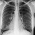
Fig. 7.1 Lung hyperinflation in asthma. Radiographs. (a) On clinical deterioration, bilateral, low-riding diaphragm as a sign of hyperinflation. (b) The findings revert to normal on clinical improvement.


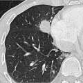
Fig. 7.2 Bronchial wall thickening (arrow) and mucoid impaction (arrowhead) in asthma. CT image.

Allergic bronchopulmonary aspergillosis progresses in five clinical stages7:


  • Stage 1: Acute stage (initial manifestation with cough, fever, transient pulmonary infiltrates, and bronchial obstruction).


  • Stage 2: Remission of clinical and immunologic parameters.


  • Stage 3: Exacerbation.


  • Stage 4: Corticosteroid dependence.


  • Stage 5: Fibrosis.

Radiologic changes may be absent in early stages. Findings include transient pulmonary infiltrates and evidence of mucoid impaction: tubular (toothpaste sign), branching consolidation (gloved finger sign), in particular, in the apical and central lung regions, and occasionally areas of lobular or segmental atelectasis.4,8 In advanced stages, central bronchiectasis and
lung fibrosis are observed as well. Radiologic findings (▶Table 7.4) can be distinguished as transient (▶Fig. 7.3) and chronic manifestations (▶Fig. 7.4).


Table 7.2 Radiologic findings in asthma


Diagnostic imaging


Findings


Radiograph (▶Fig. 7.1)


  • Signs of hyperinflation:


    – Low-riding diaphragm


    – Diaphragmatic flattening


    – Expansion of the intercostal spaces


  • Thickening of the subsegmental bronchial walls


  • Prominent hila (vascular, lymphadenopathy)


CT (▶Fig. 7.2)


  • Bronchial wall thickening


  • Linear opacities


  • Reduced lung density


  • Mucoid impaction


  • Small centrilobular opacities


  • Cylindrical bronchiectasis


  • Rarely, consolidation


Table 7.3 Diagnostic criteria for allergic bronchopulmonary aspergillosis4,6


Diagnostic examination


Diagnostic criteria


Clinical examination


  • Asthma


  • Immediate skin reaction to Aspergillus antigens


Serology


  • Blood eosinophilia


  • Precipitation antibodies against Aspergillus antigens


  • Elevated serum immunoglobulin E levels (correlates with disease activity)


Imaging


  • Pulmonary infiltrates


  • Central bronchiectasis


7.1.3 Hypersensitivity Pneumonitis

More than 200 inhalative allergens are known to trigger allergic reactions in the lung. ▶Table 7.5 gives a brief overview of these, often occupational, diseases. Pulmonary hypersensitivity involves types III (formation of immune complexes) and IV (T cell-mediated, delayed-type) allergic reactions.9

Clinical onset of hypersensitivity pneumonitis involves acute, flu-like symptoms, cough, and shortness of breath around 4 to 12 hours after contact with the allergen. Pathologic manifestations include neutrophilic and lymphocytic alveolitis as well as alveolar damage. Alveolitis may resolve or progress to lung fibrosis.

Three stages of hypersensitivity pneumonitis may be observed; they can merge or be coexisting in an individual patient. These are determined by the duration and extent of allergen exposure as well as by the immune response:


  • Acute hypersensitivity pneumonitis: Flu-like symptoms develop within a few hours after exposure to the allergen. If there is no further exposure to the allergen, the symptoms resolve within a few days. This stage is rarely seen on radiologic images since there is generally no indication for radiographic examination. If a radiograph has been obtained, it may show bilateral, more or less extensive, consolidation and small, rounded opacities(▶Fig. 7.5).11


    Table 7.4 Radiologic findings in allergic bronchopulmonary aspergillosis


    Transient findings


    Chronic findings


    • Consolidation (often recurrence at same location)


    • Mucoid impaction (toothpaste or gloved finger sign)


    • Atelectasis


    • Pleural effusion


    • Central bronchiectasis


    • Fibrosis


    • Bronchoceles


    • Mycetoma (in particular in the middle fields)


  • Subacute hypersensitivity pneumonitis: The acute phase of the disease resolves within a few days. Then, or in the intervals between several acute flares, a discrete nodular pattern is seen on the radiograph (▶Fig. 7.6). The CT findings are virtually pathognomonic: multiple centrilobular groundglass nodules and diffuse ground-glass opacity or mosaic
    pattern (▶Fig. 7.7). Occasionally, small cysts are observed as well. The findings can completely resolve in the absence of exposure to the allergen.


    Fig. 7.3 Allergic bronchopulmonary aspergillosis. CT image. Transient findings with consolidation in right upper lobe (arrowheads); mucoid impaction in the left upper lobe (arrow).


    Fig. 7.4 Allergic bronchopulmonary aspergillosis. CT images. Chronic findings. (a) Multiple areas of central bronchiectasis. (b) Besides, fibrosis (arrow) and → (arrowhead).


  • Chronic hypersensitivity pneumonitis: Long-lasting hypersensitivity pneumonitis can cause development of lung fibrosis (▶Fig. 7.8). If contact with the allergen persists, subacute and acute disease manifestations can coincide. While the latter entities are reversible, lung fibrosis secondary to chronic hypersensitivity pneumonitis is irreversible. This is characterized by restriction on pulmonary function tests. In radiologic findings, variable amounts of pulmonary fibrosis (honeycombing, intralobular lines, traction bronchiectasis) and ground-glass opacity are seen (▶Table 7.6). Unlike other forms of pulmonary fibrosis, chronic hypersensitivity pneumonitis is not associated with basal predominance. Likewise, there is little involvement of the costophrenic angles, whereas these are the main location affected by other forms of fibrosis (see Chapter 6). Imaging criteria for differential diagnosis are summarized in ▶Table 7.7.

    Only gold members can continue reading. Log In or Register to continue

    Stay updated, free articles. Join our Telegram channel

Apr 12, 2020 | Posted by in CARDIOVASCULAR IMAGING | Comments Off on Immunologic Diseases of the Lung

Full access? Get Clinical Tree
Get Clinical Tree app for offline access