Occupational Lung Diseases


Occupational Lung Diseases


Beate Rehbock


18.1 Introduction

Occupational or work-related disease is the term used to describe health disorders fully or partially attributable to work activity. These are usually caused by multiple factors, exhibit nonspecific symptoms, and very often have a chronic course. Lung diseases account for almost half of all occupational diseases and, at 87%, are statistically the leading cause of related deaths.

The most common pulmonary occupational disease is pneumoconiosis (black lung) which is caused by inorganic dust—in particular the clinical disease entities of silicosis and mixeddust pneumoconiosis as well as asbestosis and asbestos-related pleural disease. A specific feature of most occupational lung diseases is the long latency period (years to decades) between exposure to the hazardous substance and the appearance of radiographic findings or clinical symptoms. Imaging plays a key role in diagnosis and monitoring of asbestosis, asbestos-related pleural disease, and silicosis.


18.2 Imaging Modalities


18.2.1 Chest Radiography

Chest radiography is the first imaging modality used for all occupational lung diseases (posteroanterior and optional left lateral radiograph); the International Labour Organization (ILO) classification system (▶Fig. 18.1) is commonly used to report radiographic findings of pneumoconioses, especially in preventive medical checkups and medical opinions.1,2 This standardized diagnostic schema is an epidemiological instrument for internationally uniform description of radiographic changes following exposure to hazardous substances. It is not designed to provide a definitive diagnosis or information to establish compensation; instead, it is aimed at assignment of opacity types to pulmonary and pleural inhalation damage. Furthermore, the schema can be used for quantification of the disease extent and as a prognostic predictor as well as for detection of other diseases causing respiratory symptoms.3 The findings are evaluated by comparison with reference films available as hard copies and in digital format.

The ILO classification system can be used for any occupational lung or pleural disease. No image pattern is pathognomonic for dust exposure. An occupational disease can only be diagnosed by taking a detailed occupational history and through ascertainment of an occupational exposure to hazardous substances linked to a specific image pattern.


18.2.2 Computed Tomography

Since sensitivity and specificity of CT are superior to those of chest radiography, it is increasingly used for diagnostic imaging of occupational lung diseases.

In view of the fact that the majority of occupational lung diseases manifest interstitially, CT should be primarily used as a high-resolution (HR), weight-adapted, low-dose, unenhanced multidetector CT technique. Sequential thin slices should only be obtained if needed additionally on expiration (suspected air trapping) or in the prone position (for differentiation between fibrosis and dependent opacities).

Semi-quantitative classification of the CT findings is performed in accordance with the International Classification of HRCT for Occupational and Environmental Respiratory Diseases (ICOERD),4 which is similar to the ILO classification system (▶Fig. 18.2), and for which digital reference films are also available.5 Likewise, in this purely descriptive diagnostic schema, the letters and symbols merely serve as descriptors with no claim to conferring etiologic or pathogenetic insights. However, (in the “Remarks” column) a conclusive assignment should be made to a disease entity or suspected diagnosis, with reference to differential diagnosis if applicable.

IV contrast is needed for diagnosis of suspected tumors (pulmonary nodule, lung cancer, mesothelioma, pleural effusion).


18.2.3 Other Imaging Modalities

MRI has presently no established role in primary diagnosis of occupational lung diseases but can be superior to CT in resolving certain tumor-related diagnostic issues in lung cancer and pleural mesothelioma (e.g., local invasion of adjacent structures).6 MRI is reported to be advantageous in differentiating between progressive massive fibrosis and a malignant tumor (see ▶Fig. 18.11)7 as well as between rounded atelectasis and lung cancer.8 Likewise, PET-CT and ultrasonography are reserved for particular individual cases.


18.3 Disease Entities


18.3.1 Inorganic Dust-Induced Lung Diseases (Pneumoconiosis)

Pneumoconiosis presents as a result of a lung tissue reaction to inhaled dust particles that reach the alveoli. There is no clear-cut definition of pneumoconiosis or of how occupational lung diseases are classified as such. While the term coined in 1867 by Zenker also included organic dust, “pneumoconiosis” is currently generally understood to mean lung diseases caused by inorganic dust (▶Table 18.1). Pneumoconiosis may be fibrogenic or nonfibrogenic (▶Table 18.2). While berylliosis is also classified as pneumoconiosis, it occupies a special place because of its immunologic pathogenesis.3 Beryllium is a metal used, for example, in the automobile and aircraft construction industries as well as in aerospace technology but may also be found as traces in dental alloys.


Fig. 18.1 ILO classifications form—several variants of this form are available. Instructions for use and a key to the symbols are given in International Labour Office.9 (Reproduced with permission from © International Labour Organization, 2002.)


Fig. 18.2 CT classification form according to ICOERD. (Reproduced with permission from Kusaka et al.4)

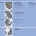
In terms of pathogenesis of pneumoconioses, it is thought that larger dust particles are eliminated by the ciliated epithelia of the tracheobronchial system or are deposited in the nasopharyngeal region. Dust particles measuring less than 5 µm in diameter and fibrogenic particles may be retained depending on the amount of dust and exposure time,
individual disposition and the integrity of the mucociliary clearance function (phagocytosis by alveolar macrophages). The mucociliary clearance function is adversely affected by cigarette smoking and toxic gases. After alveolar deposition the dust particles can trigger a chronic inflammatory reaction in the lung interstitium or be transported in the lymphatic and blood systems. Asbestos fibers can alter the pleura through pleural drift. All fibrogenic substances have the potential to cause irreversible damage to the lung parenchyma. The associated radiologic features will range from reticular (e.g., asbestosis) through reticulonodular to predominantly nodular pattern (e.g., silicosis) depending on the dust composition and severity of damage. Short-term exposure identified at an early stage often exhibits a nodular pattern with ground-glass opacity on HRCT (which applies also to rare types of pneumoconiosis). The more chronic the exposure and disease process, the more widespread the fibrosing pattern.


Table 18.1 Principal types of inorganic dust-induced pneumoconiosis


Pneumoconiosis


Occupations


Silicosis/coal workerspneumoconiosis


Stone quarry, stonemason, glass and ceramics industry, coal mining, foundry


Silicotuberculosis


Asbestosis/asbestos-related pleural disease


Roofer, fitter, insulator, etc. Currently, (generally little) exposure especially in demolition work


Aluminum-induced lung disease


Aluminum powder production (pyro grinding), aluminum welding, production of corundum for grinding wheels


Hard metal lung disease


Production of hard metal tools, e.g., drill heads


Siderofibrosis


Welding processes: with extreme and prolonged exposure to welding smoke and gases


Dental technician lung disease


With prolonged (18-year) exposure to mixed dusts; prevalence 10% (chromium, cobalt, molybdenum, beryllium, etc.)


Table 18.2 Common fibrogenic and nonfibrogenic dusts and resulting pneumoconiosis


Fibrogenic dust


Nonfibrogenic dust


Quartz, sand, and other silicic acid-containing minerals → silicosis


Iron oxide (“inert”) → siderosis


Asbestos (white and blue asbestos) → asbestosis and asbestos-related pleural disease


Carbon (soot, graphite) → anthracosis (coal workers’ lung disease)


Tin dust → stannosis


Talc soapstone (usually with quartz and/or asbestos content) → talcosis


Barium sulfate → baritosis


Aluminum dust → alumininosis


Hard metals (including titanium, tungsten carbide, cobalt) → hard metal lung disease


Table 18.3 Principal occupational causes of hypersensitivity pneumonitis


Disease entity


Antigens


Occupational risks


Farmer lung


Thermophilic actinomycetes


Agriculture, gardeners


Bird fancier lung


Animal-based antigens


Bird handlers, veterinarians


Air humidifier lung


Molds


Air-conditioning systems, air humidifiers


Baker lung


Moldy flour


Bakers, millers


Isocyanate alveolitis


Isocyanate compounds


Chemical workers


Clinically, inorganic—including fibrosing—pneumoconiosis can remain silent for a very long time. This is usually followed by onset of symptoms of restrictive lung function. In particular, in silicosis obstructive ventilation disorders are also observed.


18.3.2 Organic Dust-Induced Lung Diseases

Organic dust of animal and plant origin can trigger an allergic reaction of the airways. Hypersensitivity pneumonitis is a collective term that includes a number of disease entities with similar symptoms that may be triggered by various allergens (▶Table 18.3); for more details, please consult Section 7.1.3.

Byssinosis is a disease caused by the toxic potential of inhaled uncleaned cotton which may manifest clinically as chronic bronchitis and emphysema but is not associated with any specific radiographic features.

Pathophysiologically, hypersensitivity pneumonitis is caused by a type III and IV immunologic response. Its pathohistology is characterized by bronchocentric lymphocytic alveolitis with granulomatous inflammation that may evolve to fibrosis.10

Clinically, acute hypersensitivity pneumonitis may manifest with flu-like symptoms, including dyspnea and cough 6 to 8 hours after exposure. The acute variant is often self-limiting and reversible if allergen exposure is avoided early on. Persistent exposure can lead to interstitial fibrosis.


Table 18.4 Most common exposures that can cause a malignant tumor


Noxae


Malignant tumors


Asbestos


Asbestos-induced lung cancer


Asbestos-induced pleural mesothelioma


Lung cancer due to interaction between asbestos dust and polycyclic aromatic hydrocarbons


Ionizing radiation


Lung cancer


Silicium dioxide


Lung cancer in quartz dust-related lung disease (silicosis or silicotuberculosis)


18.3.3 Acute Inhalation Toxicity

The place and extent of damage are determined by the physical state, water solubility, dose, and pH value of the noxae. The following disease entities may result:


  • Acute toxic tracheitis and bronchitis: Water-soluble substances (e.g., ammonia, chlorine gas, hydrochloric acid, and formaldehyde) cause damage especially to the upper respiratory tract.


  • Chemical irritation or toxic asthma (reactive airways dysfunction syndrome): sulfur dioxide, sulfuric acid, isocyanate, and formaldehyde can trigger acute reflex bronchoconstriction with or without reversible obstruction.


  • Organizing pneumonia: Following inhalational injury with high doses of, e.g., nitrogen dioxide, sulfur dioxide, ammonia, or chlorine gas, organizing pneumonia can manifest after a latency period of only up to 3 weeks.


  • Pulmonary edema: Substances with poor water solubility (e.g., phosgene and ozone) as well as lipophilic substances, such as nitrogen dioxide, can cause intra-alveolar edema that manifests clinically only after a dose-related latency period. Bacterial pneumonia is common because of the damage to the immune function of the alveolar macrophages.


18.3.4 Chronic Bronchitis and Asthma

There are myriad workplace-related hazardous substances that can cause chronic as well as acute irritation disorders of the airways not amenable to primary diagnostic imaging. Exceptions to that rule are chronic obstructive pulmonary diseases like chronic bronchitis and emphysema; imaging plays an important role in these disease entities when they present in the following circumstances:


  • As a complication of silicosis and silicotuberculosis.


  • In underground coalminers exposed to a cumulative dose of more than 100 fine dust years ([mg/m3] × years).

In addition to the pathognomonic changes following quartz dust exposure, special attention should be paid on chest radiography, and possibly HRCT, and to emphysema criteria since this can present as a complication of occupational chronic bronchitis.


18.3.5 Malignant Occupational Diseases of the Lung and Pleura

Malignant tumors of the lung and pleura quantitatively account for the majority of occupational cancer diseases (▶Table 18.4). In particular, these are asbestos- and quartz dust-related lung cancers as well as asbestos-related pleural mesotheliomas. While imaging is unable to directly impute the tumor to an occupational pathogenesis, through indirect radiologic signs it can often establish a probable link to asbestosis, asbestos-induced pleural disease, or silicosis.


18.4 Diagnostic Imaging of Special Disease Entities


18.4.1 Asbestosis and Asbestos Dust-Related Pleural Disease

As per its definition, asbestosis consists of diffuse, bilateral interstitial fibrosis caused by inhalation of asbestos fibers. Asbestos-induced fibrosis can affect the pulmonary interstitium (asbestosis) as well as the parietal and/or visceral pleura.11 Histologic evidence of asbestos bodies in the lung parenchyma is only a marker of exposure and in itself does not constitute disease.

High asbestos exposure levels are generally responsible for parenchymal fibrosis with a latency of more than 20 years.3 Compared to lung fibrosis, pleural changes already appear at lower levels of asbestos exposure. Therefore, they are today more common findings than lung fibrosis because of the ban on the use of asbestos in numerous countries from its historical peak in 1980 and the introduction of extensive occupational safety measures. During the first decade of asbestos exposure, the pleura often reacts with exudation (asbestos pleuritis), only later followed by areas of discrete pleural thickening (pleural plaques) as well as pleural fibrosis (visceral pleural thickening).3


Fig. 18.3 Diffuse pleural thickening (ILO 2b) with involvement of the right recess consistent with hyalinosis complicata. (a) Magnified section of a radiograph. (b) On CT, virtually circumferential, partially calcified thickening of the costal pleura (ICOERD w [ =wall] of visceral type) with subpleural parenchymal reaction and fibrosis. Ill-defined, lung-sided delineation of the area of pleural thickening, with parenchymal involvement (arrow).


Pleural Asbestos-Related Changes

Pleural plaques are areas of discrete hyaline or calcified fibrosis of predominantly the parietal pleura (costal, diaphragmatic, and mediastinal pleura).7 The following variations in attenuation are observed:


  • Noncalcified (hyaline): probably primary.


  • Calcified: partial or complete.


  • Location of calcifications: at the center or base of the plaque or isolated pleural calcification.

A distinction must be made between the following morphologic types:


  • Table mountain-shaped (deemed pathognomonic for asbestos exposure and usually smoothly marginated [parietal type]; predominantly located in the costal pleura).


  • Hill- and spindle-shaped pleural thickening.


  • Thickening at the level of the pleura.


  • Visceral pleural thickening (with ill-defined, lung-sided margin, consistent with subpleural parenchymal changes like local subpleural fibrosis; ▶Fig. 18.3).

The following locations are characteristic:

Apr 12, 2020 | Posted by in CARDIOVASCULAR IMAGING | Comments Off on Occupational Lung Diseases

Full access? Get Clinical Tree

Get Clinical Tree app for offline access