Nonvascular Interventions
Conduct of percutaneous diagnostic and therapeutic interventions in the thorax is long established. These are carried out under the guidance of different imaging modalities (fluoroscopy, ultrasound, CT, including fluoro-CT, and MRI). The choice of modality is determined by the available equipment, location and size of the suspicious lesion, and the radiologist’s experience and preferences. These imaging techniques differ considerably in terms of costs, availability, ease of handling and control, and radiation dose (▶Table 20.1).
As for all interventions, three preconditions must be met for interventions in the chest:
Indication: The following sections give a detailed account of the indications for the various techniques.
Patient information: While severe or life-threatening complications are rare in thoracic interventions, they do occur. Therefore, on the day before the intervention by the latest a physician with experience of the proposed intervention should inform the patient about the risks involved and obtain the patient’s written informed consent.
Appropriate coagulation: Except for chest wall interventions, hemostasis through manual compression is generally not possible for the chest organs. Functional blood coagulation is therefore paramount. To ensure that this is the case, an in-depth history of blood coagulation must be taken (bleeding events and anticoagulant medication). Interventions should only be carried out if there is no clinical or paraclinical evidence of coagulation disorders or when any such existing disorder has been effectively treated.
20.1 Biopsy
Transthoracic biopsies are performed to harvest tissue samples for histologic or cytologic analysis. There are two techniques available: fine needle aspiration and core biopsy. The first lends itself only to cytological and microbiological analysis, while histologic analysis can only be performed after core biopsy. Accordingly, core biopsy should be conducted for differential diagnosis in cases of suspected lung cancer. Furthermore, novel treatment approaches to lung cancer require molecular genetic analysis of the biopsy specimen; hence, enough tissue must be available.
Table 20.1 Comparison of imaging techniques for guidance of thoracic interventions1 | ||||||||||||||||||||||||||||||||||||||||||||||||
|---|---|---|---|---|---|---|---|---|---|---|---|---|---|---|---|---|---|---|---|---|---|---|---|---|---|---|---|---|---|---|---|---|---|---|---|---|---|---|---|---|---|---|---|---|---|---|---|---|
|
20.1.1 Indications
Note 
Important indications for obtaining tissue specimens (transthoracic biopsy) from a pulmonary or mediastinal pathologic process1,2:
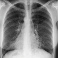
Pulmonary nodule or mass of unclear etiology.
Suspected malignant pulmonary nodule if no primary surgical treatment is prescribed or minimally invasive surgery is planned (intraoperative confirmation based on frozen section is not possible).
Nodule in patient with history of malignant tumor, tumor in clinical remission, or multiple primary tumors.
Residual nodule following radiotherapy or chemotherapy.
Tissue harvesting for molecular biology tests.
For pathogen isolation in suspected inflammatory process in immunoincompetent patients.
Other indications may apply in an individual case. Indications should always be based on multidisciplinary consultation.
While tissue sampling of centrally located lesions is a domain of bronchoscopy, lesions in the lung periphery can better be approached with transthoracic biopsy.3
20.1.2 Preprocedure Assessment
A safe venous approach is needed to treat any complications arising during the intervention. Provision should be made for pulse oximetry and a control monitor. In principle, sedation compromises the patient’s cooperation capability and should therefore be reserved for unavoidable exceptional cases.
Patient positioning will depend on the chosen access route (see below). In general, the prone position confers greatest stability since chest wall breathing movements are minimal. The supine position assures good conditions, too. However, the lateral position is the least stable because even shallow breathing causes maximum chest movements and minimal changes to the patient positioning have major effects.
20.1.3 Technique
A local anesthetic is administered before beginning the intervention, while avoiding puncture of vessels or the pleura; this is assured by means of aspiration while advancing the needle. In addition to the skin, the parietal pleura, which is very sensitive to pain, must be anesthetized.
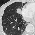
Biopsy cannulas with a diameter of more than 18 G are more likely to cause bleeding complications than are thinner cannulas. This is particularly true for interventions in the vicinity of major vessels or highly vascularized lesions. To avoid multiple pleural passes with the biopsy needle, a coaxial system is used in principle for pulmonary core biopsies. But caution is advised for lesions with air bronchogram since the use of a coaxial system increases the risk of air embolism.4
The risk of vascular injury is the chief determinant of the access route chosen. In particular, the internal thoracic vessels and intercostal arteries must be protected. The number of pleural passes should be kept to a minimum; in particular, needle pass of pulmonary fissures should be avoided. Emphysematous lung areas, especially emphysematous bullae, present a high risk of pneumothorax and should be evaded. For biopsy of mediastinal masses, preference is given to an extrapleural over a transpulmonary access approach.5 Widening of the mediastinum by injection of several 10 mL of saline solution in the mediastinal fat may establish an extrapleural access in some cases.
The biopsy cannula is inserted into the suspicious lesion under imaging guidance. Due to the risk of false-negative results, the biopsies should be taken from a peripheral region, especially in the case of large nodules with central necrosis. Several core biopsies are obtained from different parts of the lesion by using different angulations during needle insertion.
Imaging is conducted again after removal of the biopsy cannula to rule out complications. If there is evidence of pneumothorax this can be aspirated immediately after the intervention. Postinterventional radiography after a few hours is also indicated for transpleural punctures.
20.1.4 Complications
The complication incidence of fine needle aspiration is similar to that of core biopsy. There is only a loose correlation between onset of pneumothorax and the biopsy cannula gauge. Only the bleeding risk is somewhat higher when using large-gauge cannulas.4 The typical complications associated with biopsy of chest organs are summarized in ▶Table 20.2.
Related posts:

Stay updated, free articles. Join our Telegram channel

Full access? Get Clinical Tree


