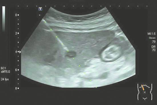
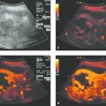
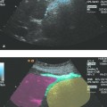
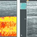
Indications for Diagnostic Interventions in the Abdomen and Thorax (Liver, Pancreas, Spleen, Kidneys, Lung, Other Sites) Focal lesions in abdominal organs are a common finding in routine ultrasound examinations. An ultrasound-guided percutaneous aspiration or biopsy is a simple and safe procedure that allows an experienced operator to obtain material for histologic or cytologic analysis with little risk of complications.1,2 Besides distinguishing between benign and malignant lesions,3 this procedure can provide material for immunohistochemical testing that can further differentiate the lesion and direct further management. In parenchymal diseases of the liver, a histologic examination is crucial for initial evaluation and follow-up (▶ Table 12.1). Neither imaging studies nor serologic tests can accurately quantify disease activity or the fibrotic and cirrhotic changes that will determine the further clinical course. For its simplicity, we prefer the Menghini needle for ultrasound-guided liver biopsies (Hepafix Luer lock, 18 gauge/1.2 mm, needle length 88 mm, 45° bevel; Braun) (see Chapters ▶ 2 and ▶ 14). Acute viral hepatitis is usually diagnosed by serologic testing (anti-HAV IgM, anti-HEV IgM; HBsAg, if positive anti-HBc IgM and HBV DNA; anti-HCV, if positive HCV RNA; anti-HDV IgM in acute hepatitis B; EBV IgM in patients with corresponding symptoms; CMV Ab and CMV DNA in immunosuppressed patients). Biopsy confirmation is necessary only if there is another presumed cause of liver damage besides acute viral hepatitis (e.g., a known history of chronic hepatitis with suspected superinfection, known alcohol abuse, autoimmune hepatitis). If the disease takes a fulminating course with impending liver failure, a prompt needle biopsy can be an important prognostic study—for if the rare necrotizing form is identified, liver transplantation will be the only remaining therapeutic option. In patients who show progressive jaundice, a rapid fall in transaminases, hepatic encephalopathy, and deteriorating clinical status, percutaneous biopsy is contraindicated in cases with coagulation system collapse (Quick value <50% and platelet count <50 × 109/L) due to the excessive bleeding risk4 (see Chapters ▶ 4 and ▶ 9). For the histologic evaluation of necrosis, transjugular liver biopsy is a safer alternative as it substantially reduces the risk of significant bleeding. Specific diseases that can be investigated by ultrasound-guided biopsy are reviewed in the paragraphs below. Relevant diagnostic tests are shown in parentheses. Hemochromatosis can be diagnosed serologically (ferritin, transferrin saturation, HFE gene mutation chromosome 6—C282Y, H63D, and S65C). The low genetic penetrance requires a liver biopsy if it is suspected that an additional agent is responsible for the liver damage (e.g., nonalcoholic steatohepatitis, NASH). The suspicion of severe fibrosis or cirrhosis should also be investigated by biopsy. This also applies to hepatic porphyria (total porphryins in the urine and stool during an acute attack) and alpha1-antitrypsin deficiency (serum electrophoresis, phenotyping if required). With Wilson disease (ceruloplasmin; may test urinary copper at baseline and with D-penicillamine administration), the diagnosis should be confirmed histologically if tests are inconclusive (▶ Table 12.1). Urinary copper excretion <100 μg/24 h confirms a positive treatment response. Later in the course of the disease, it may be necessary to perform a repeat liver biopsy to assess the degree of cirrhosis, especially as a prelude to liver transplantation. The glycogen storage diseases require biopsy with a glycogen analysis and, if necessary, an in vitro enzyme activity assay. Primary biliary cirrhosis (PBC) is diagnosed serologically (anti-mitochondrial antibodies [AMAs]). Histologic evaluation of the activity and stage of the disease has additional prognostic significance (e.g., expand pharmacologic therapy, prepare for liver transplantation) and fully justifies liver biopsy.5 Autoimmune hepatitis should be consistently investigated by liver biopsy before treatment is initiated and later to evaluate the course. Histologic evaluation is also recommended before treatment is concluded.6 While Doppler ultrasound can demonstrate occlusion of the large hepatic veins in Budd–Chiari syndrome, the diagnosis of venoocclusive disease relies entirely on histologic findings. In chronic hepatitis C and B, the histologic grading of viral hepatitis (e.g., using the Desmet–Scheuer score7) provides an additional decision-making criterion for treatment planning, since higher grades of fibrosis would predict a poorer response to interferon therapy. Moreover, evaluation by an experienced pathologist will disclose aspects that may be important in the clinical management of the patient (e.g., changes due to coexisting NASH indicating a need for weight reduction).8 In patients with nonalcoholic steatohepatitis (NASH), we routinely perform percutaneous liver biopsy as an aid to differential diagnosis and staging (▶ Table 12.1). Alcohol-induced hepatitis is usually diagnosed from the patient’s history and does not consistently require biopsy. If inflammatory hepatitis due to a different cause is suspected (e.g., hepatic involvement by sarcoidosis), liver biopsy may be helpful in exceptional cases—especially if the findings of the presumed disease are inconclusive. Focal liver lesions are a frequent indication for biopsy, due in part to the possibility of metastatic lesions from various tumors. On the other hand, the overall percentage of cases selected for biopsy has fallen by an estimated 5 to 10% due to the high diagnostic accuracy of contrast-enhanced ultrasonography.9 Hemangioma and focal nodular hyperplasia (FNH) are the most common benign liver lesions and occur predominantly in female patients.10 Imaging evaluation is adequate in most cases, making it unnecessary to proceed with biopsy (caution: FNH is difficult to distinguish from fibrolamellar liver cancer). Liver biopsy is indicated only in patients who have a history of malignancy and equivocal imaging findings. This also applies to the much rarer entity of hepatic adenoma. Larger adenomas should be resected due to the high incidence of rupture (up to 30%).11 The need for preoperative biopsy should be assessed in consultation with the surgeon. The fine needle aspiration cytology of metastatic lesions in known cancer patients is often sufficient to confirm a malignant histology. But if a primary tumor is not known or the tumor is assumed to have originated in the liver, core needle biopsy may be necessary to establish the tumor histology. On the initial discovery of liver metastases in the absence of a known primary (CUP syndrome), needle biopsy can provide essential information and should therefore be done liberally and without delay (▶ Fig. 12.1). Immunohistologic staining of the specimen is a very rewarding test. Fig. 12.1 Biopsy of a hepatic metastasis from breast cancer. Metachromatic metastasis. The patient also had a history of renal cell carcinoma. The ultrasound visualization of cholangiocellular carcinoma (CCC) is difficult and often requires the use of sonographic contrast agents. Cytologic-histologic confirmation can be obtained during ERCP (endoscopic retrograde cholangiopancreatography) in tumors that have invaded the biliary tract. Percutaneous biopsy of CCC can also be done in a palliative setting,12,13 in which case we usually perform a fine needle aspiration biopsy. The cirrhotic liver is an unusual site for metastatic lesions but is a relatively common site for hepatocellular carcinoma (HCC). The friable structure of cirrhotic liver tissue makes it more difficult to retrieve a good-quality tissue core, but it is important for the pathologist to receive an intact core due to the inherent difficulty of distinguishing among regenerative nodules, dysplastic nodules, and well-differentiated hepatocellular carcinoma11 (▶ Fig. 12.2, ▶ Fig. 12.3).
12.1 Liver
12.1.1 Diffuse Liver Diseases
Disease
Abbreviation
Indication for core histology
Acute viral hepatitis
AVH
Fulminating clinical course with impending liver failure
Chronic viral hepatitis
CVH
Grading before treatment and during further course
Autoimmune hepatitis
AIH
For confirmation and follow-up
Primary biliary cirrhosis
PBC
For staging and determining activity
Primary sclerosing cholangitis
PSC
Only in exceptional cases, as in overlap syndrome
Caroli syndrome
Assessment of fibrosis and before liver transplantation
Vanishing bile duct syndrome
VBDS
Essential for confirming diagnosis
Venoocclusive disease
VOD
To confirm diagnosis
Budd–Chiari syndrome
BCS
Biopsy generally is not indicated
Nonalcoholic steatohepatitis
NASH
May be done to confirm diagnosis
Alcoholic steatohepatitis
ASH
For grading of fibrosis and cirrhosis (in selected cases), with coexisting diseases, in preparation for liver transplantation
Hemochromatosis, porphyria, alpha1-antitrypsin deficiency
For grading of fibrosis and cirrhosis
Glycogen storage disease
Biopsy with glycogen analysis, in vitro enzyme assay if needed
Wilson disease
For copper determination in doubtful cases
Drug-induced injury
Biopsy is essential to establish diagnosis
Osteomyelofibrosis
OMF
Detection of extramedullary hematopoiesis
12.1.2 Focal Liver Lesions
Benign Lesions
Malignant Lesions
Metastases
Primary Liver Tumors
Related posts:
Stay updated, free articles. Join our Telegram channel

Full access? Get Clinical Tree


