Percutaneous Gastrostomy
A gastrostomy can be performed by surgical or nonsurgical means. Nonsurgical methods are broadly subdivided into endoscopic procedures and interventional radiology.
Endoscopic techniques are well established, and the “pull” method for percutaneous endoscopic gastrostomy tube delivery is the most commonly used. It is easily learned and has low complication rates.
Interventional radiology using fluoroscopy or ultrasound can eliminate the need for endoscopic access. The strength of imaging techniques is that they can safely avoid extraluminal structures such as the liver and transverse colon and prevent endoscopic access.
This chapter will examine the role of ultrasonography both as an adjunct and as a standalone guidance modality for percutaneous gastrostomy placement. The potential benefits of the method and its indications will be explored.
21.1 Indications
Percutaneous gastrostomy is generally done to provide intermediate- or long-term nutrition in patients who cannot take food orally. It follows the principle that, whenever possible, enteric feeding is preferable to parenteral nutrition. Thus the indications include patients on long-term ventilatory support and patients with acquired diseases or disturbances of deglutition, as in CNS diseases. A gastrostomy may also be placed to decompress the stomach contents when gastrointestinal continuity is disrupted due to a malignant bowel obstruction.
21.2 Contraindications
The only acute situation that would require an emergency gastrostomy is tube placement for gastric decompression because of bowel obstruction. This may be done for palliation in patients with, say, a malignant stricture distal to the stomach.
Feeding tube placement is contraindicated in unstable patients, who should be adequately managed by temporary parenteral nutrition.
In principle, a feeding gastrostomy is a palliative option in a terminally ill patient only if it would subjectively improve the patient’s quality of life.
21.3 Materials and Equipment
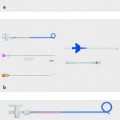
Two standard systems used for percutaneous gastrostomy placement are the Wills–Oglesby gastrostomy sets (Cook Medical Europe), which are available in a conventional version and in the Mallinckrodt Institute modification. The retention mechanism in both versions is a locking pigtail loop. The conventional system measures 12F, while the modification has a 14F catheter. All initial drainages must be exchanged. We prefer the 12F system as its smaller diameter affords a certain degree of safety, especially at the start of the learning curve. It is supplied with a long Lunderquist wire together with 7F, 10F, and 12F dilators. The Mallinckrodt system is supplied with a 100-cm Amplatz wire and only two dilator sizes (12F and 14F). With both systems, the T-fasteners that are necessary for pulling the stomach wall against the abdominal wall must be ordered separately. The fasteners are available in a twin-pack. Similar systems are offered by other companies. These techniques require the use of very stiff, torque-stable guidewires. The Lunderquist wire is even stiffer than the Amplatz wire; both are made of coated stainless steel.
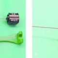
The Freka-Pexact gastrostomy tube (Fresenius Kabi) is an alternative set for the placement of a 15Ch (15F) balloon tube designed for insertion by an introducer. The manufacturer recommends the use of endoscopic guidance. Special features include placement with a special gastropexy system, which is described later in the chapter. Also, the abdominal wall is not punctured with a guidewire followed by dilators as in the traditional introducer technique. Instead, the puncture is performed directly with a 16F trocar covered with a sheath. The solid steel trocar is then removed, and the sheath provides access for placing the gastrostomy tube. The sheath is then peeled apart and removed.
21.4 Types of Gastrostomy
The following nonsurgical procedures are most widely practiced:
Percutaneous endoscopic gastrostomy (PEG) using the pull method, the push method, or the introducer method
Percutaneous radiologic gastrostomy (PRG)
Percutaneous sonographic gastrostomy (PSG)
21.4.1 Percutaneous Endoscopic Gastrostomy
Pull Method
The “pull” method is the standard technique. First the puncture site is identified endoscopically by transillumination or digital indentation and a needle is passed through the abdominal wall into the stomach. A thread is passed through the needle, grasped with an endoscopic forceps, and brought out through the mouth with the endoscope. The feeding tube is attached to the thread. The percutaneous end of the thread is then pulled to draw the PEG tube down the esophagus and against the stomach and abdominal wall, where it is fixed with a retention system. The advantage of this method is that sturdy, stable retention plates can be delivered through a normally patent esophagus.
Push Method
This method is rarely if ever used today. The technique is analogous to the pull method, except that the PEG tube is pushed into the stomach over the guidewire.
Introducer Method
In the conventional introducer method, a needle is passed into the stomach at a point identified by transillumination and/or indentation. Then a guidewire is introduced and placed in the gastric fundus. Most but not all authors perform a gastropexy. The tract is then dilated over the wire to allow the placement of an approximately 12F gastrostomy catheter. This tube must have a retaining device that can “collapse” during the insertion process and allow the tube to enter the stomach. Balloon retention catheters are most often used for this purpose. Other alternatives are locking pigtails or retention baskets.
Direct puncture systems are available in which a peel-away plastic sheath is introduced over a trocar, creating access for the placement of a 15Ch (15F) balloon catheter (Freka Pexact, Fresenius Kabi)
Percutaneous Radiologic Gastrostomy
The stomach is distended with 500 to 1500 mL of air via stomach tube and is punctured at a site located by transillumination. Some authors perform a contrast enema to opacify the transverse colon. Then a guidewire is passed into the gastric fundus. Many authors recommend performing a gastropexy. But there is no consensus on the number and positioning of gastropexy devices. Then the tract is enlarged with serial dilators, and a gastrostomy catheter is placed as in the introducer method.
21.4.2 Percutaneous Sonographic Gastrostomy
The technique is described below in the section on Ultrasound-Assisted PEG.
21.5 Advantages and Disadvantages of Different Methods
The advantages and disadvantages of the various gastrostomy methods are outlined in ▶ Table 21.1.
| Method | Advantages | Disadvantages |
| Endoscopy | The luminal view permits accurate placement of a stable retention plate and allows safe gastric distention. | Interposed liver or colon cannot be seen. The endoscope itself may cause complications in some cases. Most patients require sedation. |
| Fluoroscopy | Clear visualization; the entire stomach can be seen. The colon can be visualized. | In most cases the colon requires opacification by contrast enema. The liver is poorly visualized. Radiation exposure. |
| Ultrasonography | Needle insertion can be monitored in real time while also visualizing the transverse colon and liver. | Ultrasound does not give a clear view of the stomach. |
21.6 Success Rates of Different Gastrostomy Techniques
It is difficult to compare the success rates of different gastrostomy techniques because published patient cohorts have been heterogeneous with a range of underlying diseases. Success rates and complication rates vary widely in neurologic patients, patients with head and neck tumors, and patients with upper gastrointestinal tumors.
Endoscopic placement is frequently preferred. Nevertheless, local practices and preferences—in short, local expertise—will often dictate the selection of a particular gastrostomy method. If it is unsuccessful, an alternative procedure will be used. Thus there is a preselection of critical patients or patients with a complicated anatomy for the “second choice” method. Accordingly, local expertise will have a significant impact on complications and success rates.
The success rates stated in the literature are probably too high, and no randomized comparative studies are available. Reported success rates are generally in the range of 92 to 100% for PEG, 99 to 100% for PEG using the introducer technique, 95 to 100% for PRG, and 98 to 100% for PSG in the few studies that have been published on ultrasound-guided placement. With few exceptions, all the studies on PSG published to date have been feasibility studies.
21.7 Complication Rates of Different Gastrostomy Techniques
Complications can be classified as follows:
Endoscopy-related complications (perforation, etc.)
Sedation-related complications (allergies, respiratory arrest, etc.)
Puncture-related complications (perforation, hemorrhage, etc.)
Later complications (dislodgment, infection, peritonitis, etc.)
As far as sedation is concerned, we note that procedural sedation (brief general anesthesia) is used in most PEG studies while some radiologic studies use local anesthesia alone. Upper gastrointestinal endoscopy does not require sedation in itself, and the same is true for the abdominal intervention. Issues of comorbidity and compliance are more important factors in determining whether sedation should be used in any given case (uncooperative patients, children).
Moreover, no reliable data have been published on the incidence of complications. The reasons for this were mentioned in the preceding section. It is also difficult to distinguish between major and minor complications, as the underlying data are not presented in all studies.
The complication rates stated in the literature are summarized in ▶ Table 21.2.
| Major | Minor | Mortality | Source (reference no.) | |
| PSG with a stomach tube | 0% | 0% | 0% | 7 |
| PSG without a stomach tube | 0–3% | 0–14% | 0–2% | 7,10 |
| US-assisted PEG | 7% | 14% | 0% | 5 |
| PRG | 0–16% | 6–56% | 0–8% | 2,3,11–16 |
| PEG by pull method | 0.5–16% | 3–45% | 0–6% | 2,11,12,15–20 |
| PEG by introducer method | 0.70% | 11% | 0% | 21 |
| Abbreviations: PEG, percutaneous endoscopic gastrostomy; PRG, percutaneous radiologic gastrostomy; PSG, percutaneous sonographic gastrostomy; US, ultrasound. | ||||
An important observation is that the complication rates of endoscopic gastrostomy, especially using the pull method, are higher in patients with head and neck tumors. Alternative or supportive techniques are particularly recommended in this subset of patients. It should be possible to cross the stenosis with the thinnest available gastroscope and then place the tube percutaneously using Seldinger technique. Sonographic guidance for the puncture can provide additional safety.
In a series of 79 patients with head and neck carcinoma, Tucker and colleagues found that the pull method (n = 50) and introducer method (n = 29) had the same success rates but that the pull method was associated with a significantly higher complication rate.1
In one study of 177 gastrostomies placed endoscopically and 193 placed fluoroscopically, no significant differences were found in the success rates or complication rates.2
The studies that have been published on ultrasound-assisted gastrostomies probably do not report valid complication rates due to small case numbers and a presumed selection bias, which will be discussed later in this chapter.
21.8 Role of Ultrasonography
21.8.1 General
Related posts:
 Informed Consent
 Percutaneous Sclerotherapy of Cysts
 Contraindications, Complications, and Complication Management
 Fine Needle Aspiration Biopsy and Core Needle Biopsy
 Indications for Diagnostic Interventions in the Abdomen and Thorax (Liver, Pancreas, Spleen, Kidneys, Lung, Other Sites)
 Thoracic Interventions
Stay updated, free articles. Join our Telegram channel

Full access? Get Clinical Tree


