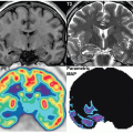
Infection and Inflammation


Infection and Inflammation


Søren Hess

Abass Alavi


INTRODUCTION

Radionuclide imaging has played a major role in the diagnostic workup of suspected infection and inflammation in the past almost 50 years since the incidental discovery that 67Ga-citrate accumulated in inflammatory tissue while investigating its use in Hodgkin lymphoma—the exact mechanism is still not fully understood, but 67Ga-citrate is taken up by transferrin and other iron-carriers ion and the 67Ga-citrate-complexes accumulate at sites of inflammation by a combination of hyperperfusion, increased vascular permeability, and direct binding to leucocytes or bacterial siderophores. Although 67Ga-citrate scintigraphy displayed good sensitivity for both acute and chronic inflammation, specificity was lacking due to widespread physiologic tracer distribution throughout the body for several days after tracer administration, which necessitated delaying imaging for as much as 72 to 96 h. Furthermore, 67Ga has an unfavorable long half-life and high energy gamma photons leading to a relatively high radiation burden. The next generation infection scintigraphy based on labeled white blood cells (WBC) was introduced in 1976 with 111In-oxine, 10 years later with 99mTc-HMPAO, and later 18F-fluorodeoxyglucose-positron emission tomography (FDG-PET) was applied to infection and inflammation (1,2).

With increasing availability, FDG-PET/CT has been applied in most common inflammatory and infectious indications. While 67Ga-citrate SPECT/CT remains an effective method for demonstrating spinal infection and active pneumonitis and nephritis and chronic infections, it is no longer considered the radionuclide gold standard for infection and inflammation. The usual clinical choices in infection imaging is now limited to WBC scintigraphy or FDG-PET/CT.

It has become apparent that the diagnostic value of FDG-PET/CT is at least comparable to WBC scintigraphy in most clinical settings. FDG-PET/CT is performed within 1 h with a contemporaneous CT, all without the task of collecting, labeling, and risk of reinjecting the patient’s blood, or waiting 24 to 48 h for the final results. Thus, FDG-PET/CT should be considered the modality of choice for most indications—this chapter outlines the most common use of radionuclide imaging in infectious and inflammatory diseases (Table 9.1).


Basic principles of imaging labeled white blood cell scintigraphy

Two inherently different agents are available for ex-vivo labeling of WBC, 111In-oxine, and 99mTc-HMPAO. Both are based on ex-vivo labeling of autologous WBC. After withdrawal of whole blood, red blood cells are sedimented and plasma is centrifuged to segregate the leucocytes, primarily neutrophils, which in turn are labeled and reinjected into the patient. 111In-oxine is lipid soluble and readily diffuses across the cellular membrane and forms stable binds to cytoplasmic components, whereas 99mTc-HMPAO is lipophilic. Isotope half-life and radiation dose is lower with 99mTc-labeled HMPAO compared to 111In-oxine, and the decay energy and image quality is also more favorable with the former than the latter (1).

Regardless of labeling method, interpretation is based on uptake patterns over time, that is, within about an hour after injection leucocytes migrate to sites of infection as a result of chemotaxis and most reports use increased uptake on sequential scans to define a positive WBC scintigraphy, whereas stable or decreasing uptake is not considered indicative of infection (3,4,5). However, several caveats and pitfalls need to be acknowledged; first of all, the rate of accumulation depends on several factors including the site (e.g., faster in vascular tissue and slower in bone due to compromised blood flow), type of pathogen, virulence, and extent (i.e., more or less chemotactic signals to accumulate leucocytes) (3). Secondly, the bio-distribution of the labeled cells is important, as both 111In-oxine-labeled and 99mTc-HMPAO-labeled WBC accumulate in the lungs rapidly after injection and subsequently lung activity clear after 3 to 4 h. Labeled WBCs also accumulate in the liver, spleen, and bone marrow as part of the reticuloendothelial system where they are retained. However, because of differences in labeling attributes, over time, 99mTc elutes the cells and is excreted in urine and stool, thus radioactivity from 99mTc-HMPAO-labeled WBC is visible in the entire urinary and gastrointestinal system, for example, kidneys, bladder, gall bladder and especially colon, which limits the usage in suspected inflammation in these organs (1,4). Thus, specific imaging protocols should be employed depending on the clinical challenge, for example, early-late phase images in suspected
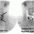
abdominal infection, especially if 99mTc-HMPAO is used, delayed images in suspected bone or prosthetic joint infection, or completely refraining from using labeled WBC if infection in the spine is suggested (5).


Basic principles of imaging FDG

As a glucose analogue, FDG in many respects imitates the distribution and cellular uptake of glucose throughout the body. A multitude of clinical settings are characterized by increased glucose uptake, most prominently exploited by FDG-PET/CT in imaging malignancies; it is well-known that cancer cells adhere to the so-called Warburg effect, that is, their metabolism is based on glucose consumption rather than the more energy-efficient oxidative phosphorylation even under aerobic conditions, so the net effect is a generally increased glucose metabolism through upregulation of GLUT. Inflammatory cells also utilize glucose; in fact, the increased uptake of FDG in inflammatory cells was initially dismissed as a nuisance leading to false-positive findings in patients suspected of malignant disease, but already in the early days of FDG-PET, it became clear that the effect was not specific to cancer (6,7). One of the first to present FDG-PET used directly in the diagnostic workup of infection was Tahara et al., who presented two case reports of abdominal abscesses with clearly increased FDG-uptake compared to adjacent soft tissue, both visually and semi-quantitatively (8). Subsequent preclinical pathophysiologic studies established that GLUT were not only upregulated in cancer cells but also by immune-mediated cytokine release in inflammatory settings; autoradiography showed increased expression of GLUT in the activated granulocytes that dominate early phases of inflammation in artificially induced septic inflammation, and in the macrophages that dominate more chronic stages of inflammation in artificial aseptic turpentine-induced inflammation (9,10). Thus, the potential use in inflammatory diseases was slowly recognized, but FDG did not become a serious contender in inflammation imaging until the recently increased availability in the past two decades.

Patient preparation before FDG-PET/CT in infection and inflammation imaging generally follows the same principles as for tumor imaging, that is, fasting for at least 4 to 6 h prior to injection to ensure optimum blood glucose levels of <150 mg/dL (8.3 mmol/L) (11,12). It seems though that the effect of relative hyperglycemia is less pronounced on inflammatory cells compared to cancer cells; several studies including meta-analysis of large populations suggest limited effect as long as blood glucose is kept below 200 mg/dL (11.1 mmol/L), and chronic hyperglycemia has less impact than acute hyperglycemia or hyperinsulinemia (13,14,15). On the other hand, prolonged fasting for up to 12 to 18 h preceded by low-carb-high-fat dietary restraints are important measures to shift the physiologically predominant glucose-driven metabolism of the heart toward free fatty acids to suppress physiologic FDG uptake sufficiently to assess suspected cardiac inflammation and infections (16).

Some medication may generally interfere with interpretation of FDG-PET/CT, for example, metformin should be discontinued for 48 to 72 h prior to the scan if pathology in the bowel is suspected, and this is also the case in infection and inflammation. More specifically, the mainstay treatment strategy of several noninfectious inflammatory disorders includes corticosteroids and the impact on sensitivity may be significant as it has been shown in large vessel vasculitis; sensitivity of visual as well as semi-quantitative assessment was not affected after 3 days but was significantly reduced after 10 days’ treatment; thus, imaging
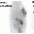
in suspected inflammatory disease should be performed before or shortly after treatment is instituted, and otherwise discontinuation should be considered (17).


Clinical imaging strategies in selected clinical settings

Radionuclide imaging is employed in a multitude of clinical settings of infectious and inflammatory diseases, and it is beyond the scope of this chapter to present an exhaustive review. In the following sections, we present some overall considerations regarding the most common or well-established indication in systemic diseases as well as focal/local organ specific affection. Generally, WBC scintigraphy is of little use in the initial diagnostic workup of suspected systemic inflammation; FDG-PET/CT provides a better solution on most accounts. On the other hand, some WBC scintigraphy is still considered routine examination or even the modality of choice in several local infections, for example, suspected prosthetic infection.

One of the issues that is often broad forth is the relatively non-specificity of FDG compared to labeled WBC, but it is important to remember that the migration of leucocytes is also governed by relatively nonspecific chemotactic signaling, adhesion molecules, and diapedesis. These are specific markers of leucocytic infiltration rather than infection per se2. There may also be differences between bacterial strains. A scintigraphic study of WBC distribution and uptake in a rabbit model also showed considerable differences between induced gram-positive and gram-negative osteomyelitis in femur and vertebra. Pathologic WBC uptake was seen in 88% of gram-positive animals (both femur and vertebra) but only in 13% of gram-negative animals (both femur and vertebra), and the authors speculated that circulating anti-chemotactic factors secreted by gram-negative bacteria could account for the lack of WBC accumulation (18).

In a study of 132 patients with suspected infection who underwent WBC scintigraphy, 62 were considered positive for infection, and 30% had positive WBC scintigraphy. They found WBC scintigraphy overall clinically useful in 48% of patients but with variations according to indications; WBC scintigraphy was most helpful in osteomyelitis (70%) or vascular graft infections (67%), and least helpful in fever of unknown origin (FUO, 34%) (19). Nonetheless, however interesting such studies are, obviously the choice of modality in clinical practice is guided very little by such post-hoc analyses in the setting of infectious and inflammatory disorders because of the often nonspecific presentation.


Systemic infection and inflammation

A major and relatively well-established indication for FDG-PET/CT is FUO, a challenging clinical condition first defined by Petersdorf et al. in 1961 as unclear diagnosis despite relevant workup in patients with fever admitted for prolonged periods. Today, the definition has been revised slightly to fit contemporary outpatient-based healthcare. Although final diagnoses may be divided into overarching groups of infection (20%-40%), noninfectious inflammation (10%-30%), cancer (20%-30%), and miscellaneous (10%-20%), most patients present with relatively few, nonspecific symptoms and diagnostic clues, and as many as one third of patients remain without a firm final diagnosis despite extensive diagnostic workup (20,21,22). Before the FDG-PET/CT era, 67Ga-citrate and WBC scintigraphy were used with the formerly considered gold standard. Currently, FDG-PET/CT offers a faster, more sensitive, and more patient convenient whole-body assessment with less radiation exposure and less expense in many settings. In the case of FUO, the relative non-specificity of FDG is an advantage in a highly heterogeneous patient population with more than 200 differential diagnoses. Positive scans may point the clinicians toward potential sites of disease regardless of the underlying etiology (Figs. 9.1 and 9.2), whereas negative scans generally exclude treatable focal disease and harbor good prognoses (23,24). On the downside, the non-specificity also gives rise to a significant proportion of false-positive findings that may lead to unnecessary diagnostic procedures. As for 67Ga-citrate and WBC scintigraphy, 67Ga-citrate is as nonspecific as FDG, while labeled WBCs may be more specific for septic inflammation and to some extent aseptic inflammation, but with less sensitivity. Labeled WBCs may also accumulate in some malignancies. This is underlined in a recent meta-analysis with sensitivities and specificities for FDG-PET/CT, FDG-PET, 67Ga-citrate, and WBC scintigraphy that strongly favor FDG-PET/CT, that is, 86% and 52%, 76% and 50%, 60% and 63%, and 33% and 83%, respectively, with an overall diagnostic yield of 58%, 44%, 35%, and 20%, respectively (25).

Several other meta-analyses also support the use of FDG-PET/CT in FUO with pooled sensitivities and specificities of 83% to 98% and 58% to 86%, respectively (26,27,28), although several caveats pertain to the underlying literature; most are retrospective, definitions of FUO is highly variable, populations are relatively small, final diagnoses was not reached in varying proportions, and some older studies use stand-alone PET only. Also, due to the heterogeneous etiologies, sensitivity and specificity may not be the best outcome measures, so most studies instead report on the proportion of patients in whom FDG-PET/CT was considered helpful in the diagnostic process, which ranged from 26% to 92% depending on patient population and selection. It is also important to realize that in many of the studies FDG-PET/CT was performed late in the diagnostic process and many cases could be considered the most challenging ones where a diagnosis had not yet been reached by other modalities (29). Accordingly, several factors may influence diagnostic yield, including inflammatory markers. Prior studies have shown a positive correlation between increased CRP and ESR and positive scans, and generally FDG-PET/CT should not be performed in patients with normal inflammatory markers (30,31).

A different but also serious systemic infectious condition is bloodstream infection. Prognosis and treatment strategy depend on whether the bacteremia is uncomplicated or complicated, that is, with metastatic foci (for instance prosthetic material or spondylodiscitis). Mortality is higher in complicated bacteremia. To ensure sufficient eradication, treatment is usually required for several weeks longer than in uncomplicated cases. Consequently, it is imperative to locate any metastatic foci to ascertain patient prognosis and facilitate a proper treatment strategy. Achieving this may be a challenge since half the patients present without any signs or symptoms related to infectious foci (32). One of the only large prospective studies on this topic demonstrated significant differences between two groups of patients with bacteremia, one who underwent FDG-PET/CT during workup, and a matched historic control group undergoing workup without FDG-PET/CT. Metastatic foci were found in significantly more cases than controls (67.8% vs. 35.7%), and relapse rate and mortality was significantly lower in cases than in controls (i.e., 2.6% vs. 7.4%, and 19.1% vs. 32.2%, respectively) (33,34). Similar to FUO, definitions are different in various studies; some studies report the number of metastatic foci, others the detection rate for infectious foci, but semantics aside the rates of detection of metastatic infectious foci are comparable, that is, 46% to 74% (33,34,35,36,37,38,39). Some

studies have found direct clinical impact of FDG-PET/CT on patient management such as changes in treatment in 47% to 74% of cases (35,36,37), and in one series relapse rates and mortality was similar in patients with uncomplicated bacteremia compared to a group with bacteremia with high risk of metastatic spread, but negative echocardiography and FDG-PET/CT who were treated with antibiotics for the same duration (40).

Only gold members can continue reading. Log In or Register to continue

Stay updated, free articles. Join our Telegram channel

Jun 23, 2022 | Posted by in NUCLEAR MEDICINE | Comments Off on Infection and Inflammation

Full access? Get Clinical Tree

Get Clinical Tree app for offline access