Infections of the Developing and Mature Nervous System


Infections of the Developing and Mature Nervous System


Gary L. Hedlund

James F. Bale Jr.

A. James Barkovich


Introduction

The clinician who cares for infants, children, and adolescents often faces the challenge of identifying the typical or uncommon manifestations of potentially life-threatening infectious diseases. Although infections of the pediatric central nervous system (CNS) constitute a small proportion of all pediatric infections, delays in the diagnosis and treatment of CNS infections can have catastrophic consequences. Despite safe vaccines that can prevent several viral and bacterial diseases and effective antimicrobial medications that can be used to treat many viral, bacterial, fungal, or parasitic disorders, infections of the developing or mature nervous system remain major causes of permanent neurodevelopmental disability and death worldwide. This chapter describes selected infections of newborns, infants, children, and adolescents, beginning with congenital and perinatal period and continuing with acquired infections (1,2). For these disorders, common or rare, we describe the epidemiology, clinical manifestations, and characteristic imaging features (3,4) in the hope that the information will aid in the early identification and treatment of these infectious disorders. Information regarding immune-mediated disorders that may mimic infectious diseases can be found in Chapter 3.


Congenital and Perinatal Infections


General Concepts

Infections of the fetus and neonate differ from those of older children and adults in that they can damage the nervous system while it is developing; the manifestations and outcomes of these infections differ depending upon the gestational stage at the time of infection, the immune status of the mother, and the neurotropism of the infectious pathogen. Clinicians must appreciate that the gestational age of the fetus at the time of the insult can be more important than the nature of the insult. In general, infections during the first two trimesters can result in congenital malformations, whereas those that occur during the third trimester or in the immediate postnatal period manifest as destructive lesions (5).


Another feature of these early infections is an altered biological response to injury. As described in Chapter 4, the immature brain does not respond to injury by astroglial reaction; instead, the immune response in the brain repairs the damage, removes abnormal cells, and compensates for missing tissue. The immune-mediated inflammatory response, which contributes to the damage produced by viral infection at later ages, is absent or less pronounced in the fetus and newborn infant (4,6).

Nearly 40 years ago, physicians at Emory University, Atlanta, Georgia, and the United States Centers for Disease Control and Prevention coined the term TORCH, an acronym that denotes Toxoplasma gondii, Rubella, Cytomegalovirus, and Herpesviruses and emphasizes these agents as important potential causes of congenital and perinatal human infections (5,7). Despite remarkable advances in vaccine prevention and antiviral therapy in the subsequent decades, several of these agents, as well as a few more recently recognized pathogens, remain major causes of permanent CNS injury in children (5,7). In addition to cytomegalovirus (CMV), Toxoplasma gondii, rubella virus, and herpes simplex virus types 1 and 2, the clinical and imaging features of other important agents, including lymphocytic choriomeningitis virus (LCMV), parechoviruses, varicella-zoster virus, and Zika virus, are chronicled in Table 11-1 and discussed in the text of this chapter.

Infectious pathogens can be transmitted to the fetus via two main pathways (5,7). Bacteria can ascend from the cervix to the amniotic fluid, whereas T. gondii, rubella, cytomegalovirus, and other viruses, including the Zika and LCMV, are generally transmitted via the transplacental route. This section describes the imaging appearance of malformations caused by these intrauterine infections, in contrast to those caused by ischemia or gene mutations, as well as the infections that occur during the third trimester or perinatally that result in destructive brain lesions (5,7,8,9).


Table 11-1 Selected Congenital and Perinatal Infections


Etiologic Agent


Clinical Manifestation


Neuroimaging Features


Cytomegalovirus


Jaundice, hepatomegaly, splenomegaly, rash microcephaly, chorioretinitis, IUGRa, SNHLb


Intracranial Ca++, microcephaly, ventriculomegaly, neuronal migration anomalies, pretemporal, germinal matrix zone and/or cerebellar cysts, WMc regions of T2 hyperintensity


Herpes simplex virus


Microcephaly, vesicular rash, cataracts, IUGR


Multifocal T2 hyperintense lesions, basal ganglia involvement, hemorrhage, watershed distribution lesions, extensive encephalomalacia


Human parvovirus B19


Anemia, hydrops fetalis


Confluent cerebral hemispheric WM T2 hyperintensity, diffusion restriction, and polymicrogyria


LCMVd


Microcephaly, hydrocephalus, chorioretinitis


May precisely mimic CMV, microcephaly, hydrocephalus, and Ca++


Rubella virus


Jaundice, rash, cataracts, congenital heart disease, microcephaly, IUGR, osteopathy, SNHL


May cause lobar destruction and extensive encephalomalacia, atrophy, lenticulostriate vasculopathy on US, may have periventricular and basal ganglia calcification. Cortex may show Ca++


Syphilis


Jaundice, hepatosplenomegaly, IUGR, osteochondritis


Causes basilar meningitis, +/− infarction


Toxoplasma gondii


Jaundice, hepatomegaly, hydrocephalus, seizures, chorioretinitis


Intracranial Ca++ less extensive than CMV, +/− hydrocephalus, lack of cortical malformations


Varicella-zoster virus


Microcephaly, cicatrix, cataracts, Horner syndrome


Hydrocephalus, cerebellar aplasia, polymicrogyria, necrosis of deep GMe and cerebellum


Zika virus


Microcephaly


Scalp rugae


Spasticity/abnormal tone


Contractures/arthrogryposis


SNHL


Microcephaly, Ca++ at gray/white matter junction, ventriculomegaly, and polymicrogyria


a Intrauterine growth retardation.

b Sensorineural hearing loss.

c White matter.

d Lymphocytic choriomeningitis virus.

e Gray matter.


Cytomegalovirus

Epidemiological studies from many different locations indicate that 0.25% to 1% of all infants shed CMV in urine or saliva at birth, a finding consistent with congenital infection. Approximately 3000 to 4000 infants are born annually in the United States with the clinical features of congenital CMV disease (10). An additional 30,000 to 35,000 infants are born annually in the United States with CMV in their urine or saliva (CMV infection), but lack clinical symptoms at birth (10). Detailed natural history studies indicate that infants with asymptomatic congenital CMV infections have very low rates of adverse sequelae except for sensorineural hearing loss (11). By contrast, infants with congenital CMV disease, particularly those with abnormal imaging studies, have high rates of neurodevelopmental sequelae (12,13,14). Transmission of CMV results from direct contact with infected secretions, especially saliva, or transfusion of blood products or transplantation of organs from CMV-seropositive persons (15).

Clinical manifestations in infants with congenital CMV disease include jaundice, hepatomegaly, splenomegaly, microcephaly, hearing loss, chorioretinitis, and petechial or purpuric skin rash; laboratory
studies can show thrombocytopenia, direct hyperbilirubinemia, and elevated serum transaminases (16). The majority of infants with congenital CMV disease have permanent neurodevelopmental sequelae, including microcephaly, cerebral palsy, cognitive impairment, sensorineural hearing loss, visual abnormalities, and seizures. Sensorineural hearing loss can progress after birth, in both infants with congenital CMV disease and those who were without symptoms at birth (17). By contrast, the vast majority of CMV-infected infants who lack signs of infection, either at birth or subsequently, have normal neurodevelopmental outcomes. Steinlin et al. (18) have described a characteristic clinical syndrome in patients who are infected with CMV during the third trimester. Characteristics of the syndrome include microcephaly with sensorineural hearing loss, hyperactivity and associated behavioral problems, reduced apprehension for pain, and, sometimes, ataxia and hypotonia.

The diagnosis of congenital CMV infection can be established by detecting CMV in the infant’s urine or saliva during the first 3 weeks of life by using cell culture or the polymerase chain reaction (PCR) (15,16). PCR analysis of newborn blood spots (Guthrie cards) can be used to establish the diagnosis retrospectively, but this method lacks sufficient sensitivity and specificity for the screening of infants for CMV for the routine diagnosis of congenital CMV infection (19,20). Treatment with ganciclovir can diminish the risk of sensorineural hearing loss and improve neurodevelopmental outcomes in infants with CMV disease (21).

The mechanism of CNS injury in congenital CMV disease has not been determined with certainty (22). Some have speculated that the virus infects and damages the rapidly growing cells of the germinal matrix, causing the deposition of calcium in the periventricular region, and perturbs the migration of neuronal cell populations, resulting in abnormalities of the cerebral and cerebellar cortices (22). Others postulate a primary vascular target by the virus, with hematogenous seeding of the choroid plexus and viral replication in the ependyma, germinal matrix, and capillary endothelia. These events result in brain injury secondary to fetal brain ischemia (23). Whatever the underlying cause, affected infants and children are typically microcephalic with diminished white matter, anterior temporal lobe cysts, astrogliosis, cerebral calcifications, delayed myelin maturation, or dysmyelination. More severe cases have cortical malformations (agyria, lissencephaly, pachygyria, and polymicrogyria) and cerebellar hypoplasia (Table 11-2) (24,25,26,27,28). These disorders can be detected by imaging, as described in the following section.


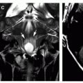
Figure 11-1 Fetal congenital CMV infection. Twenty-eight weeks of gestation. A. Axial T2-weighted fetal MRI image shows bilateral periventricular germinolytic cysts (arrowheads) and abnormal gyri (arrows). B. Coronal T2-weighted image demonstrates diffuse polymicrogyria (arrows). (Courtesy of Catherine Adamsbaum, MD, Paris, France.)


Table 11-2 Congenital CMV Infection Spectrum of Imaging Abnormalities


Calcifications


Cerebellar hypoplasia


Cerebral cortical abnormalities


  • Polymicrogyria


  • Cortical cleft dysplasia


  • Schizencephaly


  • Hippocampal dysplasia


  • Lissencephaly


Cysts


White matter abnormality


Imaging

Fetal MRI is more sensitive than is antenatal sonography in detecting the combination of malformations and destructive lesions caused by CMV. These include microcephaly, cerebral cortical abnormalities, cerebellar hypoplasia, white matter lesions, and temporal lobe or polar abnormalities such as enlarged temporal horns, abnormal white matter, and temporal polar cysts (29). Fetal brain MR imaging is able to demonstrate CMV-associated abnormalities before 24 weeks of gestation and can demonstrate evolving infection-related changes. Even when level II fetal US results have been reported as normal, performing fetal MRI in the third trimester can be helpful in the detection of new cerebral malformations and destructive lesions and can accurately monitor the evolution of previously defined lesions (Fig. 11-1).

Findings on cross-sectional imaging vary depending upon the degree of brain destruction and the timing of the injury, timing of imaging, and imaging modality. Postnatal, transfontanelle cranial ultrasound may show branching curvilinear hyperechogenicities in the basal ganglia and/or punctuate subependymal hyperechogenicities (mineralization) (Figs. 11-2 and 11-3). The pattern of basal ganglia involvement is called “lenticulostriate vasculopathy” (30,31). It is seen in children with congenital infections of many types and is also
described in children with trisomy 13, trisomy 21, prenatal drug exposure, congenital heart disease, and a variety of anoxic and toxic injuries to the developing brain (31,32). Postmortem studies have described evidence of a mineralizing vasculopathy as the cause, whereas other studies suggest altered perfusion, perhaps the result of impaired autoregulation (33,34). Lenticulostriate vasculopathy is a nonspecific finding, and the diagnosis of congenital infection should not be strongly considered unless other supportive findings (parenchymal regions of hyperechogenicity, intraventricular septations, periventricular necrosis [germinolytic cysts], anterior temporal white matter lesions, and/or abnormalities of sulcation) are also seen by fetal imaging (US or MRI) or postnatal US, CT, or MRI (Fig. 11-4) (35,36,37). Antenatal cranial sonography may offer predictive information for the fetus infected with CMV but may underestimate the presence of anterior temporal lobe or temporal polar abnormalities (38). On CT and MR, some patients, presumably infected during the first half of the second trimester, demonstrate agyria or lissencephaly with a thin cortex, cerebellar hypoplasia, delayed myelination, marked ventriculomegaly, germinal zone cysts, anterior temporal lobe cysts, and periventricular calcification (Figs. 11-3, 11-4, 11-5 and 11-6). Those injured later, presumably in the middle of the second trimester, have more typical polymicrogyria, less ventricular dilatation, and less consistent cerebellar hypoplasia; schizencephaly and infoldings of cortical dysplasia can be seen (Figs. 11-6, 11-7 and 11-8) (39,40). Patients infected near the end of gestation or in the early postnatal period have normal gyral patterns, mild ventricular and sulcal prominence, and damaged periventricular or subcortical white matter with scattered periventricular calcification or hemorrhage (Figs. 11-7, 11-8 and 11-9) (12,18,24,28,41,42). FLAIR images, particularly in the first year of life, provide relatively poor contrast between gray and white matter; therefore, T2-weighted images are essential to identify

cortical malformations (Figs. 11-6 and 11-7), the detection of which helps establish the diagnosis of congenital CMV.


Figure 11-2 Neurosonographic features of congenital CMV infection. A. Parasagittal sonogram shows linear hyperechogenicity (arrows) within the basal ganglia consistent with lenticulostriate mineralizing vasculopathy. Note the focus of periventricular hyperechogenicity (arrowhead) corresponding to calcification on CT (not shown). B. Coronal sonogram shows multiple subcortical and periventricular foci of hyperechogenicity confirmed by CT (not shown) to represent calcification (arrows). Also note the left germinal matrix cyst (arrowhead). C. Parasagittal sonogram in a microcephalic newborn demonstrates extensive linear and punctuate periventricular hyperechogenicities (arrows) representing calcification. There is moderate ventriculomegaly. D. Parasagittal sonogram shows a cyst of the caudothalamic germinal matrix zone (arrow) and focal peritrigonal hyperechogenicities (arrowheads) consistent with calcification.


Figure 11-3 Congenital cytomegalovirus infection. A. Coronal sonogram in this microcephalic newborn shows moderate ventriculomegaly and multifocal periventricular hyperechogenicities (arrows). B. Axial noncontrast CT shows extensive periventricular calcification. Note the moderate ventricular dilatation and overlap of the coronal sutures reflecting underlying brain injury and poor brain growth. C. Axial T1-weighted image at the level of the enlarged lateral ventricles demonstrates ependymal T1 shortening (arrows) representing calcification. D. Axial T2-weighted image shows less conspicuous T2 shortening at sites of known calcification (black arrows). E. Axial T2-weighted image 3 years later shows persistent ventricular enlargement and near-complete absence of periventricular T2 hypointensities. In time, the microglial cells or Hortega cells will remove calcium and hemorrhage.


Figure 11-4 Congenital CMV infection. A. Coronal cranial ultrasound shows basal ganglia linear hyperechogenicities consistent with lenticulostriate vasculopathy (arrows). Also note the focal dilation of the temporal horns (arrowheads). B. Axial T2-weighted image demonstrates bilateral cystic temporal horn dilation (curved arrows). Hyperintense white matter is identified anterior to the temporal horns.

Damage to the white matter may be seen in those infected at any gestational age. Potential causative factors include primary CMV infection, host inflammatory and immune response including cytotoxic T-cell response, and injury to oligodendroglial cells. These white matter lesions are static like those of periventricular leukomalacia. van der Knaap et al. (43) have described a distinct pattern of white matter abnormalities in neonates with congenital CMV infection. Some of these patients lack gyral anomalies; in such patients, multiple white matter lesions typically involve the deep white matter, often sparing immediate periventricular and subcortical white matter, with the largest lesions often in the parietal lobes (Figs. 11-7 and 11-8). In patients with gyral anomalies, white matter abnormalities may be diffuse or multifocal (Figs. 11-3, 11-5, 11-6, 11-7, and 11-9). Abnormalities of the temporal polar regions are strongly suggestive of congenital CMV infection. Dilation of the temporal horns may be detected as early as 20 weeks of gestation, with subsequent evolving temporal lobe white matter T2 hyperintensity (swelling) and, ultimately, the formation of temporal polar cysts (Figs. 11-4, 11-7, and 11-9) (43). The apparent selective vulnerability of the temporal lobe regions to congenital CMV infection and the pathological underpinnings remains unexplained.


Figure 11-5 Congenital cytomegalovirus infection in a neonate. A and B. Axial T1-weighted images show the agyria, markedly hypoplastic cerebellar hemispheres (white arrows), and periventricular calcifications (black arrows) common in this disorder.

In affected newborns and infants, the white matter lesions demonstrate diffusivity (also called apparent diffusion coefficient, ADC) values that are increased; fractional anisotropy (FA) values (which represent a measure the degree of diffusion anisotropy) are decreased (43).

Calcifications can be seen on CT as foci of high attenuation (Figs. 11-3, 11-5, and 11-6). Although calcifications can be detected on MR in neonates and young infants as foci of short T1 and T2 relaxation times (Figs. 11-3 and 11-6), they are much more easily and reliably

detected on CT than on MR in older infants and children. Although dogma has suggested that congenital CMV CNS infection is predictably associated with cerebral calcifications, the prevalence of calcification may be less than 70%. Differentiation of calcification from hemorrhage may be difficult by either CT or MRI (24). Including susceptibility-weighted imaging (SWI) and SWI filtered phase maps into the routine MR examination aids in the differentiation of SWI hypointensities of blood products (paramagnetic) from calcifications
(diamagnetic). The SWI filtered phase map of calcifications is hyperintense (Fig. 11-10) (44). Clinicians should recognize that calcification is not uniquely specific for congenital infection, as any injury to the brain, including those caused by ischemia, genetic syndromes, metabolic errors, and nonspecified neurodegeneration, can cause dystrophic calcification (45,46,47). MR is the imaging examination of choice for the detection of cortical malformations, myelin abnormalities (delay and demyelination), germinal zone cysts, and cerebellar hypoplasia/dysplasia. When these features are present in a child with microcephaly, developmental delay, sensorineural hearing loss, and seizures, a diagnosis of congenital CMV should be considered (24).


Figure 11-6 Cortical dysgenesis and congenital cytomegalovirus infection. A. Axial noncontrast CT image shows extensive intracranial parenchymal calcification and shallow sylvian fissures strongly suggesting an accompanying neuronal migration anomaly. B. Axial T1-weighted image demonstrates foci of T1 shortening (arrows), which correspond to calcification on CT. Note the simplified perisylvian sulcation. C. Axial T2-weighted image through the posterior fossa shows a hypoplastic cerebellum. Cystic change (arrow) and folial dysgenesis are noted. D. Axial T2-weighted image shows the cortical dysgenesis of the cerebrum to best advantage; this is most likely diffuse polymicrogyria. Note the subtle foci of T2 shortening corresponding to calcification (arrows) and the occipital periventricular cysts.


Figure 11-7 Infant with congenital cytomegalovirus infection. A. Sagittal T1-weighted image shows diffuse polymicrogyria (arrows). Also note temporal horn dilation (curved arrow) and T1 prolongation of anterior temporal lobe white matter. B. Axial T2-weighted image at the level of the temporal horns shows bilateral temporal horn dilation and T2 prolongation of temporal lobe white matter.


Figure 11-7 (Continued) C. Axial FLAIR image shows simplified sulcal gyral pattern consistent with bihemispheric polymicrogyria (arrows) and confluent regions of cerebral hemispheric white matter T2 prolongation.


Figure 11-8 Congenital cytomegalovirus infection with focal clefting and encephalomalacia. A. Axial noncontrast CT image shows bilateral frontal lobe subcortical hypoattenuating regions (black arrows) and sparse parenchymal calcification (white arrow). B. Parasagittal T1-weighted image shows several areas of subcortical encephalomalacia (black arrows), manifested as hypointensity. C. Axial T2-weighted image demonstrates the encephalomalacic regions to be continuous with the subarachnoid space as clefts (white arrows). Also note the other patchy bihemispheric regions of white matter hyperintensity. D. Axial FLAIR image shows to best advantage the confluent regions of bihemispheric white matter hyperintensity and the hypointense subcortical white matter clefts (black arrows). E. DTI-derived axial color-encoded fractional anisotropy (FA) map demonstrates a loss of coherence (hypointensity and distortion of normal tracts, as well as predominant superior-inferior orientation, manifested as blue color) in the periventricular corticospinal tracts (white arrows).


Figure 11-9 Congenital cytomegalovirus infection and white matter disease. A. Parasagittal T1-weighted image demonstrates an anterior temporal lobe subcortical cyst (white arrows). Also note confluent T1 prolongation within the parietal white matter (black arrows). B. Coronal T2-weighted image demonstrates anterior temporal cysts (black arrows). Note the multifocal areas of central and subcortical white matter T2 prolongation (white arrowheads). C and D. Axial FLAIR images at the level of the lateral ventricles and centra semiovale show bilateral asymmetric patchy regions of white matter FLAIR hyperintensity.


Herpes Simplex Virus: Congenital and Neonatal Infections

Approximately 2000 infants in the United States annually have neonatal infections with either herpes simplex virus (HSV) type 1 or type 2 (48). Approximately 2% of women in the United States acquire HSV-2 annually, and approximately 80% of these occur without maternal symptoms or awareness. Neonatal HSV infections are considered mucocutaneous (skin, eye, mouth without CNS involvement), disseminated (with or without CNS involvement), or encephalitic. Infants with isolated HSV encephalitis constitute up to 30% of all HSV-infected infants. Symptoms develop at an average age of 16 days, nonspecifically with fussiness, lethargy, or poor feeding or overtly with seizures and coma (48). Disease onset, including seizure semiology, may be very subtle (49,50). Approximately 5% of infants with HSV acquire the virus in utero and have a congenital infection syndrome with microcephaly, skin rash, or scarring and cataracts (51).

Clinicians must maintain a high index of suspicion for neonatal HSV infection; only two-thirds of the infants with perinatal HSV encephalitis have herpetic skin rashes. Infants with disseminated infections feed poorly and manifest fever, jaundice, hepatomegaly, or respiratory distress with or without CNS involvement; those with meningoencephalitis have focal or generalized seizures, lethargy, and coma. Ultimately, two-thirds of affected neonates will have some degree of CNS involvement (49,50,52). CSF typically shows a mononuclear pleocytosis, elevated protein, and decreased glucose with a negative Gram stain (53). The diagnosis is established by detecting HSV DNA in serum or cerebrospinal fluid; however, as many as 25%
of infants with neonatal HSV encephalitis will have negative HSV PCR studies (52). Therefore, a negative result should be interpreted with caution. If there is a clinical suspicion of HSV meningoencephalitis, a second lumbar puncture should be performed and acyclovir administered empirically until the results of the second PCR are known. Therapy of neonatal HSV CNS infections consists of high-dose (60 mg/kg/day) acyclovir for 28 days (54). Infants who survive perinatal HSV infections are at high risk for cerebral palsy, epilepsy, and developmental delay despite aggressive acyclovir therapy (49,52). Suppressive antiviral therapy for 6 months after the initial treatment substantially improves the long-term outcome of neonatal HSV infections (55).


Figure 11-10 Susceptibility-weighted imaging (SWI) and Ca++ detection. A. Axial NECT in a patient with tuberous sclerosis complex demonstrates focal periventricular highattenuation calcified tubers. B. Axial SWI image shows focal hypointensities where CT demonstrated calcifications. C. Axial filtered phase map of the SWI image demonstrates the calcified tubers as hyperintense foci (arrows). This demonstrates the value of SWI filtered phase maps in discriminating hemorrhage (paramagnetic and SWI-hypointense, filtered phase map hypointense) from calcification (diamagnetic and SWI hypointense, filtered phase map hyperintense).

Pathologically, HSV can infect and severely damage many brain regions, with resultant necrosis, cellular debris, macrophages, mononuclear inflammatory cells, calcification, and hypertrophied astrocytes (56). The pial-glial membrane remains intact, and the ependyma and choroid plexuses are notably spared, in contrast to their involvement in congenital CMV and toxoplasmosis infections (57).


Imaging

The radiologist must consider neonatal herpes simplex encephalitis (HSE) in the neonate whose cranial imaging in the second to third weeks of life demonstrates diffuse cerebral edema and the presence of leptomeningeal enhancement, with or without parenchymal hemorrhage.

Sonographic findings are often subtle, initially showing nonspecific diffuse parenchymal hyperechogenicity and normal ventricles; parenchymal echogenicity subsequently increases with associated ventricular compression (58,59). The ventricles eventually enlarge as encephalomalacia develops (59). MRI is the study of choice in neonates with suspected herpes encephalitis (60,61,62,63), with imaging features of HSV type 2 encephalitis consisting of multifocal lesions (67%), temporal lobe involvement (67%), deep gray matter injury (58%), hemorrhage (66%), watershed pattern of injury (40%), and occasional involvement of the brain stem and cerebellum (63). In neonatal HSV encephalitis, diffusion-weighted MR imaging is critical to making an early diagnosis, monitoring disease progression, and detecting rare CNS relapses (64,65,66). Diffusion imaging depicts early cellular necrosis, which is seen as hypointensity (reduced diffusion) on average diffusivity (Dav) images (65,66). In this acute stage of disease, standard MR imaging is often normal (Fig. 11-11) (61,63). Subtle hyperintensities may or may not be seen on T2-weighted images in the late acute and early subacute stages of infection. As the infection progresses (at the end of the first week), diffusion imaging becomes less helpful and standard spin echo imaging more helpful (61,63). Early proton MR spectroscopy shows
elevated lactate and, often, reduction of N-acetyl aspartate (NAA) in affected regions (Fig. 11-12) (61,63). After 1 or 2 days, CT and MR show patchy, multifocal areas of injury (low attenuation on CT, T1 hypointensity, T2 hyperintensity, and regions of reduced diffusion), affecting gray and white matter (Figs. 11-11, 11-12 and 11-13), which progresses in prominence and extent of involvement during the course of the next several days. Hemorrhage is a common finding in neonatal herpes encephalitis, seen in up approximately two-thirds of patients (Fig. 11-14) (63). Contrast enhancement, although minimal, occurs early in a meningeal pattern (Fig. 11-14) (63). Near the end of the first week of disease, there are often regions of cortical gray matter injury (increased attenuation on CT, T1 hyperintensity, and T2 hypointensity on MR) that persists for weeks to months(61,63,67). Loss of brain substance occurs rapidly, often as early as the second week. Eventually, severe, diffuse cerebral atrophy ensues with profound cortical thinning and encephalomalacia; in the end stage, the brain often appears multicystic (Fig. 11-15E). Punctate or curvilinear gyral calcifications may also be seen as a late finding. The cerebellum is injured in about half of the affected patients (61,63,67).


Figure 11-11 Early diffusion-weighted imaging in neonatal herpes simplex encephalitis. A. Axial noncontrast CT in a seizing 3-week-old female shows diffuse hypodensity. B. Axial T2-weighted image shows normal gray matter and white matter differentiation without cortical blurring. Cortical blurring is an indicator of early peripheral edema. C. Axial apparent diffusion coefficient image shows reduced diffusion within the left temporal lobe (arrows) and occipital poles. Diffusion-weighted imaging is particularly valuable in the early phase of neonatal herpes encephalitis where injury of tissue is manifested as regions of reduced diffusion.

Although the white matter changes in neonatal herpes encephalitis are nonspecific (Figs. 11-12, 11-13, 11-14 and 11-15), early reduced diffusion, parenchymal hemorrhage, and a meningeal pattern of enhancement in the proper clinical setting should lead to a suggestion of the diagnosis of neonatal HSE. It is important for the radiologist to remember that neonatal herpes simplex encephalitis demonstrates nonpatterned MR abnormalities. The clinical presentation and imaging appearance of herpes encephalitis in older children and adults (HSE) (usually caused by herpes simplex virus, type I) is quite different than neonatal HSV infection, primarily reflecting infection of the temporal lobe and insular cortex, limbic structures (61).


Lymphocytic Choriomeningitis Virus

Congenital infections with LCMV, a rodent-borne Arenavirus, have been reported in infants from several regions, but the overall incidence of congenital LCMV infection remains undefined (68,69,70,71). Humans appear to become infected through contact with aerosols or fomites that contain the infectious virus. In contrast to other congenital infections, hepatosplenomegaly, jaundice, and petechial/purpuric rash are uncommon in infants with congenital LCMV infections. However, such infants commonly have hydrocephalus and chorioretinitis, thus closely resembling infants with congenital toxoplasmosis (71a). The diagnosis can be established by detecting LCMV-specific serologic responses (IgG and IgM); since seroprevalence for LCMV is low among adults, detection of LCMV-specific-IgG strongly suggests congenital infection. No specific therapy exists for congenital LCMV infections; most infants have substantial long-term sequelae consisting of cerebral palsy, hydrocephalus, vision loss, and developmental delays/mental retardation.


Figure 11-12 Multifocal cerebral injury pattern in neonatal herpes encephalitis. A. Parasagittal T1-weighted image shows swelling of the right parietal cortex (arrow). B. Axial T2-weighted image demonstrates T2 prolongation (arrows). C. Coronal FLAIR image shows right thalamic hyperintensity (arrowhead). Also note the focus of right opercular cortical hyperintensity (arrow). D. Axial apparent diffusion coefficient image shows multiple sites of reduced diffusion within the cerebral hemispheres (hypointensities). E. Proton MR spectroscopy (TE = 288 ms) of the thalamus shows diminished NAA peak. Note the elevation of lactate (Lac). Along with diffusion-weighted imaging, magnetic resonance spectroscopy gives insight into early tissue injury.

LCMV infection should be considered among infants who have chorioretinitis in association with congenital hydrocephalus or microcephaly, lack hepatosplenomegaly, and have negative microbiologic studies for the more common pathogens such as T. gondii and CMV (71). As with most congenital infections, the severity of the infection is related to the fetal age at the time of infection, with earlier infections being more severe (72). First trimester infection typically results in spontaneous abortion, whereas second and third trimester infections strongly resemble those of toxoplasmosis and CMV, with predominant disease being located in the CNS (73). The most prominent clinical feature is chorioretinitis, seen in about 90% of infected neonates, which is lacunar and resembles the retinal lesions in Aicardi syndrome

(see Chapter 5) and toxoplasmosis. Hydrocephalus, seen in more than 50% of affected neonates, results from necrotizing ependymitis and aqueductal obstruction (70,71). Other infants with congenital LCMV virus infections can have microcephaly and intracranial calcifications (70,71). Seizures commonly develop during the first year of life. Longterm outcome is generally poor, with mortality as high as 35% and severe neurologic sequelae in more than 60% of survivors (71).


Figure 11-13 Leukotropic neonatal herpes simplex encephalitis. A. Axial T1-weighted image shows bilateral frontal white matter foci of T1 shortening (arrows) likely representing coagulative necrosis of the white matter. There was no corresponding GRE blooming. Also note the focal right germinal zone hemorrhage (arrowhead). B. Axial T2-weighted image demonstrates corresponding T2 shortening (arrows). C. Coronal T2-weighted image shows more hypointense foci within the centrum semiovale of the cerebral hemispheres (arrows). D. Axial diffusion-weighted image shows bifrontal and left peritrigonal foci of hyperintensity (arrows). E. Axial apparent diffusion coefficient image confirms the bright diffusion-weighted signal abnormalities to represent sites of reduced diffusivity (arrows). As the encephalitis becomes less acute, standard spin echo images become more useful and diffusion-weighted imaging less useful. F. Axial color-encoded fractional anisotropy image (F) at the location of previously described reduced diffusion shows disturbance of white matter fiber track coherence: increased anisotropy in corpus callosum but decreased in the frontal and occipital white matter (arrows).


Figure 11-14 Hemorrhagic neonatal herpes simplex encephalitis. A. Axial T1-weighted image in a 3-week-old neonate demonstrates right thalamic and left temporal lobe T1 shortening confirmed on GRE imaging to represent hemorrhage. The deep cerebral nuclei and temporal lobes are common sites of injury in neonatal herpes encephalitis. B. Coronal T2-weighted image depicts the multifocality of brain parenchymal involvement that occurs in 67% of neonatal herpes encephalitis. Note the bilateral frontal, right basal ganglia, and left parafalcine parenchymal hemorrhages (T2 shortening) and associated edema (T2 prolongation). C. Coronal GRE shows cortical, subcortical, and basal ganglia regions of blooming hypointensity representing multifocal hemorrhage. D. Axial T1-weighted contrast enhanced image at the level of the lateral ventricles demonstrates bilateral subinsular foci of T1 shortening (arrows). Prior to contrast, no corresponding hyperintense lesions were seen.


Imaging

Imaging studies of neonates and infants with LCMV may overlap in neonates and infants with congenital toxoplasmosis and CMV infections (7). Timing of this congenital infection dictates the pattern of CNS injury. Hydrocephalus can be seen by sonography, CT, or MR (Fig. 11-17) (71,74,75). Periventricular calcifications, when present, are most easily seen by CT as high attenuation punctate periventricular lesions (Fig. 11-16) (69,72). Calcifications on MRI may be very subtle, showing punctate hyperintensity from T1 shortening when calcium molecules are less densely packed (and the paired outer shell electrons of water can transiently bind to them), hypointensity from T2 shortening or absence of water in tightly packed crystals, and hyperintensity on SWI filtered phase maps. MR may also show a cortex composed of multiple small, shallow sulci, the appearance of which suggests polymicrogyria (Fig. 11-17). These findings strongly suggest congenital infection, but the final diagnosis must be made by serologic and microbiologic studies.


Rubella Virus

Prior to widespread implementation of the measles-mumps-rubella vaccine, epidemics of rubella (German measles) occurred worldwide at 6- to 9-year intervals. During the 1962-1965 pandemic, the last major US rubella outbreak, approximately 20,000 infants were affected by congenital rubella syndrome (CRS); there were more than 10,000 fetal deaths and 2000 neonatal deaths (76). CRS largely disappeared from the US and other developed nations by the late 1980s because of immunization programs (77) but remains a major health concern in many regions


of the world, with more than 100,000 cases estimated by the Centers for Disease Control and Prevention (CDC) to have occurred worldwide annually during the first decade of the current millennium. Humans represent the only reservoir of rubella virus, and transmission results from contact with virus-contaminated respiratory secretions (76).


Figure 11-15 Progression of neonatal herpes encephalitis to cystic encephalomalacia. A. Axial T1-weighted image in a preterm neonate with new-onset seizures shows a focus of hemorrhage within the right parietal lobe (arrow). There are multiple bilateral centrum semiovale foci of T1 shortening. GRE imaging confirmed hemorrhage in multiple white matter sites. The simplified sulcation and gyration pattern was attributed to prematurity. B. Axial T2-weighted image shows a focal hemorrhage with hematocrit effect in the right parietal white matter. T2 signal is prolonged throughout the white matter. C. Coronal T2-weighted image shows symmetric temporal and frontal white matter T2 hyperintensity. Note the subtle hypointensity within the right temporal white matter (arrow) that corresponded to hemorrhage on GRE. D. Axial apparent diffusion coefficient image at the level of the temporal lobes 14 days after symptom onset shows increased diffusivity throughout the white matter (regions of hyperintensity). E. Axial FLAIR image at 5 years of age shows extensive cystic encephalomalacia.


Figure 11-16 Lymphocytic choriomeningitis virus (LCMV). Axial noncontrast CT images (A-C) show a diffuse array of small calcifications involving subcortical and periventricular white matter and internal capsules. This microcephalic infant with chorioretinitis had a “negative” clinical/laboratory evaluation for TORCH infection. The imaging studies of neonates and infants with LCM may be identical to those of neonates and infants afflicted with congenital CMV and toxoplasmosis.


Figure 11-17 Lymphocytic choriomeningitis virus. A. Axial noncontrast CT image shows marked hydrocephalus and hypodensity of the white matter, but no calcifications. B. Axial T2-weighted image shows abnormal sulcation (arrows) of the frontal lobes.

Infants with CRS differ from those with congenital infections with either T. gondii or CMV by lower rates of hepatosplenomegaly and jaundice, high rates of cataracts, and the presence of congenital heart disease, typically patent ductus arteriosus (78). Microcephaly, chorioretinitis, sensorineural hearing loss, and the classic “blueberry muffin” rash, a sign indicating extramedullary hematopoiesis, are common in infants with CRS. The diagnosis can be established serologically, virologically, or molecularly, using the PCR. As no specific therapy for CRS currently exists, survivors have high risks of neurodevelopmental sequelae, including microcephaly, vision loss, cognitive impairment, and sensorineural hearing loss (79).

On pathologic examination, brains of affected infants are microcephalic with ventriculomegaly resulting from loss of brain tissue. Multiple small areas of liquefactive necrosis and gliosis with calcification are seen in the periventricular white matter, basal ganglia, and brain stem; these result from a prominent vasculopathy (78). Myelination is typically impaired (78,80), but the encephalitis is usually not progressive after infancy.


Figure 11-18 Congenital rubella. A. Axial noncontrast CT demonstrates subtle cerebral calcifications (arrows). B and C. Axial T2-weighted images show patchy regions of periventricular white matter hyperintensity (arrows) most consistent with regions of demyelination and/or gliosis. (Courtesy of Majda M. Thurner, MD, and Elsevier.)


Imaging

The appearance of the brain on imaging studies varies depending upon the timing of in utero infection. Early infection will result in congenital anomalies, whereas late infection will result in a nonspecific generalized edema, gliosis, and loss of brain tissue. Ultrasound may show nonspecific lenticulostriate vasculopathy (see discussion under CMV) (30,31). CT typically shows ventriculomegaly, multifocal regions of hypodensity throughout the cerebral white matter, often in association with periventricular and basal ganglia calcification and cysts (Fig. 11-18) (81,82). Calcification may also be seen within the cortex. In severe cases, nearly total brain destruction and microcephaly are present (83). MR imaging may show ventriculomegaly, multifocal regions of white matter prolonged T2 relaxation (T2 hyperintensity), or myelination delay/destruction (82,84,85). Frontal dominant white matter lesions showing T2 hyperintensity have been reported following congenital rubella and CMV infections (28). High-resolution CT of the temporal bones may show malformations of the inner ear structures. Enhanced MR imaging may show enhancement of the cochlea (cochleitis) (86).


Treponema pallidum (Syphilis)

Congenital infection with Treponema pallidum occurs by transplacental transmission during maternal infections (spirochetemia), principally in the second and third trimesters of gestation (87). Infection results in 25% to 80% of children of untreated mothers with syphilis, with symptoms and signs of congenital syphilis occurring in up to 16%. Congenital syphilis is unlikely to cause neurological symptoms in the neonatal period (88), however; instead, early signs include failure to thrive, jaundice, hepatosplenomegaly, and rash (small blisters on palms of hands and soles of feet). Later signs (older infants and young children) include saber shins, saddle nose deformity, mulberry molars, frontal bossing, rhagades (scars around the mouth or nose), and Hutchinson triad (sensorineural deafness, interstitial keratitis, and Hutchinson teeth—peg-shaped upper incisors). Abnormalities of the bones (osteochondritis and periostitis) are common (60%-80%). Neurologic signs and symptoms may develop in the first 2 years of life, reflecting meningovascular and perivascular space mononuclear cellular infiltration and resulting in seizures, stroke, cranial nerve palsies, and signs of increased intracranial pressure (ICP) (56,89).


Imaging

The most common imaging findings are leptomeningeal enhancement, hydrocephalus, and infarction (Fig. 11-19). Cisternal inflammatory exudative involvement (inflammation, fibrosis, and gumma formation) investing the pituitary gland and infundibulum may lead to persistent hypoglycemia, diabetes insipidus, and hypopituitarism (90,91).


Toxoplasma gondii

Congenital toxoplasmosis, the result of intrauterine infection with the ubiquitous intracellular parasite, Toxoplasma gondii, is the second most common congenital infection, after CMV, in many regions of the world. Rates of congenital toxoplasmosis range from less than 0.1 in many areas to 1 per 1000 live births in highly endemic regions. The seroprevalence of toxoplasmosis among adults, a measure of acquired infection with T. gondii, is highest in France; intermediate in Latin American, sub-Saharan Africa, and central Europe; and lowest in North America, Southeast Asia, and Oceana (92). Toxoplasma gondii infects birds and many mammals, especially felines, worldwide (93). Infected domestic cats, a major source of human disease, excrete vast quantities of oocysts; human infection results from ingesting undercooked meats that contain viable T. gondii tissue cysts or foods contaminated with infectious oocysts.


Figure 11-19 Congenital syphilis. Axial T2-weighted image shows watershed ischemia in the right cerebral hemisphere and severe ischemic damage in the left cerebral hemisphere. Ischemic injury is due to invasion of perivascular spaces by an inflammatory infiltrate. (Courtesy of Robert A. Zimmerman, MD, Philadelphia, PA.)

The clinical and laboratory manifestations of congenital toxoplasmosis, as implied by the TORCH acronym, resemble those of congenital CMV disease and consist of hepatosplenomegaly, jaundice, chorioretinitis, petechial or purpuric rash, thrombocytopenia, elevated serum transaminases, and hyperbilirubinemia (94). Chorioretinitis is very common. In contrast to CMV, macrocephaly (a sign of intrauterine or postnatal hydrocephalus) and chorioretinitis (a potential cause of visual impairment) are common in infants with congenital toxoplasmosis. The diagnosis is established by detecting T. gondii-specific IgM or IgA in the infant’s serum. Analysis of paired sera from the infant and the infant’s mother can be useful; the absence of T. gondii-specific IgG or IgM in the infant’s serum and the absence of T. gondii-specific IgG in the mother’s serum argues strongly against congenital toxoplasmosis. Prolonged postnatal therapy with pyrimethamine and sulfadiazine and early shunting of T. gondii-induced hydrocephalus substantially improve the long-term prognosis for infants with congenital toxoplasmosis (95,96). As many as 85% of untreated infants with congenital toxoplasmosis have chorioretinitis, and mortality in untreated infants can be as high as 15% (97). Survivors of congenital toxoplasmosis are at risk for cerebral palsy, cognitive impairment, and epilepsy (96); the risk of sequelae is reduced when antimicrobial therapy is initiated early (96,98,99).

Pathologically, a diffuse inflammatory infiltration of the meninges is found, with large and small granulomatous lesions or a diffuse inflammation of the brain. Hydrocephalus is frequent, most often caused by an ependymitis occluding the cerebral aqueduct (46,100). Porencephaly or hydranencephaly may occur if the disease is severe and occurs in the second trimester (101). In contrast with congenital CMV infection, malformations of cortical development, such as lissencephaly, polymicrogyria, and schizencephaly are not typical features of congenital toxoplasmosis.


Imaging

The findings on cross-sectional imaging may be similar to those in CMV. As in congenital CMV, fetal brain imaging often detects early destructive changes (102). Calcifications are common; they usually involve the basal ganglia, periventricular regions, cerebral cortex, and subcortical white matter (more scattered in distribution than CMV) (Figs. 11-20 and 11-21). It is important to recognize that the predilection for calcium in the periventricular regions is less common than with congenital CMV infection. Fetal imaging (US and MRI) reveals ventriculomegaly, echodense (US) and T2 hypointense (MRI) calcifications of the cerebral parenchyma, and parenchymal cysts (102). In the neonate, cranial sonography may show shadowing parenchymal hyperechoic foci, which correspond to calcifications detected by CT (103). Due to the inflammatory nature of this disease and aqueductal obstruction, hydrocephalus is more common than in CMV (Figs. 11-20 and 11-21). As with CMV, the spectrum of potential abnormalities ranges from relatively mild disease, with a few periventricular calcifications and mild atrophy, to severe disease with near-total destruction of the cerebrum accompanied by diffuse cerebral calcifications (Fig. 11-20). Diebler et al. (104) have related the severity of the brain involvement to the date of maternal infection, noting that infection before 20 weeks is generally accompanied by severe neurological findings, including microcephaly, hydrocephalus, tetra- or diplegia, seizures, mental retardation, and blindness. CT revealed dilated ventricles, areas of porencephaly, and extensive calcifications, particularly in the basal ganglia (Fig. 11-20), that may exhibit a tram-track morphology (105). Infection between 20 and 30 weeks leads

to a more variable outcome; on CT, sparse (Fig. 11-21) or multiple periventricular calcifications and ventricular dilation are typical findings (Fig. 11-20). Infection after the 30th week of gestation is generally associated with mild clinical and imaging abnormalities, with CT findings of small periventricular and intracerebral calcifications that are only rarely accompanied by ventricular dilatation (104). An important differentiating feature between congenital toxoplasmosis and congenital CMV infection is the absence of cortical malformations in toxoplasmosis (a common occurrence in congenital CMV). The brain calcifications of congenital toxoplasmosis may resolve slowly in some infants after antitoxoplasma therapy (106). Therefore, the disappearance of calcification over time should not cause confusion if the diagnosis was established at birth on the basis of characteristic ocular findings and serologic testing.


Figure 11-20 Congenital toxoplasmosis. A and B. Axial noncontrast CT images in a newborn show extensive parenchymal calcifications that are predominantly cortical and subcortical in location. Also note moderate ventriculomegaly. Hydrocephalus is more common in toxoplasmosis than CMV infection. C and D. Axial and coronal T2-weighted images show multiple foci of T2 hypointensity corresponding to CT confirmed calcification (black arrows). Also note the subcortical cysts (c). Ventriculomegaly is moderate.


Figure 11-21 Congenital toxoplasmosis with minimal cerebral calcification. A and B. Noncontrast axial CT images show sparse parenchymal calcification (arrows). Calcification was not present on the initial CT studies in the neonatal period. Note the presence of ventricular shunt catheters. Congenital toxoplasmosis is often associated with ependymitis and resultant hydrocephalus.


Varicella-Zoster Virus

In unimmunized populations, the rate of varicella infection (chickenpox), acquired through contact with the respiratory secretions of infected children, ranges from less than 1 to 3 per 1000 pregnancies, and very few (<2%) of these pregnancies result in infants with the congenital varicella syndrome (107,108). Infants with this disorder, like those with congenital HSV or LCMV infections, generally lack typical signs of congenital infection, such as jaundice, hepatosplenomegaly, or petechial/purpuric rash. Infants with congenital varicella infected before 20 postconceptional weeks may suffer spontaneous abortion or embryopathy with microcephaly (resulting from cerebral destruction), cataracts or chorioretinitis, limb or digit hypoplasia, and a characteristic pattern of skin scarring known as a cicatrix (108,109).


Figure 11-22 Congenital varicella-zoster virus infection. A. Axial T1-weighted image in a microcephalic term newborn shows extensive bilateral periventricular T1 shortening consistent with coagulative necrosis. SWI showed no evidence of hemorrhage or calcification. B. Axial T2-weighted image shows no significant abnormality. Note the normal cortical ribbon at this level, unlike polymicrogyria often observed in congenital CMV infection. C. Axial ADC map demonstrates scattered foci of reduced diffusion (arrows).


Imaging

Polymicrogyria involving the cerebral hemispheres and necrosis of white matter and deep gray nuclei and cerebellum have been reported in autopsy studies (110). Two MR studies have been reported, emphasizing variable findings likely reflecting the timing and severity of infection. One report describes hydrocephalus and cerebellar aplasia, while the other describes destruction of the temporal and occipital lobes with marked ventricular dilation in those areas, but normal cerebellum, basal ganglia, and frontal/parietal lobes (59,111). As with many of the previously described congenital CNS infections, MRI may lack specificity but does allow a full appraisal of brain injury and malformations (Fig. 11-22). Although retrospective diagnosis can be challenging, intrauterine infections with VZV can be established by serological or virological methods.


Zika Virus

First isolated from a rhesus monkey in the Zika Forest of Uganda in the late 1940s, Zika virus belongs to the Flaviviridae, a family of RNA arboviruses that includes West Nile, St. Louis encephalitis, and yellow fever viruses. Prior to 2000, human infections were rare (112). In 2007, Zika virus appeared in Micronesia, and in the fall of 2013, an outbreak of Zika virus affected nearly 30,000 persons or more than 10% of the population of French Polynesia. Most infected persons had a mild viral syndrome with low-grade fever, conjunctivitis, arthralgias, and a maculopapular rash, but some experienced Guillain-Barré syndrome approximately 1 week after Zika virus infection (113,114). The virus continued eastward, and in early 2015, cases of Zika virus appeared in Brazil, transmitted to humans by Aedes aegypti, a mosquito species prevalent throughout the Americas.

Approximately 80% of cases in the Brazil outbreak occurred asymptomatically. In contrast to the Polynesian outbreak, the Brazilian epidemic was associated with a marked increase in the numbers of infants born with microcephaly and intracranial calcifications, compatible with intrauterine transmission of Zika virus (115). In addition to microcephaly and intracranial calcifications, congenital Zika virus syndrome can be associated with lissencephaly, polymicrogyria, passive ventriculomegaly, arthrogryposis, sensorineural hearing loss, fetal brain disruption, optic nerve hypoplasia, and abnormalities of the retinal pigment (116,117,118,119,120).

Zika virus infection should be suspected in infants with microcephaly and intracranial calcifications who lack microbiological evidence for the agents commonly associated with congenital infection, especially CMV. The diagnosis of Zika virus infection can be established by serologic testing of maternal and infant sera for Zika virus-specific IgM and neutralizing antibodies (plaque reduction neutralization test) and RT-PCR testing of the infant’s serum, urine, or CSF for Zika virus RNA (121). Because there is no available vaccine or antiviral treatment, Zika virus represents an ominous, potential threat to the offspring of pregnant women who visit or live within the host range of Aedes aegypti mosquitoes, including a considerable portion of the United States.


Imaging

The scope of brain abnormalities and corresponding imaging findings resulting from congenital Zika virus infection are protean, the most severe manifestation being fetal brain disruption sequence (120,122). As with other congenital CNS infections, the unique qualities of the viral pathogen and the timing of fetal infection can lead to a range of possible injury patterns and malformations; however, postmortem histological findings have revealed gaps in the pial limiting membrane in affected (personal communication, Dr. Fernanda Tovar Moll, Rio de Janeiro). As discussed in Chapter 5, the cobblestone malformations (a form of polymicrogyria) that result from these gaps may look like lissencephaly, pachygyria, or polymicrogyria depending upon the number and size of the gaps, and that is what is seen in children with congenital Zika infection. MRI remains the most useful imaging tool to fully appraise the scope of CNS abnormalities (116,120).

In their retrospective review of neuroimaging and autopsy data of 45 neonates with proven or presumed congenital Zika virus infection, Soares de Oliveira-Szejnfeld and colleagues chronicled the brain imaging findings associated with congenital Zika infection (120). Cerebral calcifications were reported in all 45 neonates. Calcification located at the gray matter—white matter junction was the most common location followed by periventricular and deep cerebral nuclear (basal ganglia and thalami) regions (Fig. 11-23). In our experience, the precise locations of the calcifications are difficult to define in many patients because the volume of white matter can be extremely diminished and the cortex, therefore, very close to the cerebral ventricles. Calcifications are also seen within the brain stem and cerebellum in about 10% of patients.


Figure 11-23 Congenital Zika virus infection. Axial noncontrast CT in a microcephalic newborn shows bilateral cerebral hemispheric calcifications predominantly at the gray matter/white matter junctions. Also note the simplified sulcal gyral pattern and widened lateral cerebral fissures consistent with extensive bilateral polymicrogyria (arrows). Overlapping of the cranial sutures reflects micrencephaly. (Courtesy of Andres Pessoa, MD, Fortaleza, Brazil.)

Cranial ultrasound reveals calcifications as shadowing or twinkling hyperechoic foci, nonenhanced CT (NECT) of the brain shows highattenuation punctate or chunky calcium deposits (Fig. 11-23). On MRI, calcium in a loose hydration matrix demonstrates T1 (hyperintense) and T2 (hypointense) shortening (Fig. 11-24). Susceptibility-weighted MR imaging (SWI) demonstrates focal hypointensities at sites of calcium deposition. To distinguish calcification from blood products, the SWI filtered phase map shows calcification as focal hyperintensity. Blood products (paramagnetic) appear hypointense (see discussion under congenital CMV).

Ventriculomegaly was identified in all patients, many of these affected neonates showing ventricular septa. The causal factors for ventriculomegaly in these microcephalic patients likely include both impaired CSF absorption and parenchymal volume loss; one of the authors (AJB) has seen sequential studies where the ventricles enlarge as the volume of white matter diminishes in the absence of head growth (excluding hydrocephalus from impaired absorption of CSF). Abnormalities of the corpus callosum ranging from dysgenesis to absence was observed in 94% of the neonates; as the partial or complete callosal absence is usually associated with worse cortical disease and decreased white matter, it is possible that the callosal anomaly is secondary to axonal injury/regression. Cortical abnormalities occurred with a similar prevalence. As discussed above, the cortex may appear as localized cortical dysgenesis (˜30%), polymicrogyria (˜55%), or agyria (12%) depending on the number and size of defects in the pial limiting membrane; these are cobblestone malformations (see Chapter 5), a form of polymicrogyria (Fig. 11-24) (120). Cerebellar parenchymal abnormalities were noted in 82% of patients spanning a spectrum from hypoplasia to severe dysgenesis involving the cerebellar hemispheres and vermis (120).


Figure 11-24 Congenital Zika virus infection. A. Sagittal T1-weighted image demonstrates decreased cranial-to-facial ratio consistent with microcephaly. Also note the frontal lobe region of T1 shortening consistent with calcification at the gray matter/white matter junction (arrow). B. Axial T1-weighted image confirms regions of T1 shortening consistent with calcifications (arrows). C. Axial T2-weighted MR image shows diffuse bilateral cerebral hemispheric simplified sulcal gyral pattern consistent with polymicrogyria (arrows). (Courtesy of Lara Brandao, MD, Rio de Janeiro, Brazil.)


Parechovirus

Neonatal infections with parechovirus, a picornavirus with at least 16 types, can produce neonatal encephalitis and permanent injury to the developing CNS. The cases reported to date suggest to us that parechovirus infection should be considered in infants who have sepsis-like illnesses, especially when accompanied by rash, irritability, and seizures. CSF pleocytosis appears to be an uncommon feature of this infection, although cerebrospinal fluid should be obtained to exclude other conditions, particularly neonatal HSV infection (123,124,125,126). Parechovirus infection can be confirmed by reverse transcription PCR analysis of blood or cerebrospinal fluid (using enterovirus-specific primers) or by culturing the virus from cerebrospinal fluid, stool, or nasopharyngeal secretions. At present, no specific antiviral therapy exists for this disorder.


Imaging

The imaging abnormalities reported have shown bilateral confluent regions of cerebral hemispheric white matter abnormality (125). Cranial sonography may show nonspecific increased echogenicity of the central white matter, NECT demonstrates diminished attenuation of the white matter, and MRI shows diffuse confluent T1 and T2 prolongation and diminished diffusivity on diffusion-weighted imaging (DWI) and average diffusivity maps (Fig. 11-25) (124,125,126). Amarnath and colleagues have pointed out that MRI abnormalities in neonatal parechovirus leukoencephalopathy may be misinterpreted as hypoxic ischemic injury of the brain (127).


Human Immunodeficiency Virus

Pediatric cases of the acquired immune deficiency syndrome (AIDS) appeared in the US in the early 1980s, and the causative agent, the human immunodeficiency virus type 1 (HIV-1), a novel retrovirus, was identified in the mid-1980s (128). Currently, nearly 40 million people are living with HIV worldwide; two-thirds reside in sub-Saharan Africa, and more than two million of these are children (129). More than 25 million persons have died since the first cases of HIV were identified in 1981 (129,130).

Without antiretroviral therapy, infants and children with HIV encephalopathy have progressive motor dysfunction, cognitive abnormalities, developmental delay, and acquired microcephaly (130). Typical clinical manifestations consist of apathy, dementia, ataxia, hyperreflexia, weakness, seizures, or myoclonus. Infants with vertical HIV infections can become symptomatic after the third month of life, manifesting hepatomegaly, lymphadenopathy, failure to thrive, interstitial pneumonitis, opportunistic infections (especially with Pneumocystis jiroveci or cytomegalovirus), or have neurologic disease. HIV infection can also cause aseptic meningitis, meningoencephalitis, myopathy, and Group B streptococcal-like conditions. Secondary CNS complications include stroke, primary CNS lymphoma, and
opportunistic infections with T. gondii, cytomegalovirus, varicellazoster virus (VZV), Mycobacterium tuberculosis, fungi, and JC virus, the cause of progressive multifocal leukoencephalopathy (PML) (131).


Figure 11-25 Neonatal parechovirus encephalitis. A. Axial T2-weighted image at the level of the centrum semiovale shows patchy regions of T2 shortening and prolongation (arrows). B. Axial diffusion-weighted image demonstrates confluent areas of subcortical and central white matter signal hyperintensity. C. Axial apparent diffusion coefficient image at the level of the frontal horns shows cortical and subcortical white matter regions of bihemispheric hypointensity (white arrows), including the left caudate head (black arrow), indicating reduced diffusivity. (These images courtesy of Linda S. de Vries, MD, PhD, Utrecht, Netherlands.)

HIV infection in infants can be confirmed by serial serum PCR assays with the first test in the immediate newborn period, a second test during the first or second month of age, and a third test after 4 months of age. If two samples are positive for HIV, the infant is considered infected; two successive negative tests make infection unlikely. In children and adolescents, serologic studies using ELISA and western blotting can identify HIV infection, and PCR monitoring of virus load guides the treatment of HIV. Current antiretroviral therapy relies upon combinations of nucleoside/nucleotide analogue reverse transcriptase inhibitors, protease inhibitors, and nonnucleoside analogue reverse transcriptase inhibitors. These antiretroviral strategies and refined treatments for the infectious or neoplastic complications improve the survival and the quality of life of pediatric patients with HIV. Combined drug regimens, using antepartum and intrapartum therapy of the HIV-infected woman and postpartum therapy of exposed infants, can prevent perinatal HIV transmission (132).

Pathologically, the brains of affected children show atrophy (decreased brain weight), infiltration of microglial nodules, multinucleated giant cells containing viral particles, and calcification. Calcium is found both in the cerebral parenchyma and in small and medium-sized blood vessels; inflammation is always present in the surrounding tissue (133).


Imaging

Neuroimaging recapitulates the pathological findings of meningoencephalitis, atrophy, and calcific vasculopathy (134). The most common finding on imaging studies of affected patients is lymphadenopathy in the head and neck, found in greater than 95%, and lymphoepithelial cysts, most commonly in the parotid gland (135). The most prominent intracranial findings are atrophy, with consequent prominence of the subarachnoid spaces and ventricles, and calcification of the basal ganglia and subcortical white matter (Fig. 11-26) (136). Calcification is seen in patients who were infected in utero with the encephalopathic form of the disease (137); children with the highest HIV viral load have the most marked calcification on CT (138). Subcortical calcification is most common in the frontal lobes but can occur in other areas of the cerebrum, as well. Myelopathy is often present in children with AIDS; affected children present with spasticity (139). Pathologic findings consist of corticospinal tract degeneration, myelin pallor, and sparing of the posterior columns (140). The majority of affected patients have
associated encephalopathy; therefore, imaging studies of the spine in children with HIV myelopathy are uncommon.


Figure 11-26 Encephalitis secondary to congenital AIDS. A. Axial noncontrast CT image at age 1 year shows mild prominence of the ventricles and subarachnoid spaces with mildly increased attenuation in the basal ganglia (arrows). B. Follow-up axial noncontrast CT image at age 2 years shows marked calcification of the lentiform nuclei (straight arrows) and subcortical frontal white matter (curved arrows).

Other CNS pathology in pediatric HIV infection includes intracranial hemorrhage (from immune thrombocytopenia and aneurysmal arteriopathy) and infarction (136,141,142,143). Clinical stroke occurs in about 1% of children with congenital HIV infection; however, some autopsy series have found infarction in as many as 30% of affected children (144,145). Aneurysmal arteriopathy (Fig. 11-27), primarily involving large vessels, seems to be a common cause of infarction in these patients (141,145,146). Arteriopathy may result from the HIV virus itself or from superinfecting organisms, such as cytomegalovirus or varicella-zoster (146).

Proton MRS in pediatric HIV infection shows the NAA/Cr ratio to be normal in children with static disease; in patients with progressive encephalopathy NAA/Cr is significantly lower than in controls or nonencephalopathic HIV patients (147,148).


Figure 11-27 Aneurysmal arteriopathy in an HIV-infected child. A. Axial time-of-flight (TOF) MRA source image at the level of the anterior clinoids shows enlarged supraclinoid carotid arteries (arrows). B. Axial TOF MRA image at the level of the pentagonal cistern shows fusiform aneurysms of the middle cerebral artery M1 segments (arrows).

Rarely, intracranial neoplasms develop in pediatric patients with HIV. The most common intracranial neoplasm is lymphoma, reported in less than 5% of affected patients; the basal ganglia and thalami are the intracranial structures most commonly involved (139,142,149,150). Leiomyomas and leiomyosarcomas are also common and should be considered when extraparenchymal masses are seen in the patients with AIDS (151). CNS infections are quite rare in pediatric patients with HIV infection; toxoplasmosis, the most common infecting organism in adults with AIDS, is conspicuously uncommon in affected children. In our experience, CMV and PML are the most common infections in pediatric AIDS; PML is being seen more commonly, probably because infected children are surviving longer. The appearance of PML in these children is identical to that in adults, with areas of low attenuation on CT and prolonged T1 and T2 signal on MRI without significant mass effect or enhancement (152).


Other Congenital or Perinatal Infections

Human parvovirus B19 infection is the cause of fifth disease, a mild childhood illness associated with low-grade fever and an erythematous, “slapped cheek” rash, that can be transmitted to the fetus during infections of seronegative women and lead to fetal anemia, hydrops fetalis, or CNS disease (153). Outcomes of this infection can include spontaneous resolution in utero or occasionally fetal death, but CNS lesions are rare. Infants with congenital Chagas disease, a disorder due to intrauterine infection with Trypanosoma cruzi and endemic in Latin America, resemble those with congenital toxoplasmosis (154,155,156). In rare instances, infants can be infected in utero with arthropod-borne viruses, including West Nile virus (WNV) or Venezuelan encephalitis virus, and have CNS or ophthalmological abnormalities at birth.


Disorders Mimicking Congenital Infections

Several different disorders, mostly genetic in origin, have clinical or neuroradiologic manifestations that mimic congenital infections. Congenital hyperthyroidism, a condition occasionally affecting the offspring of women with Graves disease, can produce intrauterine growth retardation, neonatal hyperbilirubinemia, splenomegaly, hepatomegaly, petechiae and thrombocytopenia, clinical features common in infants with congenital toxoplasmosis, or CMV disease (157). However, neonatal Graves disease spares the CNS.


Figure 11-28 Genetic syndrome that mimics congenital infection. Aicardi-Goutieres syndrome. A. Axial CT through the posterior fossa shows pontine calcifications (arrows). B. Axial CT at the level of the third ventricle demonstrates bilateral thalamic calcifications centrally and numerous peripheral calcifications. C. Axial CT at the level of the lateral ventricles shows extensive periventricular calcification. Also note the passive ventricular dilation secondary to periventricular white matter volume loss. The brain stem and basal ganglia calcifications are not common in TORCH infections.

Infants with Aicardi syndrome (also discussed in Chapter 5), a disorder of girls or, rarely, boys with 47 XXY karyotype, have a characteristic lacunar retinopathy that can be confused with congenital toxoplasmosis or LCMV infection (158). Aicardi syndrome can be distinguished from the latter conditions by an absent or dysmorphic corpus callosum, vertebral anomalies, and lack of intracranial calcifications. Aicardi-Goutières syndrome (ACS, discussed more extensively in Chapter 3), a distinct disorder that results from autoimmune responses induced by genetic mutations involving proteins involved in immune system regulation, can produce intracranial calcifications, progressive cerebral atrophy, and, occasionally, thrombocytopenia and blueberry muffin rash (159). The majority of infants and young children with a disorder that has been labeled pseudo-TORCH syndrome, as well as those with Cree encephalitis, have ACS (160). Aicardi-Goutières displays genetic heterogeneity, resulting from mutations in exonuclease gene TREX1 at 3p21.31, genes encoding any of the three subunits of the ribonuclease H2 enzyme complex, the SAMHD1 gene at 20q11.23, ADAR1 gene at 1q21.3, and IFIH1 gene at 2q24.2 (Fig. 11-28) (160,162).
Unlike previously discussed infections such as CMV, which reflect a static pattern of brain injury and/or malformation, the infant and young child with ACS exhibit progressive neurodegenerative clinical and neuroimaging findings.


Figure 11-29 Genetic syndrome that mimics congenital infection. Adams-Oliver syndrome. Axial noncontrast CT image demonstrates extensive periventricular calcification and ventriculomegaly in this microcephalic infant.

Fetal brain disruption sequence, a prominent complication of congenital Zika virus syndrome (116,120), represents a severe destructive (“encephaloclastic”) process of the developing brain. Cases have also been linked to in utero twin demise, placental infarction, maternal cocaine or alcohol use, and vascular events (163). Tuberous sclerosis complex, an autosomal dominant disorder due to mutations in the TSC1 or TSC2 gene (see Chapter 6), commonly produces periventricular calcifications that are sometimes mistaken for the calcifications of congenital CMV, LCMV, or toxoplasmosis infections (164) but can be distinguished from those with congenital infections by the presence of hypopigmented skin lesions (“ash leaf spots”) and imaging features, such as cortical tubers and subependymal giant cell tumors, which are not observed in infants with congenital infections. When taken by pregnant women, isotretinoin, an analogue of vitamin used to treat severe, cystic acne, can produce several CNS defects, including hydrocephalus, microcephaly, cortical dysplasia, and intracranial calcifications that may mimic those due to intrauterine infections (165). Adams-Oliver syndrome, a rare autosomal recessive or dominant disorder due to mutations in several genes (ARHGAP31, DLL4, NOTCH1, RBPJ, DOCK6, or EOGT), causes aplasia cutis congenita of the scalp, terminal transverse limb defects, and intracranial calcifications mimicking those of congenital CMV infections (Fig. 11-29) (166).


Bacterial, Spirochetal, and Rickettsial Infections


Bacterial Meningitis


General Concepts: Clinical Features and Causes of Bacterial Meningitis

The agents causing bacterial meningitis in pediatric populations differ considerably according to the age of the child and child’s immune status. Meningitis is more common in premature than in full-term newborns and more common in the first months of life than later in infancy (4,88,167). In older children, meningitis is uncommon even when predisposing factors are present, because the subarachnoid space tends to resist infection in normal children; the exceptions are those few organisms, such as Citrobacter spp. and Cronobacter (Enterobacter) sakazakii, which display a tropism for the CNS and particularly the white matter (168,169). Immunization with vaccines for Haemophilus influenzae, Neisseria meningitidis, and Streptococcus pneumoniae has altered dramatically the epidemiology of bacterial meningitis in many regions of the world (1,167).


Neonatal Meningitis

In the neonate, a large number of gram-positive and gram-negative organisms, including Streptococcus agalactiae (group B streptococcus), Escherichia coli, Staphylococcus species, Listeria monocytogenes, and Pseudomonas species, cause bacterial meningitis and sepsis (Table 11-3) (170). In many hospitals, group B Streptoccocus is the most common organism associated with neonatal meningitis (170,171,172). Citrobacter species cause less than 5% of cases of neonatal meningitis but produce brain abscesses in approximately 75% of Citrobacter-infected infants (173). The early signs of neonatal bacterial meningitis are often subtle, consisting of low-grade fever, poor feeding, somnolence, or “fussy” behavior; later, vomiting, lethargy, and seizures can ensue. The physical examination can be nonspecific, showing somnolence, irritability, hyperreflexia, and a full or bulging fontanelle. Meningeal signs are usually absent. This is particularly the case with early-onset neonatal meningitis where clinical manifestations may include hypotension, apnea, and/or jaundice. Systemic manifestations can also include shock and signs of disseminated intravascular coagulopathy (DIC) (2,173).


Meningitis in Infants, Children, and Adolescents

Two organisms, Streptococcus pneumoniae, a gram-positive Diplococcus, and Neisseria meningitidis, a gram-negative Diplococcus, account for the majority of cases of bacterial meningitis among immunocompetent children and adolescents living in regions with compulsory immunization for Haemophilus influenzae type b (Hib) (167). However, the overall incidence of meningitis has declined considerably among persons
of all ages in the United States and other regions with widespread use of the Hib vaccine and newer vaccines for both S. pneumoniae and N. meningitidis (167). Nontypeable Haemophilus influenzae or non-b subtypes have emerged as clinically important agents causing meningitis among healthy or immunocompromised children in many regions, including those with compulsory immunization against H. influenzae (Table 11-4) (4,174,175,176). Subtypes of H. influenzae remain important causes of bacterial meningitis with subtype F representing an aggressive, regional, circum-polar infection with reports often originating in remote northern latitudes. Subtype A and nontypeable H. influenzae are often associated with milder cases of meningitis. Among unimmunized children, H. influenzae meningitis occurs more commonly in persons with sickle cell anemia, asplenia, and HIV infection, and generally in older infants. Other risk factors for S. pneumoniae meningitis also include nephrotic syndrome, cochlear implantation, and CSF leaks. Unimmunized college students and persons with inherited complement deficiencies have an increased risk of contracting N. meningitides meningitis. Children or adolescents with congenital or acquired immunodeficiencies, including disorders of cell-mediated immunity or granulocyte function, can experience meningitis due to Pseudomonas aeruginosa, Staphylococcus epidermidis, Staphylococcus aureus, Listeria monocytogenes, and enteric organisms (174). Meningitis also represents one of the most common extrapulmonary manifestations of infection with Mycobacterium tuberculosis (177).


Table 11-3 Selected Causes and Imaging Features of Neonatal Bacterial Meningitis


Organism


Imaging Features


Group B Streptococcus


Leptomeningeal enhancement, ischemic/infarctive injuries in perforator distributions, +/− vascular distribution infarction, + white matter lesions (scattered or confluent)


Citrobacter species


Rapidly cavitating lesions of the cerebral white matter, “squared” rim enhancing abscesses


Enterobacter species


Like Citrobacter exhibits a tropism for cerebral white matter. Large rim enhancing cavitary lesions


Escherichia coli


Basal meningitis, ventriculitis is common, hydrocephalus


Listeria monocytogenes


Granulomatous involvement of meninges, choroid plexus, and subependymal regions


The child or adolescent with bacterial meningitis typically has fever, headache, somnolence, and manifestations of meningeal irritation, such as the Kernig sign (involuntary spasm of the hamstring muscle provoked by knee extension with the patient supine) and the Brudzinski sign (flexion of the legs provoked by flexion of the neck). Systemic signs can include pneumonia in children with pneumococcal or H. influenzae meningitis and septic arthritis, petechiae, purpura, or signs of DIC in children with meningococcal or H. influenzae meningitis Management of bacterial meningitis in infants, children, and adolescents consists of treatment with organism-specific antibiotics and management of complications, such as seizures, increased ICP, subdural effusions, subdural empyema, hydrocephalus, stroke, or brain abscess (4). In rare instances, bacterial meningitis can recur. Factors that predispose to recurrent meningitis include immunodeficiency, such as HIV infection; complement deficiencies or agammaglobulinemia; anatomical defects, such as basal skull fracture or dermal sinuses; or abnormalities of the cochlea, such as Mondini dysplasia (178).


Table 11-4 Common Causes of Bacterial Meningitis in Infants, Children, and Adolescents


Infants


Gram Positive


Group B streptococcus (Streptococcus agalactiae)


Staphylococcus aureus


Staphylococcus epidermidis


Gram Negative


Escherichia coli


Citrobacter species


Listeria monocytogenes


Pseudomonas aeruginosa


Children and Adolescents


Haemophilus influenzae type b


Mycobacterium tuberculosis


Neisseria meningitis


Nontype b or nontypeable Haemophilus influenzae


Streptococcus pneumoniae


Pathophysiology of Meningitis

Bacterial meningitis results from hematogenous seeding of the choroid plexus, a highly vascular, intraventricular structure that produces cerebrospinal fluid (CSF, see Chapter 8), or from the direct inoculation of bacteria into the CSF via penetrating trauma, CSF leak, or a bacterially contaminated ventriculoperitoneal shunt (179,180,181). Rarely, meningitis may develop secondary to bacterial invasion via a dermal sinus tract (see Chapter 9). When organisms enter via the choroid plexus, they typically spread into the ventricles, causing ventriculitis; then to the subarachnoid spaces, causing meningitis; and subsequently into the perivascular spaces causing vasculitis (181). Because the CSF generally lacks intrinsic cellular or humoral immune response elements, bacteria can multiply unchecked, leading to the clinical signs of headache, fever, stiff neck, vomiting, and somnolence or coma.

Inflammatory cytokines, especially tumor necrosis factor, participate in the host responses to bacterial infection and contribute to the pathogenesis of meningitis and, sometimes, complications such as effusion, empyema, ventriculitis, hydrocephalus, sensorineural hearing loss, venous thrombosis/infarction, and arterial spasm/occlusion/infarction (181). The endotoxins that are released as a result of bacterial cell death and the resultant cytokine-driven meningeal inflammation can cause diffuse cerebral edema, ICP, and altered cerebral blood flow (CBF). Liposaccharides from the bacterial cell walls disrupt the blood-brain barrier, allowing invasion of microorganisms into deeper cerebral structures (4,181,182). The pyogenic infection spreads along the leptomeningeal sheaths of penetrating cortical vessels in the perivascular spaces; endothelial cells swell, proliferate, and narrow the vessel lumen within 48 to 72 hours of the initial infection. Moreover, inflammatory cells infiltrate the vessel wall, allowing foci of necrosis to develop in the arterial walls and, occasionally, induce arterial thrombosis (183). A similar process occurs in the veins, where mural necrosis and thrombi can partially or totally occlude the lumen. Venous thrombosis, more frequent than arterial thrombosis, is particularly common when meningitis is associated with a subdural empyema; the vein thromboses along its course through the infected subdural space (184,185). Overall, cerebral infarctions (arterial and venous) can be seen in up to 30% of neonates with bacterial meningitis (184,186). Moreover, extension of the infectious process through obstructed vessels into brain parenchyma can result in cerebritis and abscess formation.

Fibrinopurulent inflammatory exudate can accumulate within the ventricular system, throughout the basal cisterns, and around the spinal cord, resulting in obstruction of normal CSF circulation and resultant hydrocephalus. Proliferation of ependymal or glial cells within the cerebral aqueduct and involvement of the arachnoid villi exacerbate the imbalance between CSF production and absorption, as does thrombosis of subependymal veins. Hydrocephalus may spontaneously resolve after resolution of the infection; it must be differentiated from passive ventricular enlargement that results from injury to, and resorption of, cerebral white matter, a process that evolves over several weeks (4).


Ventriculitis occurs in 30% of affected patients and is especially common in neonates, with a prevalence as high as 92% (187). Ependymal changes are minimal early in the course of the infection; however, in severe or prolonged meningitis, cellular infiltration of the subependymal perivascular spaces and glial proliferation may occur, resulting in overgrowth of the ependymal lining and obliteration of the aqueduct (46). Thrombosis commonly develops in the subependymal and periventricular veins, resulting in periventricular white matter injury that may be very severe (46).

The CSF in bacterial meningitis shows a neutrophilic pleocytosis with elevated protein content and reduced glucose content; the Gram stain reveals leukocytosis and gram-positive or gram-negative bacteria. The etiologic diagnosis of bacterial meningitis is confirmed by culturing a specific bacterial pathogen, but cultures can be negative when children receive oral antibiotics prior to CSF sampling. Partial treatment can sterilize the CSF and modify the white blood cell count, but the protein and glucose content usually remain abnormal. In children with imaging or clinical signs of increased intracranial pressure (ICP), antibiotics must be initiated empirically and lumbar puncture deferred until the intracranial pressure normalizes (4).


Imaging Manifestations of Meningitis

Except for rare occasions, the diagnosis of meningitis is routinely made from clinical signs and symptoms and the results of a lumbar puncture. Neuroimaging is indicated if the clinical diagnosis is unclear, the child fails to respond as expected to appropriate therapy, neurologic deterioration occurs, or signs or symptoms of ICP develop, or if meningitis is associated with persistent seizures or focal neurologic deficits (180,185).


Figure 11-30 Uncomplicated neonatal Group B Streptococcus (S. agalactiae) meningitis. A. Coronal cranial sonogram shows echogenic widening of the sulci (arrows). Also note the expansion of the subarachnoid spaces. B. Sagittal cranial sonogram also shows echogenic widening of the sulci (arrow). Normally, the pia-arachnoid membrane should not exceed 2 mm in thickness. This thickened sulcus measured 4 mm. Note the expanded subarachnoid space. Color Doppler ultrasound (not shown) is useful in assigning the location of the extra-axial fluid (subdural vs. subarachnoid). C. Postcontrast coronal T2 FLAIR image shows extensive leptomeningeal enhancement. This is particularly well seen within the lateral cerebral fissures (arrows). Note the expanded convexity subarachnoid spaces. No complications of meningitis were encountered.

In neonates and young infants, cranial sonography may play an initial role in the evaluation of suspected or confirmed meningitis and monitoring for complications. Echogenic widened sulci, meningeal thickening, and hyperemia may be detected in uncomplicated meningitis (Fig. 11-30A and B) (188). CT and MR performed in uncomplicated cases of purulent meningitis may be normal. Unexplained expansion of the ventricular and extraventricular CSF spaces reflects early impedance to normal CSF circulation. Some enhancement of the meninges may be seen on postcontrast CT and MRI (Figs. 11-30C and 11-31F) (83). In granulomatous meningitis, enhancement is most typically seen in the basal meninges, whereas bacterial meningitis typically shows enhancement over the cerebral convexities. In either type of meningitis, contrast-enhanced MR is more sensitive than contrastenhanced CT in detecting inflammatory changes in the meninges, particularly if fat suppression is used to reduce signal of the white matter, bone marrow, and overlying subcutaneous tissues (189,190). Contrast-enhanced FLAIR may be even more sensitive in detecting leptomeningeal enhancement (191). Diffusion tensor imaging (DTI) and quantification of mean FA of the leptomeningeal-cortical-subcortical white matter (LCSWM) in meningitis gives insights into the state of inflammatory activity and treatment response. Compared to normal
controls, neonates with meningitis have increased FA values in the LCSWM, likely reflecting the presence of inflammatory adhesion molecules within the subarachnoid space (192).


Imaging the Complications of Meningitis


Effusions and empyemas

Effusions represent proteinaceous fluid that has escaped from vessels or lymphatics and has collected within anatomic spaces or tissues. In the setting of bacterial meningitis and in the postmeningitic phase of recovery, these collections may be observed within the subarachnoid, subdural, or (rarely) the epidural space (193). It is estimated that 15% to 39% of infants and children with bacterial meningitis will develop effusions (194). With the widespread use of H. influenzae conjugate vaccine, postmeningitic subdural effusions or hygromas (proteinaceous collections) are now more commonly seen with infections caused by Group B Streptococcus, Escherichia coli, Streptococcus pneumoniae, N. Meningitides, and non-type b encapsulated or nontypeable H. influenzae (193). Effusions rarely require neurosurgical intervention (195). In the neonate and young infant, cranial sonography can play a role in detecting and localizing extra-axial fluid collections, detecting meningeal thickening, and characterizing ventricular debris (188). Effusions are iso- or slightly hyperdense with respect to CSF on NECT and hyperintense to CSF on T1, T2 FLAIR, and SWI MR sequences and typically isointense to CSF on T2-weighted imaging (Figs. 11-31 and 11-32) (191,196). The most common locations are the frontal, parietal, and temporal convexities. Occasionally, the medial surface (cerebral surface) of an effusion will enhance on postcontrast images, presumably from an inflammatory membrane or underlying cortical infarction (83,197). Effusions typically regress over several days in parallel with the signs and symptoms of meningitis (4,6,194).


Figure 11-31 Bacterial meningitis and effusions (subarachnoid and subdural). A. Axial noncontrast CT image of a 4-year-old male with Non-type b encapsulated Haemophilus Influenzae meningitis. Symmetric frontal extra-axial fluid collections similar to CSF in attenuation (arrows). Subdural effusions were favored. B. Axial T1-weighted image shows frontotemporal fluid collections isointense to ventricular CSF. A membrane with increased signal intensity traverses the left frontal subarachnoid space (arrow). C. Axial T2-weighted image shows numerous veins coursing through the expanded subarachnoid spaces (arrowheads). This supports that the effusion is primarily subarachnoid in location. A thick hypointense membrane is seen in the left subarachnoid space (arrow). D. Axial FLAIR image shows to better advantage several membranes within the left frontotemporal extra-axial space (arrows).

Empyemas should be suspected when the pediatric patient being treated for meningitis presents with bulging fontanelle, prolonged fever, or seizures despite adequate antibiotic therapy; rarely hemiparesis or altered consciousness is noted (195,198). Subdural empyemas are typically unilateral, whereas effusions are often bilateral; demonstrate T1, T2 FLAIR, and SWI hyperintensity compared to CSF; and show endosteal and meningeal dural and pial enhancement following intravenous contrast. As a result of their viscous nature, empyemas demonstrate reduced diffusion (Figs. 11-33 and 11-34), whereas effusions do not (Fig. 11-31E) (199,200,201).


Figure 11-31 (Continued) E. Axial diffusion-weighted image shows subtle signal increase at the site of previously described membranes (arrows), but the effusions show no evidence of diffusion restriction. F. Postcontrast coronal T1-weighted image shows numerous enhancing veins coursing through the supratentorial subarachnoid spaces (subarachnoid effusions). Note the medial displacement of the left frontal veins (arrows) by a mildly compressive subdural effusion.


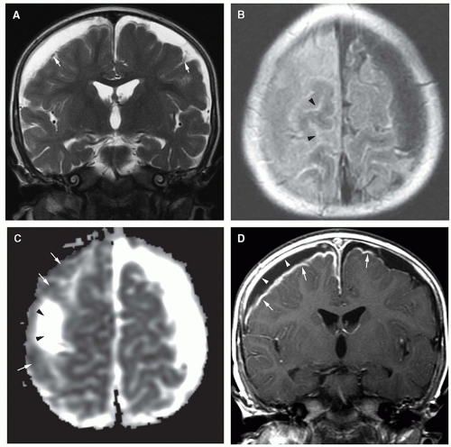
Figure 11-32 Subdural effusions and empyema in type F Haemophilus influenzae meningitis. A. Coronal T2-weighted image shows compressive bilateral frontal subdural fluid collections. Note the hypointense cortical veins adjacent to the brain surface (arrows). B. Axial FLAIR image through the cerebral convexities shows increased FLAIR signal within the right frontal subdural collection. Signal within the left subdural collection approximates CSF. Note the increased signal within the sulci reflecting leptomeningeal inflammation (arrowheads). The patient was not sedated under general anesthesia or with the use of fentanyl, both of which can produce increased FLAIR signal within the subarachnoid space. C. Axial average diffusivity image (ADC) through the convexities shows regions of hypointensity within the right subdural collection indicating reduced diffusion (empyema) (arrows). Note the component of the right subdural collection that shows increased diffusivity (more rapid water movement, arrowheads). D. Coronal postcontrast T1-weighted image shows mass effect of the convexity subdural collections. Note the thick right endosteal dural (arrowheads), and meningeal dural and leptomeningeal (arrows) enhancement. Due to seizures and persistent fever, the patient required right frontal subdural drainage.


Figure 11-33 Subdural empyema complicating Streptoccocus pneumoniae meningitis. A. Axial noncontrast CT image shows a diminished attenuation left parafalcine subdural fluid collection (white arrows). B. Axial T2-weighted image performed within 24 hours of the CT shows bilateral hyperintense parafalcine collections (white arrows). C. Axial apparent diffusion coefficient (ADC) image shows hypointense signal (white arrows) within the subdural fluid confirming reduced diffusion secondary to viscous purulent material. D. Axial postcontrast T1-weighted image shows marginal enhancement of the dural borders and prominent leptomeningeal enhancement secondary to meningitis. Postcontrast MR imaging is very useful in detecting small empyemas of the convexities, within the middle cranial fossa, and adjacent to the tentorium.


Ventriculitis

Ventriculitis is a common early complication of neonatal meningitis, particularly that caused by Escherichia coli. Bacteria enter the ventricles via the choroid plexuses and multiply as a result of diminished CSF flow, reduced secretion of CSF by the choroid plexus, and the relatively feeble intrinsic immune responses of CSF (4,6,194,196). The presence of intraventricular debris, which often layers dependently within the occipital horns of the lateral ventricle, is the best imaging sign of ventriculitis (Fig. 11-35). High-resolution cranial sonography will show hyperechogenic intraventricular debris and ependymal thickening (Fig. 11-35A and B) (188). The viscous dependent pus exhibits reduced diffusion on DWI (Fig. 11-35D) (199,202). After administering intravenous contrast, enhancement of the inflamed ependyma is seen on both CT and MR, with MR exhibiting greater sensitivity (Fig. 11-35E) (203). The ventricles are nearly always dilated due to reduced CSF absorption. A more ominous complication of ventriculitis is necrosis of periventricular white matter (Fig. 11-36), due to obstruction of subependymal and periventricular veins or necrosis of white matter from the release of (a) endotoxins, lipopolysaccharides from gram-negative bacteria, (b) teichoic acid from gram-positive organisms, and (c) cytokine-induced inflammation (6,204). Ultimately, multiple loculations may form in the ventricles, as a result of septa caused by astroglial response to infection; ultrasound and MR detect the septa with greater sensitivity than does CT (Fig. 11-35A and B) (188,203).


Hydrocephalus

Hydrocephalus is evaluated well by all imaging modalities; MRI, however, is most effective at localizing the level of obstruction and full appraisal of associated complications (see Chapter 8). Contrast-enhanced CT or MR is required when ventricular enlargement is unexplained, the diagnosis


is uncertain, or a complication of meningitis is suspected (Fig. 11-37). Hydrocephalus complicating meningitis indicates altered CSF circulation and/or resorption, which may occur at several levels (Fig. 11-38) (4,6,194), reflecting the intense inflammatory reaction and purulent exudates within the interventricular communications pathways, basal cisterns, sulci, and perivascular spaces (Figs. 11-37 and 11-38).


Figure 11-34 Subdural empyema, a complication of sinusitis. A and B. Axial T1 (A) and T2 (B) weighted images show a left parafalcine and left frontal subdural empyema (arrows). C. Axial diffusion-weighted image demonstrates hyperintensity (arrows) of the empyema secondary to the viscous nature of the purulent material. D. Coronal postcontrast T1-weighted image shows the empyema fluid (e) to be slightly hyperintense to CSF. There is underlying meningeal enhancement and outer dural enhancement. Note the moderate mass effect and left to right subfalcial herniation. Intracranial empyemas may evolve remote from the infected paranasal sinus. Thus, whole brain imaging is warranted when evaluating for suspected complications of sinusitis. E. Axial postcontrast FLAIR image clearly demonstrates the meningeal (inner) and endosteal (outer) margins of subdural compartment empyema enhancement.


Figure 11-35 Ventriculitis secondary to Escherichia coli. A and B. Coronal (A) and parasagittal (B) cranial sonograms show septated material filling and expanding the lateral ventricles (arrows). Note the hyperechoic thickening of the frontal horn ependyma (ependymitis, arrowheads in B). C. Axial T2-weighted image shows dependent debris (d) within the atria of the lateral ventricles. There is evidence for early ventricular obstruction. D. Axial diffusivity image shows hypointense signal within the occipital horns (arrows) and the right sylvian cistern subarachnoid space (arrowheads) consistent with reduced diffusion from highly viscous purulent material. E. Axial postcontrast T1-weighted image demonstrates dependent ventricular debris (d) and the thickened enhancing ependyma (white arrowheads) in ventricular trigones and occipital horns. Also note the prominent leptomeningeal enhancement within the sylvian cisterns.


Figure 11-36 White matter necrosis secondary to ventriculitis. A. Axial T1-weighted image shows proteinaceous debris (white arrowheads) layering in the posterior portion of the lateral ventricles. Early white matter necrosis is seen as mixed areas of hyper and hypointensity (small white arrows) in the frontal lobes. Poor gray matter-white matter contrast is seen in the entire cerebrum. B. Axial T2-weighted image shows extensive white matter necrosis (white arrows) seen as hypointensity of the white matter with some surrounding hyperintense edema. C. Postcontrast T1-weighted image shows better, the extent (small white arrows) of frontal necrosis. Some of the necrosing tissue (white arrowheads) is enhancing. The walls of the lateral ventricles (black arrowheads) are enhancing posteriorly. D. Follow-up postcontrast T1-weighted image 6 weeks later shows hydrocephalus and cavitation of the frontal lobes. Note that proteinaceous debris (white arrowheads) layering dependently within the ventricles. Some loculations (L) have formed adjacent to the ventricles.


Venous thrombosis/venous infarction

Thrombosis of deep veins, cortical veins, and venous sinuses represents an uncommon, but potentially fatal, complication of bacterial meningitis. Affected patients are initially detected as a result of seizures, coma, motor weakness, cranial nerve palsy, or headaches (205). Factors such as superimposed dehydration predispose to thrombogenesis (205,206). In the acute phase (when the clot is dense), thrombus in the sagittal sinus can be seen on CT as high density on the noncontrast scan (Fig. 11-39). Subacute sinus thrombosis is recognizable on CT by the so-called empty delta sign, which is a triangle of decreased density in the posterior portion of the affected sinus on a contrastenhanced scan. This sign is only visible after the clot becomes less dense than the contrast enhanced blood flowing around it (207,208).

On MR, sinus thrombosis is readily diagnosed when the thrombus is subacute and, therefore, hyperintense on T1-weighted images (Figs. 11-39 and 11-40). The best sequence is a 3D T1-weighted (gradient or spin echo) sequence examined in three planes with 1 mm


partition size (158) (see discussion on the following page). This observation is most easily made on midline sagittal images encompassing the sagittal sinuses and straight sinus/vein of Galen complex and on parasagittal images for the transverse and sigmoid sinuses. Other than in the subacute phase, the MR diagnosis of sinus thrombosis is difficult. Pronounced hypointensity seen in a venous sinus on sagittal T1-weighted images or in the superior sagittal sinus on coronal FLAIR images mitigates against thrombosis. However, in the absence of pronounced hypointensity on T1-weighted images, the patency of the sinus remains uncertain on routine MR images (Figs. 11-39B and 11-40A). The acutely thrombosed sinus is isointense to the brain on T1-weighted images and hypointense to the brain on T2-weighted images. This appearance cannot be distinguished from slow flow or pseudogating (in which the sinus is imaged during diastole). In this situation, evaluating the SWI images and reformations is very helpful to look for the hypointensity (paramagnetic effect) of thrombosis. Overall, as mentioned above, intravenous contrast-enhanced 3D MRV techniques are superior in the detection of cerebral sinovenous thrombosis as well as cortical and bridging vein thromboses (Figs. 11-40E and 11-41B) (158). In the clinical setting where contrast is contraindicated, robust noncontrast time-of-flight (TOF) 3D MRV contributes useful information.


Figure 11-37 Early hydrocephalus secondary to non-type b Haemophilus influenzae meningitis. A and B. Axial noncontrast CT images demonstrate infectious debris adjacent to the falx cerebelli (arrows) and dilation of the temporal horns (arrowheads) and third ventricle. The cerebrum is low in attenuation and there is absence of gray matter-white matter contrast. C. Coronal postcontrast T1-weighted image from MR 48 hours following the CT shows progressive dilation of the lateral and third ventricles. D. Axial postcontrast FLAIR image at the level of the basal cisterns demonstrates diffuse enhancement of the basal cisterns (white arrows). CSF flow is obstructed at this level.


Figure 11-38 Hydrocephalus following Escherichia coli meningitis. A. Four weeks following the completion of therapy for E. coli meningitis, Axial T2-weighted image in this infant with a bulging anterior fontanelle shows focal encephalomalacia of the brain stem secondary to remote brain stem infarction (arrow). There is moderate dilation of the temporal horns. B. Axial T2-weighted image at the level of the third ventricle demonstrates moderate dilation of the lateral ventricles and third ventricle. There is stretching of the massa intermedia (black arrowhead). Also note the lack of normal hypointense signal within the cerebral aqueduct (black arrow), indicating disturbance of normal CSF flow. This represents postmeningitic noncommunicating hydrocephalus.


Figure 11-39 Sagittal sinus thrombosis secondary to meningitis. A. Noncontrast CT scan shows high attenuation in the torcular herophili (arrows) at the junction of the straight sinus (also hyperdense and thrombosed) and the superior sagittal sinus. The presence of high density within blood vessels in a child beyond the first few months of life is extremely suspicious for sinus thrombosis. B. Sagittal T1-weighted image shows high signal intensity in the posterior half of the superior sagittal sinus (black arrows). The straight sinus (white arrowheads) also appears thrombosed. C. Coronal FLAIR image shows high signal intensity (arrow) within the superior sagittal sinus, supporting the diagnosis of sagittal sinus thrombosis. D. Axial T2-weighted image shows hyperintensity in the sagittal sinus (white arrows), suggesting the presence of extracellular deoxyhemoglobin, present in subacute clot. E. 2D time-of-flight MR venogram shows absence of flow-related enhancement in the superior sagittal sinus, confirming the diagnosis of thrombosis.


Figure 11-40 Streptococcus pneumoniae meningitis, venous thrombosis and cerebral ischemia. A. Axial T2-weighted image shows right cerebral convexity regions of subcortical T2 prolongation (edema) (arrows). B. ADC map shows reduced diffusion involving the right frontoparietal convexity (arrow). C. Axial SWI shows numerous right convexal serpentine hypointensities consistent with slow flow or thrombus within convexity veins (arrows). D. Axial T1-weighted following intravenous contrast shows filling defects (clot) within the right convexity veins (arrows). E. Sagittal post-IV contrast 3D SPGR image demonstrates clot within the right transverse venous sinus (arrow).


Figure 11-41 Cortical vein thrombosis and cerebral ischemia secondary to Neisseria meningitides meningitis. A. Coronal GRE image shows several convexity cortical veins demonstrating diminished signal indicating cortical vein thrombosis or slow venous flow. B. Axial postcontrast T1-weighted image shows thick enhancement within the left intraparietal sulcus (large white arrows). Note the linear hypointense filling defect (small black arrow) consistent with thrombosed cortical vein. The adjacent cortex shows T1 hypointensity consistent with edema. Also note the bilateral frontal effusions. C. Axial average diffusivity image shows evidence of increased diffusivity (interstitial edema, white arrows). There is reduced diffusivity (hypointensity, white arrowheads) adjacent to the lateral intraparietal sulcus, suggesting injured tissue. Cortical venous occlusion leads to regional venous hypertension, breakdown of the blood-brain barrier and, sometimes, ischemic injury. D. DTI-derived color fractional anisotropy image through the cerebral convexities shows a loss of coherence (decreased anisotropy, arrows) of water motion in the region of the cortical occlusion and in the subcortical white matter tracts, reflecting interstitial edema and, possibly, microstructural white matter injury.

Venous ischemia and infarcts are diagnosed by their characteristic location and appearance. Typically, ischemia and infarcts that arise
from sagittal sinus thrombosis are parasagittal (Fig. 11-40); infarcts from thrombosed internal cerebral veins, straight sinus, and vein of Galen involve the thalami. Infarcts from vein of Labbé, transverse sinus, or sigmoid sinus thrombosis involve the temporal lobe. Smaller cortical/subcortical infarcts are often seen over the cerebral convexities (Fig. 11-41). On CT, venous infarcts are usually poorly delimited, hypoattenuating, or mixed attenuation areas involving the subcortical white matter and producing a slight mass effect on ventricular structures. The low attenuation is probably due to localized cerebral edema (vasogenic and cytotoxic), whereas high attenuation areas within the brain parenchyma usually represent hemorrhage. Following intravenous contrast administration, a lack of expected venous opacification is seen (venous clot may be detected), adjacent sulcal/gyral enhancement is noted, and adjacent parenchymal hypoattenuation (edema) is appreciated (209). On MR, early venous infarcts may be identified by visualization of prolonged T1 and T2 relaxation times in characteristic regions of the brain parenchyma, SWI hypointensity within the adjacent venous structure(s), and low ADC values in regional parenchyma (Fig. 11-41). Another early and important imaging sign of venous infarction is visualization of thrombus in the deep medullary veins with surrounding cavitation. Of note, reduced parenchymal diffusivity in the setting of venous thrombosis lacks sensitivity in predicting venous infarction. With venous ischemia and infarcts, diffusion appears to be heterogeneous, with areas of increased, normal, and decreased diffusion (Fig. 11-41) (210). The variable diffusion characteristics are likely related to the combination of interstitial and cytotoxic edema found in venous infarcts. In addition, part of this heterogeneity may be due to the frequent presence of hemorrhagic tissue, which makes diffusivity values unreliable. Twenty-five percent of venous infarcts are hemorrhagic and have an imaging appearance that varies from large subcortical hematomas to petechial hemorrhages admixed with edematous brain parenchyma (Fig. 11-40) (207,209). The hemorrhages are generally subcortical and often multifocal with irregular margins. They are occasionally linear in nature, indicating hematoma in and around the vein; this appearance is quite specific.

Advances in MR venography (Fig. 11-41) have greatly aided the diagnosis of venous sinus thrombosis by MR (211,212,213,214). Gadoliniumenhanced 3D gradient echo techniques, reformatted as thin sections in multiple planes, are superior to 2D TOF venography in the detection of intracranial venous thrombosis (see Chapter 1, Fig. 11-41) (214). False-negative and false-positive pitfalls exist with 2D TOF and to a lesser extent 3D non-contrasted MRV techniques including “flow gaps,” which may simulate venous thrombosis (215). Additionally, it is important to remember that time-of-flight MR angiography is acquired using T1-weighted images. Both moving blood and subacute clot are hyperintense; thus, the results of a time-of-flight venogram may be equivocal. Patients with hyperintensity of an intracranial venous sinus detected on T1-weighted images should, therefore, have either contrast-enhanced MR venography, phase-contrast venography, novel non-contrast fluid-suppressed methods for flow-independent venography, or CT venography (Fig. 11-41) (212,216). CT venography techniques are comparable to contrast-enhanced 3D MR venography for the detection of intracranial venous thrombosis (212,217,218) but are less desirable in the pediatric population because of the ionizing radiation (see Chapter 1).

Cavernous sinus thrombosis is an uncommon sequela of meningitis; it is more commonly the result of paranasal sinus, dental, or ocular infection (219,220,221). CT of cavernous sinus thrombosis shows an enlarged, outwardly convex cavernous sinus with enhancing adjacent meninges (222). Ipsilateral or contralateral orbital veins may be enlarged and thrombosed (Fig. 11-42) (223). On MR, signal intensity of cavernous sinus thrombus varies depending on the state of infection, inflammation, and clot evolution (therefore, the signal intensity of the sinus alone should not be relied upon to make the diagnosis). Direct visualization of nonenhancing clot confirms the diagnosis (Fig. 11-42). The cavernous sinus may be enlarged and the cavernous carotid artery narrowed or occluded (224,225). T2 prolongation may be seen in the adjacent clivus or petrous apex (225). With infection by aggressive organisms, a mycotic aneurysm of the cavernous carotid artery may develop. Therefore, it is useful to acquire an MR arteriogram of the cavernous carotid arteries if septic thrombophlebitis of the cavernous sinus is suspected.


Arterial infarction

Focal cerebral parenchymal abnormalities seen in patients with bacterial meningitis are typically attributable to ischemia, infarctions, and/or cerebritis (226). Arterial infarctions in the setting of meningitis usually result from arteritis secondary to involvement of the perivascular spaces and, subsequently, the arterial walls by the infection; this process is likely mediated by endotoxins, cytokine driven, or immunologically mediated (226). Widespread vasculopathy may lead to extensive white matter cytotoxic edema, acute demyelination and, ultimately, to necrosis (226). Although both CT and MR can diagnose the arterial complications of bacterial meningitis, contrastenhanced MR with DWI is the examination of choice. The regions of ischemia and infarction may be in the distribution of perforating arteries, widespread throughout the cerebral hemispheric white matter, or sharply marginated and confined to a specific vascular territory. Diffusion-weighted MR imaging is mandatory in this setting, as it will detect infarctions earlier than CT or conventional MRI (Figs. 11-43, 11-44 and 11-45) (227). Large or small vessels may be affected. When major vessels, such as the middle or anterior cerebral arteries are involved, large cortical infarctions result (Fig. 11-45). Use of arterial spin labeling to assess regional perfusion may supply additional information regarding areas at risk due to low perfusion. Group B Streptococcus, Streptococcus pneumoniae, Escherichia coli, tuberculous, and syphilitic meningitis can be associated with infarction including lacunar-type infarcts in the distribution of perforating vessels involving the brain stem, basal ganglia, white matter, and vascular territorial infarctions (Figs. 11-19, 11-43, and 11-44) (227,228). DTI may give insight into white matter structural injury that may accompany meningitis. This is particularly relevant for the neonate with meningitis where there is known vulnerability of periventricular white matter to oxidative and hypoxic/ischemic injury that may occur in the context of meningitis (229). Magnetic resonance proton spectroscopy (MRS) may be useful to assess for evidence of anaerobic metabolism within injured tissue, demonstrating lactate and necrosis (Fig. 11-43E). Mitochondrial impairment is demonstrated by reduction of NAA (Fig. 11-43E).


Tuberculous Meningitis

Meningitis is one of the most common extrapulmonary manifestations of infection with Mycobacterium tuberculosis and closely associated with miliary tuberculosis. CNS mass lesions (“tuberculomas”), meningovasculitis, and miliary involvement of the CNS can all occur in children with tuberculosis (230,231). Risk factors for tuberculous CNS disease in children and adolescents include exposure to a family member with tuberculosis, birth or extended travel in regions endemic for tuberculosis, exposure to a (former) prison inmate, and HIV infection. CNS involvement usually becomes clinically apparent within 6 months of the initial infection (232,233). Meningitis in childhood due to M. tuberculosis begins with headache, vomiting, irritability, or lethargy, and, when untreated, affected patients develop cranial nerve palsies (particularly CNs II, IV, and VII), focal deficits, or signs of ICP (177). Fever is not always present. Examination of the CSF reveals a mixed
or predominant lymphocytic pleocytosis, elevated protein (usually between 100 and 500 mg/dL), and mildly reduced glucose (234). The diagnosis of tuberculous meningitis can be established by PCR detection of mycobacteria in the CSF and supported by a positive tuberculin skin test or detection of interferon-gamma release in whole blood samples exposed to M. tuberculosis antigens (232,234). Treatment consists of combined antituberculous therapy with a four-drug regimen and administration of corticosteroids; even with therapy, death or significant morbidity is possible, especially when treatment is delayed.


Figure 11-42 Cavernous sinus thrombosis in the setting of sphenoid sinusitis/meningitis. A. Axial contrast-enhanced CT shows enlarged tubular serpentine thrombosed superior ophthalmic veins (white arrows). Note the thrombus within the cavernous sinuses (arrowheads) and the extensive preseptal and postseptal edema. B. Axial T2-weighted fat-saturated image confirms enlarged tortuous thrombosed superior ophthalmic veins (arrows). C. Axial postcontrast axial fat-saturated T1-weighted image shows the thrombosed orbital venous structures as nonenhancing elements. Enhancement is due to multiple small collateral veins. Note the diffuse pre- and postseptal edema. D. Axial postcontrast T1-weighted image through the cavernous sinus demonstrates thrombus as large hypointense filling defects (black arrows).

The meningovascular manifestations of tuberculosis, such as cerebral infarction, have a substantial impact upon neurodevelopmental outcome (235). Acute hydrocephalus, caused by the intense meningeal reaction associated with tuberculous meningitis, requires ventriculoperitoneal shunt placement. If untreated, tuberculous meningitis rapidly progresses to death, with average disease duration of only 3 weeks (236). Even with current treatment regimens, tuberculous meningitis has considerable morbidity and mortality (234).

The bacilli are distributed within the brain and meninges throughout the CNS but do not multiply as readily as they do in other organs. Meningitis probably results from rupture of small tuberculomas in the cortex, spinal cord, leptomeninges, or choroid plexus (180,237). CNS manifestations usually become apparent as tuberculous meningitis, tuberculoma, tuberculous abscess, or spinal leptomeningitis within days after miliary spread; a gelatinous exudate fills the pia-arachnoid along the basal cisterns, particularly the prepontine cistern, where it infiltrates and produces inflammation along the walls of the meningeal blood vessels. The small cortical blood vessels and perforating vessels become affected as the exudate spreads into the Virchow-Robin spaces,

causing infarction of surrounding brain tissue. Outcome is directly related to the location and extent of these infarctions (238). The basal ganglia and thalami are affected in almost half of cases (239,240); suprasellar cisternal inflammation and involvement of the posterior hypophysis may lead to the syndrome of inappropriate antidiuretic hormone secretion (241). The thick exudate blocks the subarachnoid spaces, causing hydrocephalus. Infiltration of the perineurium of the cranial nerves causes neuropathies, particularly of cranial nerves II, VI, and VII. Small tubercles over the convexity of the brain may involve the leptomeninges, while deeper lesions may infiltrate the periventricular area.


Figure 11-43 Multiple perforator arterial infarctions from Group B Streptococcus (S. agalactiae) meningitis. A. Axial T2-weighted image shows innumerable basal ganglia foci of T2 prolongation. Note the subinsular regions of T2 hyperintensity and heterogeneous left perifrontal white matter signal (coagulation necrosis of white matter, black arrow). B. Axial T2-weighted image at the level of the centrum semiovale demonstrates heterogeneous white matter signal intensity (arrows). C and D. Average diffusivity images corresponding to (A and B) show markedly reduced diffusivity corresponding to tissue injury. Note the confluent reduced diffusivity within the centrum semiovale. E. Proton spectroscopy (TE = 35 ms) of the right basal ganglia demonstrates a large lactate/lipid (L/L) and lipid (Li) peaks. Lactate is an indicator of anaerobic metabolism and lipid results from cell necrosis. There is mild decline of NAA and elevation of the excitatory neurotransmitter glutamate and glutamine (Glx).


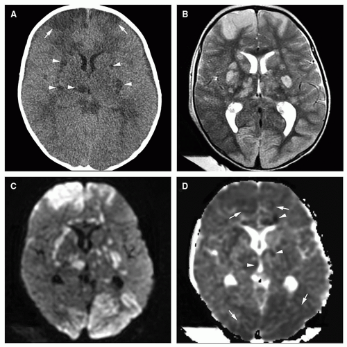
Figure 11-44 Multiple arterial infarctions from Streptococcus pneumoniae meningitis. A. Axial noncontrast CT shows multiple wedge-shaped cortical and subcortical regions of diminished attenuation (white arrows). Numerous basal ganglia/thalami foci of low attenuation are also seen (arrowheads). B. Axial T2-weighted image shows better the regions of peripheral and central T2 hyperintensity representing edema (cytotoxic and vasogenic in type). C. Axial diffusion-weighted image shows multiple sites of ischemic injury as areas of hyperintensity. D. Axial average diffusivity image 6 days after initial diagnosis demonstrates evidence of early pseudonormalization. Relatively few areas of subtle frontal and occipital reduced diffusivity (white arrows) and central basal ganglia and periventricular foci of reduced diffusion (arrowheads) are still seen.


Imaging

It serves the radiologist well to remember that the imaging manifestations of CNS Mycobacterium tuberculosis result from the military spread of disease and are as diverse as the clinical presentation; manifestations range from focal and/or diffuse basilar leptomeningeal exudate to more localized abnormalities (granulomatous meningitis, tuberculoma, cerebritis, abscess, and stroke) anywhere within the cerebrum or cerebellum. More specifically, ventriculomegaly is present in 50% to 77% of affected patients, most often secondary to hydrocephalus (237,242). In more advanced disease, the basal cisterns are partially or completely filled by the purulent tuberculous exudate, which can extend into the spinal subarachnoid space (Fig. 11-46). On noncontrast CT or MRI, the exudate appears higher in attenuation (CT) and signal intensity (T1, T2 FLAIR, and SWI sequences) than CSF; it may be overlooked on T2-weighted MR images, because the high-intensity CSF signal will obscure the cisternal disease unless small field-of-view (FOV) MR techniques are used (Fig. 11-47C). Following intravenous contrast, the involved cisterns variably enhance (Figs. 11-46 and 11-47B) (238,242,243,244,245). Although the exudate usually fills the basilar cisterns bilaterally and symmetrically, about 10% of affected children have more regional disease, typically affecting the sylvian fissure, and less commonly the ambient or quadrigeminal plate cisterns (246). FLAIR imaging (particularly delayed postcontrast FLAIR) improves conspicuity of cisternal and leptomeningeal pathology (191). Tuberculous cranial pachymeningitis has been reported but is very rare (247).

Only gold members can continue reading. Log In or Register to continue

Stay updated, free articles. Join our Telegram channel

Nov 11, 2018 | Posted by in NEUROLOGICAL IMAGING | Comments Off on Infections of the Developing and Mature Nervous System

Full access? Get Clinical Tree

Get Clinical Tree app for offline access