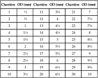
CHAPTER 4 After completing this chapter, the reader will be able to perform the following: Catheters are simply tubes of varying lengths and inside diameters with holes in each end that allow the contrast agent to flow through. Originally, catheters were adaptations of ureteral catheters that were constructed of rubber, but this soon gave way to the various thermoplastic catheters manufactured today. Catheters are manufactured out of Teflon, polypropylene, polyurethane, and polyethylene. Each of these materials possesses different characteristics that make them suitable for different procedures (Box 4-1). One major advance in catheter manufacturing was the development of the radiopaque polyethylene catheter, which allowed the radiologist to follow the path of the catheter through the body to its destination. Radiopaque catheters usually have different characteristics than the nonopaque varieties. In some research institutions catheters are custom made by the physician to meet the specifics of the procedure being studied. Catheter tubing is available from certain manufacturers and can be custom formed. Appendix 1 illustrates the method of specialized catheter formation from standard catheter tubing. Catheter sizes are expressed in inches or millimeters or by French number. The gauge scale known as the French scale was developed by Charrière, a French instrument maker (1803-1876). On Charrière’s gauge scale, 1 Charrière (or 1 French [1F]1) is equal to a diameter of ⅓ mm, with each consecutive Charrière differing from the previous one by ⅓ mm. Table 4-1 lists the millimeter conversions for French numbers from 1 to 30. TABLE 4-1 French Gauge Scale Conversions From Instruments and Catheters for Radiography. Solna, Sweden, Firma AB Kifa, 1970. OD, outside diameter. Figure 4-1 illustrates a gauge scale that was used which listed size specifications for cardiovascular catheters, facilitating the conversion from inches to millimeters or to French size. The gauge scale also shows the relative sizes of various catheters from 3 French to 34 French. Table 4-2 lists the size specifications for a selection of both standard and thin-walled cardiovascular catheters. TABLE 4-2 Size Specifications for Cardiovascular Catheters
Instrumentation and Accessories
 Identify the need for radiographic catheters
 Identify how catheters are sized using the Charrière gauge scale
 List the reasons for various shaped catheters
 Describe the guide wire used to move and place the catheter in the correct location
 Describe the use of needles during advanced procedures
 Identify the accessory items that might be found in the interventional suite
 Define the use of prepackaged and sterilized procedure trays
CATHETERS
Size
Charrière
OD (mm)
Charrière
OD (mm)
Charrière
OD (mm)
1
⅓
11
3⅔
21
7
2
⅔
12
4
22
7⅓
3
1
13
4⅓
23
7⅔
4
1⅓
14
4⅔
24
8
5
1⅔
15
5
25
8⅓
6
2
16
5⅓
26
8⅔
7
2⅓
17
5⅔
27
9
8
2⅔
18
6
28
9⅓
9
3
19
6⅓
29
9⅔
10
3⅓
20
6⅔
30
10
Standard Wall
Thin Wall
Inside Diameter
Outside Diameter
Needle Equivalent
Inside Diameter
Outside Diameter
Needle Equivalent
French Size
in
mm
in
mm
ID
OD
French Size
in
mm
in
mm
ID
OD
3
0.014
0.36
0.039
1.00
23+
20+
4
0.018
0.46
0.052
1.33
22+
18+
4
0.023
0.58
0.052
1.33
20
18+
5
0.026
0.66
0.065
1.67
19
16
5
0.034
0.86
0.065
1.67
19+
16
6
0.036
0.91
0.078
2.00
18+
15+
6
0.046
1.17
0.078
2.00
17+
15+
7
0.046
1.17
0.091
2.33
![]()
Stay updated, free articles. Join our Telegram channel

Full access? Get Clinical Tree

 Get Clinical Tree app for offline access

