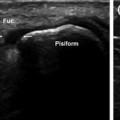
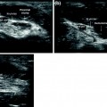
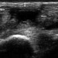
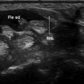
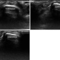
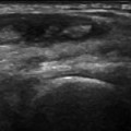
Fig. 5.1
The proximal intersection (schematic diagram) is located 3.5–4.8 cm (average 4.18 cm) from the Lister tubercle. Here, the abductor longus and extensor brevis tendons of the thumb (Abd l-Estb) cross over the extensor carpi radialis longus and brevis tendons (Est rlc-rbc)
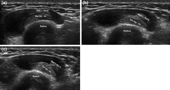
Fig. 5.2
Relations between first- and second-compartment extensor tendons during wrist movement. During wrist movements associated with daily activities, the tendons of the first compartment (Abd l-Est b) shift laterally over those of the second compartment (Est rlc-rbc) (a–c). The movement is accentuated during certain sports-related activity (e.g., weight lifting, rowing, basketball), and the friction generated at the crossover point causes the symptoms of the intersection syndrome
The intersection syndrome is rare. In most cases, it presents with fluid in the sheath of the radial extensor tendons, somewhere between the proximal and distal intersections [5]. Less commonly, a smaller effusion may be seen in the tendon sheath of the first compartment. Other presentations that are even less frequent include the presence between the two tendon groups of soft tissue edema or a serous bursa with bursitis (Table 5.1).
Table 5.1
Intersection syndrome: possible presentations
Effusion within the sheath surrounding the extensor carpi radialis longus and brevis tendons located between the proximal and distal intersections; sometimes smaller effusions are also present within the first osseofibrous tunnel |
Edema of the soft tissues between the two groups of tendons |
Presence of a bursa/bursitis between the two groups of tendons |
Stay updated, free articles. Join our Telegram channel

Full access? Get Clinical Tree


