A wire is placed through the stenosis in the common iliac artery. A balloon is inflated to dilate the narrowing (arrowheads). Note: contrast material is black
48.2 IVC filter
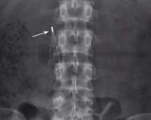
A self-expanding umbrella-like structure or IVC filter (arrow) is placed in the IVC to prevent passage of large emboli to the lungs
48.3 CT-guided biopsy
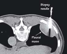
With the patient lying prone a biopsy needle is placed to gain a tissue sample of a large pleural mass
48.4 Colonic stent
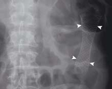
A self-expanding wire-walled stent (arrowheads) is placed via the rectum across an inoperable tumour of the sigmoid colon
48.5 Percutaneous transhepatic cholangiogram (PTC) with biliary stent insertion
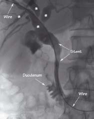
A wire is passed via the liver through a dilated biliary system (*) and into the duodenum. A stent is placed to relieve an obstruction caused by a pancreatic mass
48.6 JJ-ureteric stent
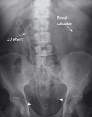
A double pigtail catheter or ‘JJ stent’ is positioned within the ureter passing from the renal pelvis to the bladder. The patient had recurrent bilateral renal calculi/stones. Multiple phleboliths/venous calcifications (arrowheads) are also seen in the pelvis outside the path of the ureters
Vascular intervention
Related posts:


Stay updated, free articles. Join our Telegram channel

Full access? Get Clinical Tree


