Interventional Urology
The first report on an interventional urologic procedure performed under continuous sonographic guidance was published in 1979. The procedure was successfully accomplished under dynamic ultrasound guidance without radiation exposure to the patient or staff.
Since our readers may be less familiar with urologic interventions than with other procedures described in this book, our discussions of interventional urology will include a review of some basic principles.1
26.1 Transrectal Ultrasonography of the Prostate
26.1.1 Introduction
Transrectal ultrasonography of the prostate (TRUS) has become an established diagnostic and therapeutic tool in urology. The close proximity of the rectum to the prostate permits the use of high-frequency transducers in the 6- to 10-MHz range, resulting in higher spatial resolution of the imaged structures.
Histologic studies of the prostate2 have identified the presence of three glandular zones (peripheral, central, and transitional) and one stromal zone (anterior segment). This subdivision can also be appreciated on ultrasound images.3
26.1.2 Equipment Requirements
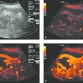
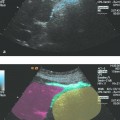
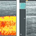
TRUS should be performed with a high-frequency transducer (6–10 MHz) in at least two standard planes. Also, the transducer should have a biopsy channel or needle guide to allow for ultrasound-guided biopsy, antibiotic injection, abscess drainage, or interstitial brachytherapy. Most modern ultrasound scanners have color Doppler and duplex capabilities, and some systems allow for contrast agent-specific imaging and elastography.
26.2 Diseases of the Prostate
26.2.1 Prostate Cancer
Etiology and Incidence
Prostate cancer is the most common visceral cancer affecting males in the Western population. The incidence in the United States is 241,740 new cases per year.4
A variety of etiologic factors have been discussed: ethnographic (incidence is 30 times higher in African Americans than in Japanese); dietary (high-fat, low-fiber foods increase the cancer risk, soy consumption reduces it); genetic (positive family history is linked to higher risk); environmental (occupational cadmium exposure); and hormonal (androgenic stimulation).
Diagnosis
The diagnosis of prostate cancer is based on digital rectal examination, serum PSA (prostate specific antigen) level, and TRUS of the prostate. Most prostate cancers develop in the peripheral zone of the gland, and tumors with a volume >0.2 mL are palpable at that location. Accordingly, a suspicious palpable nodule is an absolute indication for prostatic biopsy.
TRUS, with a specificity of approximately 36%, is considered an adjunctive test in the diagnosis of prostate cancer. Several studies have shown a detection rate of approximately 20% and a positive predictive value between 30% and 58%. The negative predictive value is 50 to 65%. Several studies have also investigated the sensitivity of TRUS, especially in the important preoperative detection of capsular involvement and seminal vesicle invasion. Extracapsular extension can be detected with a sensitivity of 83% and a specificity of 67%, seminal vesicle invasion with a sensitivity of 43% and a specificity of 86%. Today the undisputed domain of TRUS is the ultrasound-guided transrectal prostate biopsy. The transperineal approach is used mainly in patients who have had a proctectomy.5
Prostate cancer usually appears as a hypoechoic area, and this finding should be reproducible in both axial and sagittal planes. In 35% of cases, however, the lesion appears isoechoic or even hyperechoic. Conversely, not every hypoechoic lesion is prostate cancer. Based on studies comparing TRUS findings with histology, it is known that slightly hyperechoic areas in the peripheral zone of the gland may represent normal prostate tissue, acute or chronic prostatitis, atrophy, prostatic infarction, or prostatic intraepithelial neoplasia (PIN).
Accordingly, the following sonographic criteria are considered suggestive of prostate cancer:
Hypoechoic area with irregular margins located in the peripheral zone
The abnormal area penetrates through the bright rim of the prostate capsule or extends to the rectal wall.
Asymmetry of the prostate lobes
Obliteration of the angle between the prostate and seminal vesicle. Portions of the seminal vesicles not invaded by cancer may show asymmetrical dilatation.
Treatment
The treatment of prostate cancer depends on patient age and health status, PSA, histologic stage (TNM), and tumor grade (Gleason score). Localized prostate cancer is usually treated by primary radical prostatectomy. Other options are modern radiotherapy regimens (high-dose rate [HDR] and low-dose-rate [LDR], brachytherapy with or without percutaneous radiation, or even active surveillance. Metastatic prostate cancer is treated primarily with various forms of antiandrogen therapy. If the tumor becomes resistant to hormone therapy (hormone-refractory prostate cancer, HRPC), various chemotherapeutic agents (taxanes, mitoxantrone) may be administered with palliative intent.6
26.2.2 Prostatic Abscess
Diagnosis
Abscess formation in the prostate disrupts its normal zonal architecture. As in other organs, the abscess typically appears hypoechoic with ill-defined margins. The hypoechoic areas sometimes contain internal echoes.
Treatment
Percutaneous needle aspiration can be performed by the transrectal or transperineal route under TRUS guidance. If the aspiration yields putrid fluid, an abscess that is larger than 2 cm should be drained. This is done by dilating the needle tract with Teflon dilators over a guidewire using Seldinger technique. Next, a pigtail catheter is introduced, and 40 mg of gentamicin is instilled into the abscess cavity through the catheter and left there for 1 hour. The contents of the abscess cavity are then drained into a collecting bag. When the drain output reaches zero, the catheter is removed and the site is assessed by endosonography.
An alternative treatment is to open the abscess during a transurethral resection of the prostate (TURP).
26.3 Prostate Biopsy
26.3.1 Introduction
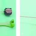
Core needle biopsy of the prostate is usually performed through a transrectal approach. The transperineal approach is less commonly used for prostate biopsy, although it is used routinely for brachytherapy. The biopsy is performed with an 18-gauge needle and a spring-loaded biopsy gun (e.g., Biopty device [Bard]). There is no evidence that biopsy needles cause tumor seeding in patients with prostate cancer. A distinction is made between targeted biopsies, which sample selected areas that have been identified by ultrasound or palpation, and systematic biopsies, which sample all portions of the prostate according to a standard scheme. The classic sextant biopsy described by Hodges has been abandoned in favor of volume- and age-dependent biopsy strategies such as the Vienna nomogram.
26.3.2 Indications
Prostate biopsy should be performed only if a possible diagnosis of prostate cancer would have therapeutic implications. The correlation between PSA levels and relative risk of prostate cancer was shown to be 6.6 for a PSA level of 0 to 0.5 and 26.9 for a PSA level of 3.1 to 4.7
26.3.3 Informed Consent and Preparation
The patient is informed about the risks and benefits of the procedure, and written informed consent is obtained (e.g., Diomed consent form).
Stay updated, free articles. Join our Telegram channel

Full access? Get Clinical Tree


