Introduction
Clinical positron emission tomography (PET) and single-photon emission computed tomography (SPECT) and integrated imaging of the brain combining PET or SPECT with magnetic resonance imaging (MRI) or computed tomography (CT) by software-based image fusion have been around for a long time, and one of the early successes of PET imaging was in imaging of the brain. That integration was possible in the brain is due to the relatively simple operations needed to coregister molecular and anatomic data. Some of the basic advantages of PET over other methods have initially been understood in the brain, notably that fluorodeoxyglucose (FDG) frequently shows the highest accumulation in areas where a tumor is most malignant. Many quantitative concepts have also been developed in brain PET and are still best applied when imaging the brain. In addition, PET is an excellent imaging modality for studying various brain functions relatively noninvasively and quantitatively, and some of these research applications, such as quantitative brain perfusion imaging, have gained clinical importance as well.
Related posts:
 PET Radiopharmaceuticals: Perfusion Compounds [13N]Ammonia, [15O]Water, [82RB]Rubidium, and Other Generator Products of Clinical Relevance
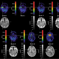 Brain Tumors: Astrocytoma, Oligodendroglioma, and Glioblastoma Images with PET and SPECT
 Sentinel Node Imaging in Head and Neck Tumors
 PET-CT and SPECT-CT of Bone Metastases
 SPECT and PET in Benign Thyroid and Parathyroid Disease
Stay updated, free articles. Join our Telegram channel

Full access? Get Clinical Tree


