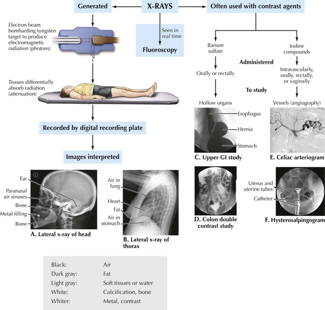
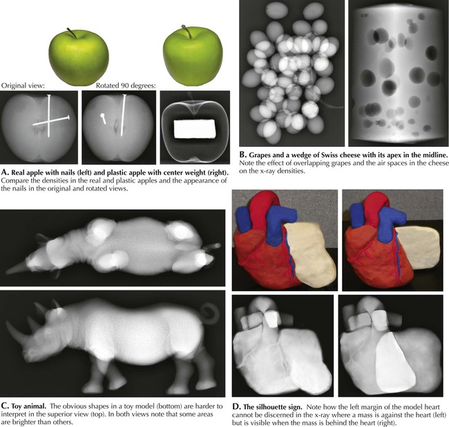
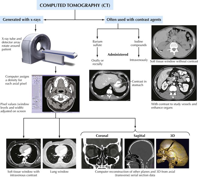
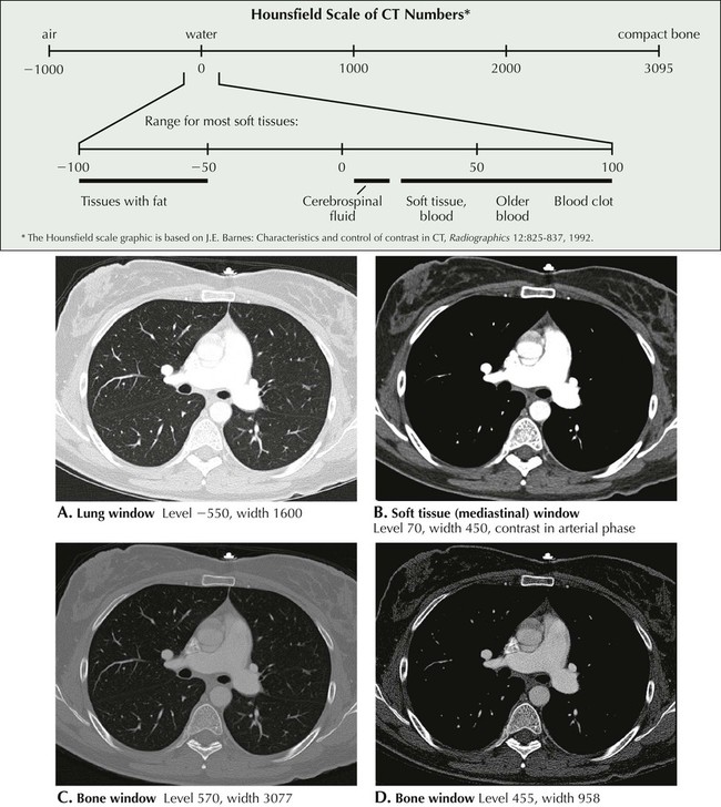
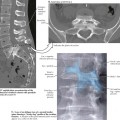
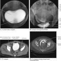
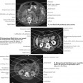
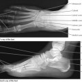
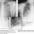
1 Introduction to Imaging Modalities 1.1. X-RAY OVERVIEW 1.2. INTERPRETATION OF X-RAY DENSITIES 1.3. COMPUTED TOMOGRAPHY OVERVIEW 1.4. THE HOUNSFIELD SCALE: CT WINDOW LEVELS AND WINDOW WIDTHS 1.5. CT USES, ADVANTAGES, AND DISADVANTAGES 1.6. MAGNETIC RESONANCE IMAGING OVERVIEW 1.7. MRI USES, ADVANTAGES, AND DISADVANTAGES 1.8. MRI PULSE SEQUENCES 1.9. NUCLEAR MEDICINE IMAGING 1.10. FLUOROSCOPY 1.11. ULTRASOUND 1.12. ANGIOGRAPHY: COMPUTED TOMOGRAPHY ANGIOGRAM VS. MAGNETIC RESONANCE ANGIOGRAM AND VOLUME RENDERING VS. MAXIMUM INTENSITY PROJECTION 1.13. ANGIOGRAPHY: DIGITAL SUBTRACTION ANGIOGRAPHY 1.14. ARCHIVING AND COMMUNICATION SYSTEM 1.15. FUTURE DEVELOPMENTS IN IMAGING 1.1 X-Ray Overview This concept map is an overview of how x-ray images are acquired and interpreted. X-rays (photons) from the tungsten target pass through the body to expose the recording plate (what used to be film). The greater the exposure, the darker the density will be. The greater the attenuation or absorption of the photons by tissues, the whiter the density will appear. Organs with air will appear dark; bone will appear white. Soft tissues and water have intermediate density. A greater thickness of bone or soft tissue results in a whiter density. In A, compare the bone densities at the periphery of the neurocranium, the interior of the neurocranium, and the dense cortical bone of the temporal bone at the base of the neurocranium. In B, compare the soft tissue densities of the heart and the abdomen. Barium contrast agents are used to study hollow organs (C and D). Water-soluble iodine compounds are used for vascular studies (E) or where contrast might enter a body cauity (F). 1.2 Interpretation of X-Ray Densities The interpretation of x-ray densities is demonstrated with x-rays of common objects. In A, densities ranging from the metal nails to the air in the plastic apple are seen. Different views of the apple (or human body) are required to evaluate the location and shapes of the nails (or anatomical structures or pathological processes—also see C). Like a thin neurocranial bone or a membranelike pleura, the thin shell of the plastic apple is much denser seen on edge than en face. For equivalent densities of objects, the x-ray image is denser for larger or thicker or overlapping objects (B and C). D illustrates the loss of a boundary of an object or structure if it is against a structure or fluid of similar density. This is called the silhouette sign. The boundary is visible if the object is against air and the similar density is behind or in front of the object. 1.3 Computed Tomography Overview Current multidetector computed tomography (MDCT) images are generated with x-rays passing through the body in a helical fashion as the patient moves through a gantry containing a rotating x-ray tube. Detectors on the opposite site of the tube collect the x-rays that have passed through the body. Mathematical algorithms are used to reconstruct axial (transverse plane) images of the body from the data collected by the detectors. Images in the sagittal and coronal planes and three-dimensional renderings can be reconstructed by computer from the serial slices of axial data. The gray-scale image can be manipulated on the monitor. 1.4 The Hounsfield Scale: CT Window Levels and Window Widths Computed tomography (CT) density numbers are attenuation units measured by what is called the Hounsfield scale, named after the British engineer who developed the first practical CT scanner in the 1970s. The density of water is set at zero, air (as in the lung or bowel) is −1000, and compact bone is +3095. Most soft tissues in the body have CT numbers between −100 and +100. Computer monitors show 256 levels of gray; thus only a portion of the Hounsfield scale can be displayed, and this “window” can be adjusted on the screen. The number on the Hounsfield scale set to middle gray is referred to as the window level, and the range of the gray scale mapped onto the Hounsfield scale is called the window width. All CT numbers below the window width display as black; CT numbers above the window width are white. A wide window width is good for imaging bone; a narrow window is better for soft tissue. 1.5 CT Uses, Advantages, and Disadvantages Since CT is based on x-rays, CT studies are especially good for evaluating bone and structures containing air, as in the bowel (D). The high speed of acquisition is good for use in the thorax and abdomen since motion artifact is limited. A bone window has excellent discrimination between compact and trabecular bone (A) and is useful throughout the body in detecting and evaluating fractures. The majority of CT studies use contrast, and vascular studies (angiography) are commonly done with CT. Vascular contrast also enhances the boundaries between organs and fat or air, and the window levels can be adjusted on the screen. The major disadvantage is the radiation dose. There is increasing concern over the amount of radiation that the U.S. population is being exposed to because of the increased use of CT and nuclear medicine in medical diagnosis. The ALARA principal (As Low As R Only gold members can continue reading. Log In or Register to continue Share this: Share on X (Opens in new window) X Share on Facebook (Opens in new window) Facebook Related posts: Back and Spinal Cord Pelvis and Perineum Abdomen Lower Limbs Thorax Head and Neck Stay updated, free articles. Join our Telegram channel Join Tags: Netters Introduction to Imaging Jan 10, 2016 | Posted by admin in RADIOGRAPHIC ANATOMY | Comments Off on Introduction to Imaging Modalities Full access? Get Clinical Tree
1 Introduction to Imaging Modalities 1.1. X-RAY OVERVIEW 1.2. INTERPRETATION OF X-RAY DENSITIES 1.3. COMPUTED TOMOGRAPHY OVERVIEW 1.4. THE HOUNSFIELD SCALE: CT WINDOW LEVELS AND WINDOW WIDTHS 1.5. CT USES, ADVANTAGES, AND DISADVANTAGES 1.6. MAGNETIC RESONANCE IMAGING OVERVIEW 1.7. MRI USES, ADVANTAGES, AND DISADVANTAGES 1.8. MRI PULSE SEQUENCES 1.9. NUCLEAR MEDICINE IMAGING 1.10. FLUOROSCOPY 1.11. ULTRASOUND 1.12. ANGIOGRAPHY: COMPUTED TOMOGRAPHY ANGIOGRAM VS. MAGNETIC RESONANCE ANGIOGRAM AND VOLUME RENDERING VS. MAXIMUM INTENSITY PROJECTION 1.13. ANGIOGRAPHY: DIGITAL SUBTRACTION ANGIOGRAPHY 1.14. ARCHIVING AND COMMUNICATION SYSTEM 1.15. FUTURE DEVELOPMENTS IN IMAGING 1.1 X-Ray Overview This concept map is an overview of how x-ray images are acquired and interpreted. X-rays (photons) from the tungsten target pass through the body to expose the recording plate (what used to be film). The greater the exposure, the darker the density will be. The greater the attenuation or absorption of the photons by tissues, the whiter the density will appear. Organs with air will appear dark; bone will appear white. Soft tissues and water have intermediate density. A greater thickness of bone or soft tissue results in a whiter density. In A, compare the bone densities at the periphery of the neurocranium, the interior of the neurocranium, and the dense cortical bone of the temporal bone at the base of the neurocranium. In B, compare the soft tissue densities of the heart and the abdomen. Barium contrast agents are used to study hollow organs (C and D). Water-soluble iodine compounds are used for vascular studies (E) or where contrast might enter a body cauity (F). 1.2 Interpretation of X-Ray Densities The interpretation of x-ray densities is demonstrated with x-rays of common objects. In A, densities ranging from the metal nails to the air in the plastic apple are seen. Different views of the apple (or human body) are required to evaluate the location and shapes of the nails (or anatomical structures or pathological processes—also see C). Like a thin neurocranial bone or a membranelike pleura, the thin shell of the plastic apple is much denser seen on edge than en face. For equivalent densities of objects, the x-ray image is denser for larger or thicker or overlapping objects (B and C). D illustrates the loss of a boundary of an object or structure if it is against a structure or fluid of similar density. This is called the silhouette sign. The boundary is visible if the object is against air and the similar density is behind or in front of the object. 1.3 Computed Tomography Overview Current multidetector computed tomography (MDCT) images are generated with x-rays passing through the body in a helical fashion as the patient moves through a gantry containing a rotating x-ray tube. Detectors on the opposite site of the tube collect the x-rays that have passed through the body. Mathematical algorithms are used to reconstruct axial (transverse plane) images of the body from the data collected by the detectors. Images in the sagittal and coronal planes and three-dimensional renderings can be reconstructed by computer from the serial slices of axial data. The gray-scale image can be manipulated on the monitor. 1.4 The Hounsfield Scale: CT Window Levels and Window Widths Computed tomography (CT) density numbers are attenuation units measured by what is called the Hounsfield scale, named after the British engineer who developed the first practical CT scanner in the 1970s. The density of water is set at zero, air (as in the lung or bowel) is −1000, and compact bone is +3095. Most soft tissues in the body have CT numbers between −100 and +100. Computer monitors show 256 levels of gray; thus only a portion of the Hounsfield scale can be displayed, and this “window” can be adjusted on the screen. The number on the Hounsfield scale set to middle gray is referred to as the window level, and the range of the gray scale mapped onto the Hounsfield scale is called the window width. All CT numbers below the window width display as black; CT numbers above the window width are white. A wide window width is good for imaging bone; a narrow window is better for soft tissue. 1.5 CT Uses, Advantages, and Disadvantages Since CT is based on x-rays, CT studies are especially good for evaluating bone and structures containing air, as in the bowel (D). The high speed of acquisition is good for use in the thorax and abdomen since motion artifact is limited. A bone window has excellent discrimination between compact and trabecular bone (A) and is useful throughout the body in detecting and evaluating fractures. The majority of CT studies use contrast, and vascular studies (angiography) are commonly done with CT. Vascular contrast also enhances the boundaries between organs and fat or air, and the window levels can be adjusted on the screen. The major disadvantage is the radiation dose. There is increasing concern over the amount of radiation that the U.S. population is being exposed to because of the increased use of CT and nuclear medicine in medical diagnosis. The ALARA principal (As Low As R Only gold members can continue reading. Log In or Register to continue Share this: Share on X (Opens in new window) X Share on Facebook (Opens in new window) Facebook Related posts: Back and Spinal Cord Pelvis and Perineum Abdomen Lower Limbs Thorax Head and Neck Stay updated, free articles. Join our Telegram channel Join Tags: Netters Introduction to Imaging Jan 10, 2016 | Posted by admin in RADIOGRAPHIC ANATOMY | Comments Off on Introduction to Imaging Modalities Full access? Get Clinical Tree