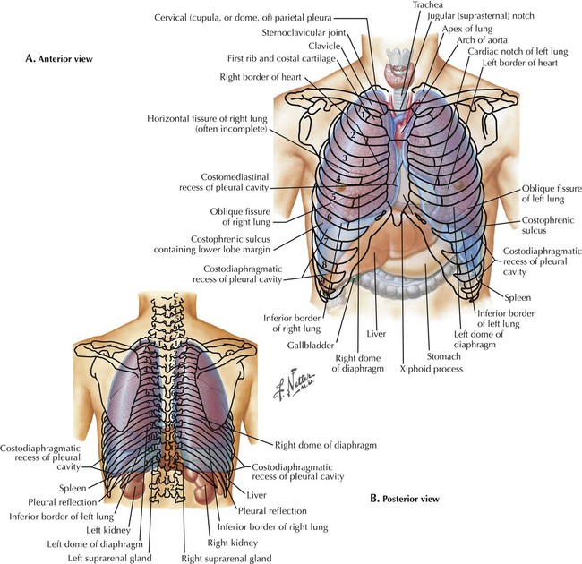
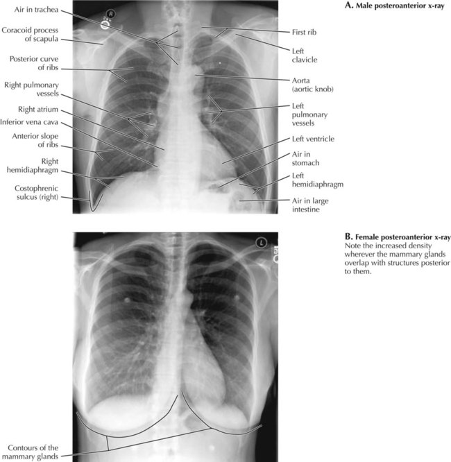
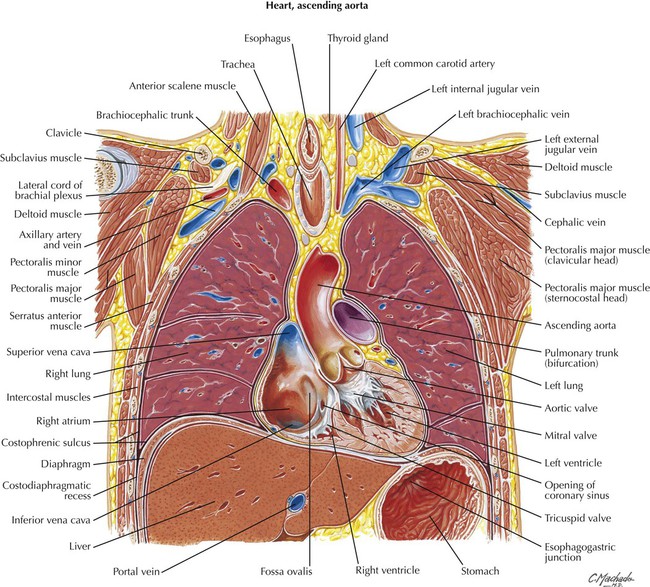
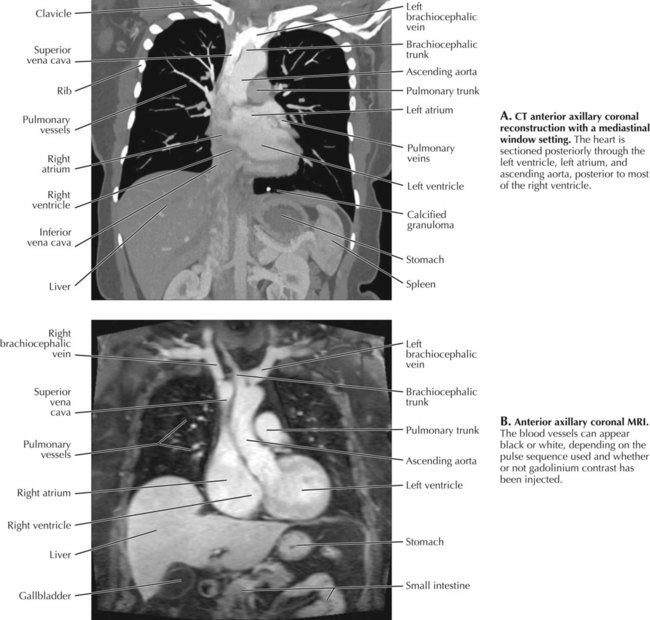
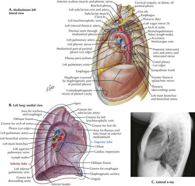
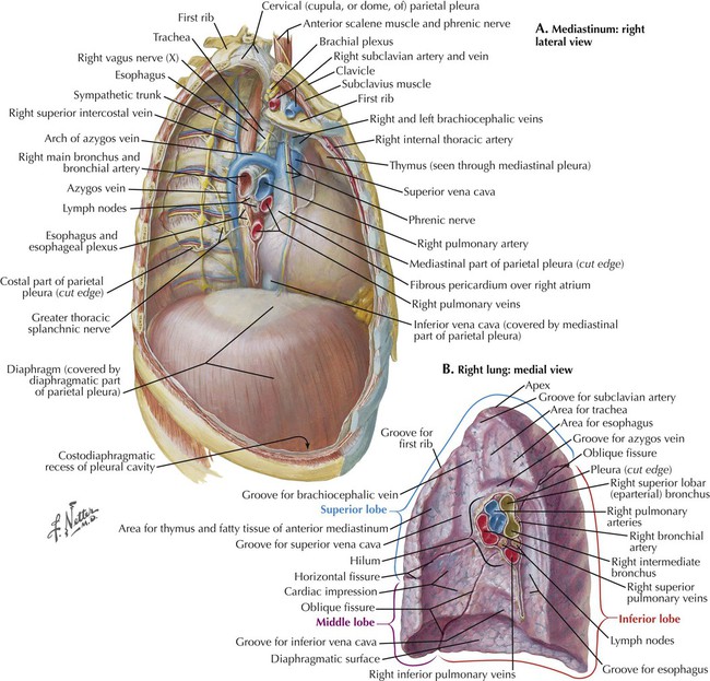
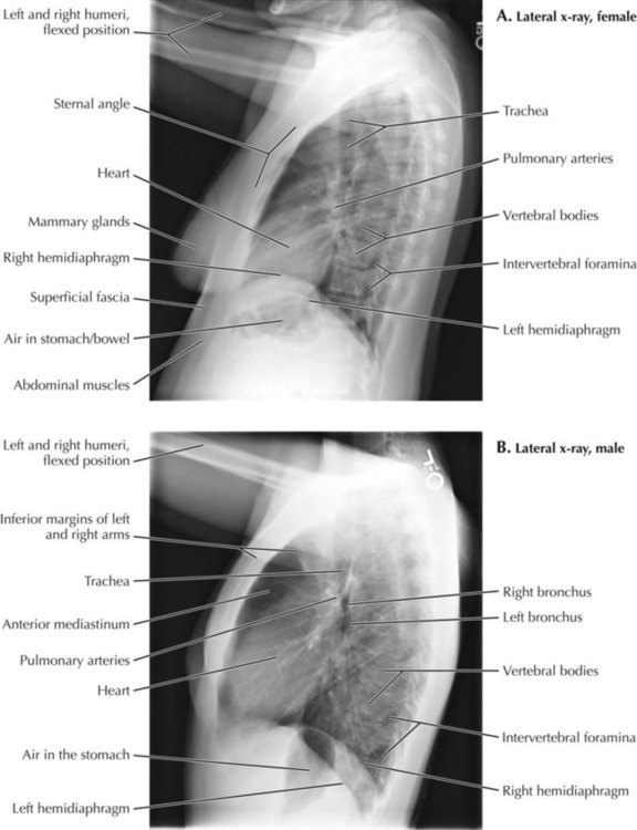
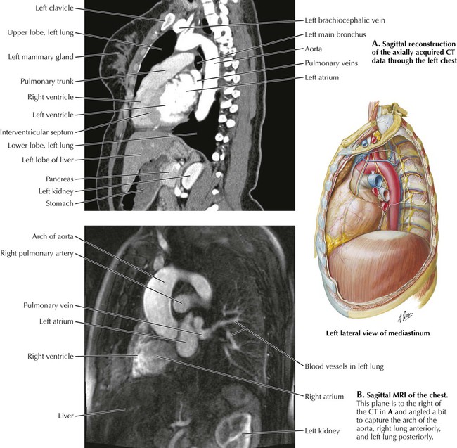
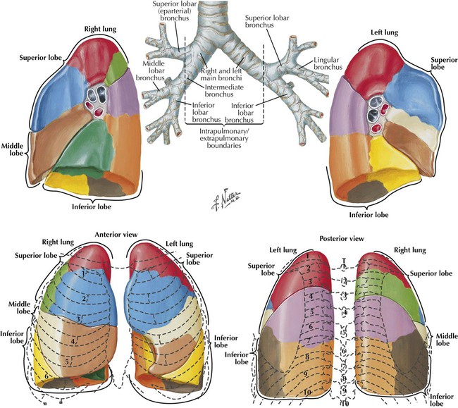
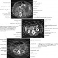
3 Thorax 3.1. THORACIC TOPOGRAPHY: ANTERIOR AND POSTERIOR VIEWS 3.2. POSTEROANTERIOR AND CHEST X-RAY (MALE AND FEMALE) 3.3. MIDAXILLARY CORONAL SECTION 3.4. ANTERIOR AXILLARY CORONAL SECTION 3.5. ANTERIOR AXILLARY CT AND MRI 3.6. MEDIASTINUM: LEFT LATERAL VIEW AND LEFT MEDIAL LUNG 3.7. MEDIASTINUM: RIGHT LATERAL VIEW AND RIGHT MEDIAL LUNG 3.8. LATERAL CHEST X-RAY 3.9. SAGITTAL CT AND MRI 3.10. LUNG ANATOMY 3.11. POSTEROANTERIOR AND LATERAL X-RAYS: SUPERIMPOSED OUTLINES OF LUNG LOBES 3.12. CT AIRWAY STUDIES 3.13. ANTERIOR AND POSTERIOR VIEWS OF THE HEART 3.14. POSTEROANTERIOR AND LATERAL X-RAYS: VIEWS OF SUPERIMPOSED HEART CHAMBERS 3.15. T8 MEDIASTINUM CROSS SECTION WITH T8 CT AND MRI 3.16. ATRIA, VENTRICLES, AND INTERVENTRICULAR SEPTUM 3.17. RIGHT CORONARY ARTERY STUDY 3.18. LEFT CORONARY ARTERY STUDY 3.19. HEART IMAGING STUDIES 3.20. ECHOCARDIOGRAPHY 3.21. SINGLE PHOTON EMISSION COMPUTED TOMOGRAPHY 3.22. COMPARISON OF CARDIAC IMAGING MODALITIES 3.23. VERTEBRAL LEVELS IN THE THORAX 3.24. T3 CROSS SECTION with T3 CT 3.25. T3-T4 CROSS SECTION with T3-T4 CT 3.26. T4-T5 CROSS SECTION WITH T4-T5 CT 3.27. T7 CROSS SECTION WITH T7 CT 3.28. SYSTEMATIC CHEST X-RAY EVALUATION 3.29. SEARCH STRATEGY: IDENTIFYING VIEWS 3.30. SEARCH STRATEGY: TECHNICAL QUALITY OF IMAGES 3.31. SEARCH STRATEGY: TUBES, LINES, AND SUPPORT DEVICES 3.32. SEARCH STRATEGY: THORACIC WALL SOFT TISSUES (E.G., AIR, CALCIFICATION, FOREIGN BODIES) 3.33. SEARCH STRATEGY: BONES 3.34. SEARCH STRATEGY: PLEURAL SPACES AND DIAPHRAGM 3.35. SEARCH STRATEGY: PLEURAL SPACES AND DIAPHRAGM (CONT’D) 3.36. SEARCH STRATEGY: UPPER ABDOMEN 3.37. SEARCH STRATEGY: MEDIASTINUM AND HILA 3.38. SEARCH STRATEGY: HEART AND VASCULATURE 3.39. SEARCH STRATEGY: LUNGS—SILHOUETTE SIGN 3.40. SEARCH STRATEGY: LUNGS, ATELECTASIS 3.41. SEARCH STRATEGY: LUNGS, ALVEOLAR VS. INTERSTITIAL OPACITY 3.1 Thoracic Topography: Anterior and Posterior Views The thorax is the part of the trunk between the neck and the abdominal cavity. It extends from the first rib to the diaphragm and is bounded by the sternum, twelve thoracic vertebrae, the twelve pairs of ribs, and the muscles that attach to these bones. The thorax contains the lungs surrounded by pleural spaces. It also contains the mediastinum, the block of tissue between the lungs consisting of the heart, esophagus, trachea, vessels, lymph nodes, connective tissue, and nerves. The heart is the approximate length of the body of the sternum. The great vessels and bifurcation of the trachea are behind the manubrium. The dome-shaped diaphragm extends over the liver on the right and spleen and stomach on the left. The apices of the lungs are above the first rib and clavicle. The parietal pleura that lines the pleural cavities extends more inferiorly than the lungs themselves in the recess between the diaphragm and the rib cage. The bases of the lungs during quiet respiration are near the seventh rib, whereas the costodiaphragmatic recess of the parietal pleura extends down to the ninth rib laterally. The right and left hemidiaphragms, pleural cavities, and lungs extend to a lower level in the posterior thorax than they do anteriorly. 3.2 Posteroanterior and Chest X-Ray (Male and Female) For a standard posteroanterior (PA) chest x-ray the patient “hugs” the x-ray recording plate. This protracts the shoulder girdles and moves the scapulae off the lungs. Having the x-ray beam pass from posterior to anterior through the patient, with the anterior chest adjacent to the recording plate, minimizes the magnification of the heart by the divergent beam. The lung fields appear dark because of their high air content. The larger pulmonary vessels (arteries and veins) are the white tubular densities near the lung roots. Note the air (darker area) in the midline trachea and in the stomach and splenic flexure of the colon under the left hemidiaphragm. Also note the clavicles, scapulae, and arch of each hemidiaphragm. The heart borders are seen clearly against the air-filled lungs. The right margin of the heart on the PA view is the right atrium. The left margin is the left ventricle. The right ventricle and left atrium do not contribute to the heart borders on a PA x-ray. They are better seen in a lateral view. 3.3 Midaxillary Coronal Section Most of the heart is anterior to the midaxillary coronal plane. This section contains the arches of the aorta and azygos vein and the division of the trachea into the primary bronchi. Cervical and lumbar vertebrae are visible. The normal thoracic kyphosis (curvature) is out of the plane of section. The esophagus follows the contour of the vertebral column behind the heart. It is visible above the trachea and below the left atrium. The pulmonary arteries are above the level of the left atrium and the pulmonary veins that drain into it. 3.4 Anterior Axillary Coronal Section This more anterior coronal section passes through the middle of the left ventricle and right atrium of the heart. The aortic semilunar valve, ascending aorta, and superior and inferior vena cavae are also seen. The pulmonary trunk is visible under the arch of the aorta. The stomach is visible in the patient’s left upper abdomen. The apices of the lungs are close to the brachiocephalic vessels. 3.5 Anterior Axillary CT and MRI Both computed tomography (CT) and magnetic resonance imaging (MRI) are used for chest studies, although CT is more common. MRI is used for some heart studies and in cases in which patients may have allergies or other problems with receiving iodinated contrast used in CT. On the CT examination (A), the contrast was introduced into a left arm vein, and it is just entering the heart and lungs. The superior vena cava is bright white, and the white profiles in the lungs are blood vessels with some contrast. The very bright nodule at the left lung base medially is a calcified granuloma. The window width and level of images can be adjusted on the computer screen to visualize either the soft tissue structures or the lung parenchyma. On this mediastinal window setting, the soft tissue structures of the mediastinum are visualized. The details of the lung parenchyma are poorly seen, and the lungs appear predominantly black. The window width and level can also be adjusted to make the white contrast in the blood vessels look brighter or less intense. 3.6 Mediastinum: Left Lateral View and Left Medial Lung The heart on the left is bounded by the lingula segment of the left upper lobe. There is no middle lobe in the left lung. Most of the left lower lobe is either below or behind the heart. A portion extends superior and posterior to the heart. The arch of the aorta courses over the root of the lung to the left of the trachea. The descending thoracic aorta is posterior to the esophagus. The right main stem bronchus is above the right pulmonary artery (eparterial), whereas the left main stem bronchus is below the left pulmonary artery (hyparterial). The lungs do not extend all the way into the costodiaphragmatic recess, the inferior boundary of the pleural cavity. This recess is a potential space that is not visible on an x-ray. 3.7 Mediastinum: Right Lateral View and Right Medial Lung The heart is anteriorly located in the thorax. It is bounded laterally on the right by the middle lobe of the right lung. The right lower lobe extends upward and posterior to the right middle lobe and the heart. The esophagus is posterior to the heart against the left atrium. The right main stem bronchus is above the right main pulmonary artery, unlike the left side where the left main stem bronchus is below the left main pulmonary artery. The arch of the azygos vein passes over the right primary bronchus and pulmonary artery in the root of the right lung. 3.8 Lateral Chest X-Ray Routinely a left lateral chest radiograph (x-ray beam passing from right to left) is obtained to keep the heart closest to the image receptor. The patient’s arms are elevated to move the humeri and soft tissues of the arms out of the field of view. Lateral radiographs are used in conjunction with the PA view to evaluate the thorax in three dimensions and better localize any pathology that may be present. The roots of both lungs are superimposed on each other in the middle mediastinum. The right upper lobe bronchus is higher than the left upper lobe bronchus. Each is seen on end where the trachea ends. The distal arch of the aorta is seen posterior to the trachea. The clear space behind the sternum superiorly corresponds to the anterior mediastinum. The right ventricle is the most anterior heart chamber and makes up the anterior superior margin of the heart on the lateral view. The left atrium comprises the superior posterior border of the heart, and the left ventricle is the inferior posterior border of the heart. The contour of the left hemidiaphragm is not seen anteriorly because it is silhouetted by (against) the heart. The right hemidiaphragm contour can normally be followed along its entire course. 3.9 Sagittal CT and MRI In the CT reconstruction in A, the descending aorta, clavicle, and breast indicate that the section is to the left of the midline. Iodinated intravenous contrast is seen in the left ventricle, aortic arch, and descending thoracic aorta and two of the branch arteries of the aortic arch. The lungs appear black on this image viewed with a mediastinal window. The anterior clear space is the upper lobe of the left lung projecting over the anterior mediastinum. The heart and aorta are in the middle mediastinum, and the thoracic vertebral column (spine) is in the posterior mediastinum. In the upper abdomen portions of the left lobe of the liver, stomach, pancreas and left kidney are seen. B is a corresponding MRI sagittal section of the chest. Blood vessels can appear white on MRI without the injection of gadolinium contrast, depending on the pulse sequence used. On this image the ascending aorta and aortic arch are seen clearly. The heart is located anteriorly. Some blood vessels are seen in the left lung, and the left kidney is seen in the upper abdomen. 3.10 Lung Anatomy A primary (main stem) bronchus supplies each lung. Secondary (lobar) bronchi supply lobes, and tertiary (segmental) bronchi supply bronchopulmonary segments, the subdivisions of each lobe that are colored in the figures. The right primary bronchus is shorter than the left and has a more vertical orientation in line with the trachea. The right lung has three lobes, with a horizontal (minor) fissure separating the superior and middle lobes and an oblique (major) fissure separating the upper and middle lobes from the inferior lobe. The left lung has only two lobes separated by an oblique fissure. The lingual segment (lingula) of the left upper lobe is equivalent to the middle lobe of the right lung. Note that the inferior lobes of both lungs extend posteriorly to the superior lobes behind the middle lobe of the right lung and lingula of the left lung. At the root of the lungs the pulmonary arteries are above the pulmonary veins. 3.11 Posteroanterior and Lateral X-Rays: Superimposed Outlines of Lung Lobes Only gold members can continue reading. Log In or Register to continue Share this: Share on X (Opens in new window) X Share on Facebook (Opens in new window) Facebook Related posts: Introduction to Imaging Modalities Back and Spinal Cord Upper Limbs Abdomen Lower Limbs Head and Neck Stay updated, free articles. Join our Telegram channel Join Tags: Netters Introduction to Imaging Jan 10, 2016 | Posted by admin in RADIOGRAPHIC ANATOMY | Comments Off on Thorax Full access? Get Clinical Tree
3 Thorax 3.1. THORACIC TOPOGRAPHY: ANTERIOR AND POSTERIOR VIEWS 3.2. POSTEROANTERIOR AND CHEST X-RAY (MALE AND FEMALE) 3.3. MIDAXILLARY CORONAL SECTION 3.4. ANTERIOR AXILLARY CORONAL SECTION 3.5. ANTERIOR AXILLARY CT AND MRI 3.6. MEDIASTINUM: LEFT LATERAL VIEW AND LEFT MEDIAL LUNG 3.7. MEDIASTINUM: RIGHT LATERAL VIEW AND RIGHT MEDIAL LUNG 3.8. LATERAL CHEST X-RAY 3.9. SAGITTAL CT AND MRI 3.10. LUNG ANATOMY 3.11. POSTEROANTERIOR AND LATERAL X-RAYS: SUPERIMPOSED OUTLINES OF LUNG LOBES 3.12. CT AIRWAY STUDIES 3.13. ANTERIOR AND POSTERIOR VIEWS OF THE HEART 3.14. POSTEROANTERIOR AND LATERAL X-RAYS: VIEWS OF SUPERIMPOSED HEART CHAMBERS 3.15. T8 MEDIASTINUM CROSS SECTION WITH T8 CT AND MRI 3.16. ATRIA, VENTRICLES, AND INTERVENTRICULAR SEPTUM 3.17. RIGHT CORONARY ARTERY STUDY 3.18. LEFT CORONARY ARTERY STUDY 3.19. HEART IMAGING STUDIES 3.20. ECHOCARDIOGRAPHY 3.21. SINGLE PHOTON EMISSION COMPUTED TOMOGRAPHY 3.22. COMPARISON OF CARDIAC IMAGING MODALITIES 3.23. VERTEBRAL LEVELS IN THE THORAX 3.24. T3 CROSS SECTION with T3 CT 3.25. T3-T4 CROSS SECTION with T3-T4 CT 3.26. T4-T5 CROSS SECTION WITH T4-T5 CT 3.27. T7 CROSS SECTION WITH T7 CT 3.28. SYSTEMATIC CHEST X-RAY EVALUATION 3.29. SEARCH STRATEGY: IDENTIFYING VIEWS 3.30. SEARCH STRATEGY: TECHNICAL QUALITY OF IMAGES 3.31. SEARCH STRATEGY: TUBES, LINES, AND SUPPORT DEVICES 3.32. SEARCH STRATEGY: THORACIC WALL SOFT TISSUES (E.G., AIR, CALCIFICATION, FOREIGN BODIES) 3.33. SEARCH STRATEGY: BONES 3.34. SEARCH STRATEGY: PLEURAL SPACES AND DIAPHRAGM 3.35. SEARCH STRATEGY: PLEURAL SPACES AND DIAPHRAGM (CONT’D) 3.36. SEARCH STRATEGY: UPPER ABDOMEN 3.37. SEARCH STRATEGY: MEDIASTINUM AND HILA 3.38. SEARCH STRATEGY: HEART AND VASCULATURE 3.39. SEARCH STRATEGY: LUNGS—SILHOUETTE SIGN 3.40. SEARCH STRATEGY: LUNGS, ATELECTASIS 3.41. SEARCH STRATEGY: LUNGS, ALVEOLAR VS. INTERSTITIAL OPACITY 3.1 Thoracic Topography: Anterior and Posterior Views The thorax is the part of the trunk between the neck and the abdominal cavity. It extends from the first rib to the diaphragm and is bounded by the sternum, twelve thoracic vertebrae, the twelve pairs of ribs, and the muscles that attach to these bones. The thorax contains the lungs surrounded by pleural spaces. It also contains the mediastinum, the block of tissue between the lungs consisting of the heart, esophagus, trachea, vessels, lymph nodes, connective tissue, and nerves. The heart is the approximate length of the body of the sternum. The great vessels and bifurcation of the trachea are behind the manubrium. The dome-shaped diaphragm extends over the liver on the right and spleen and stomach on the left. The apices of the lungs are above the first rib and clavicle. The parietal pleura that lines the pleural cavities extends more inferiorly than the lungs themselves in the recess between the diaphragm and the rib cage. The bases of the lungs during quiet respiration are near the seventh rib, whereas the costodiaphragmatic recess of the parietal pleura extends down to the ninth rib laterally. The right and left hemidiaphragms, pleural cavities, and lungs extend to a lower level in the posterior thorax than they do anteriorly. 3.2 Posteroanterior and Chest X-Ray (Male and Female) For a standard posteroanterior (PA) chest x-ray the patient “hugs” the x-ray recording plate. This protracts the shoulder girdles and moves the scapulae off the lungs. Having the x-ray beam pass from posterior to anterior through the patient, with the anterior chest adjacent to the recording plate, minimizes the magnification of the heart by the divergent beam. The lung fields appear dark because of their high air content. The larger pulmonary vessels (arteries and veins) are the white tubular densities near the lung roots. Note the air (darker area) in the midline trachea and in the stomach and splenic flexure of the colon under the left hemidiaphragm. Also note the clavicles, scapulae, and arch of each hemidiaphragm. The heart borders are seen clearly against the air-filled lungs. The right margin of the heart on the PA view is the right atrium. The left margin is the left ventricle. The right ventricle and left atrium do not contribute to the heart borders on a PA x-ray. They are better seen in a lateral view. 3.3 Midaxillary Coronal Section Most of the heart is anterior to the midaxillary coronal plane. This section contains the arches of the aorta and azygos vein and the division of the trachea into the primary bronchi. Cervical and lumbar vertebrae are visible. The normal thoracic kyphosis (curvature) is out of the plane of section. The esophagus follows the contour of the vertebral column behind the heart. It is visible above the trachea and below the left atrium. The pulmonary arteries are above the level of the left atrium and the pulmonary veins that drain into it. 3.4 Anterior Axillary Coronal Section This more anterior coronal section passes through the middle of the left ventricle and right atrium of the heart. The aortic semilunar valve, ascending aorta, and superior and inferior vena cavae are also seen. The pulmonary trunk is visible under the arch of the aorta. The stomach is visible in the patient’s left upper abdomen. The apices of the lungs are close to the brachiocephalic vessels. 3.5 Anterior Axillary CT and MRI Both computed tomography (CT) and magnetic resonance imaging (MRI) are used for chest studies, although CT is more common. MRI is used for some heart studies and in cases in which patients may have allergies or other problems with receiving iodinated contrast used in CT. On the CT examination (A), the contrast was introduced into a left arm vein, and it is just entering the heart and lungs. The superior vena cava is bright white, and the white profiles in the lungs are blood vessels with some contrast. The very bright nodule at the left lung base medially is a calcified granuloma. The window width and level of images can be adjusted on the computer screen to visualize either the soft tissue structures or the lung parenchyma. On this mediastinal window setting, the soft tissue structures of the mediastinum are visualized. The details of the lung parenchyma are poorly seen, and the lungs appear predominantly black. The window width and level can also be adjusted to make the white contrast in the blood vessels look brighter or less intense. 3.6 Mediastinum: Left Lateral View and Left Medial Lung The heart on the left is bounded by the lingula segment of the left upper lobe. There is no middle lobe in the left lung. Most of the left lower lobe is either below or behind the heart. A portion extends superior and posterior to the heart. The arch of the aorta courses over the root of the lung to the left of the trachea. The descending thoracic aorta is posterior to the esophagus. The right main stem bronchus is above the right pulmonary artery (eparterial), whereas the left main stem bronchus is below the left pulmonary artery (hyparterial). The lungs do not extend all the way into the costodiaphragmatic recess, the inferior boundary of the pleural cavity. This recess is a potential space that is not visible on an x-ray. 3.7 Mediastinum: Right Lateral View and Right Medial Lung The heart is anteriorly located in the thorax. It is bounded laterally on the right by the middle lobe of the right lung. The right lower lobe extends upward and posterior to the right middle lobe and the heart. The esophagus is posterior to the heart against the left atrium. The right main stem bronchus is above the right main pulmonary artery, unlike the left side where the left main stem bronchus is below the left main pulmonary artery. The arch of the azygos vein passes over the right primary bronchus and pulmonary artery in the root of the right lung. 3.8 Lateral Chest X-Ray Routinely a left lateral chest radiograph (x-ray beam passing from right to left) is obtained to keep the heart closest to the image receptor. The patient’s arms are elevated to move the humeri and soft tissues of the arms out of the field of view. Lateral radiographs are used in conjunction with the PA view to evaluate the thorax in three dimensions and better localize any pathology that may be present. The roots of both lungs are superimposed on each other in the middle mediastinum. The right upper lobe bronchus is higher than the left upper lobe bronchus. Each is seen on end where the trachea ends. The distal arch of the aorta is seen posterior to the trachea. The clear space behind the sternum superiorly corresponds to the anterior mediastinum. The right ventricle is the most anterior heart chamber and makes up the anterior superior margin of the heart on the lateral view. The left atrium comprises the superior posterior border of the heart, and the left ventricle is the inferior posterior border of the heart. The contour of the left hemidiaphragm is not seen anteriorly because it is silhouetted by (against) the heart. The right hemidiaphragm contour can normally be followed along its entire course. 3.9 Sagittal CT and MRI In the CT reconstruction in A, the descending aorta, clavicle, and breast indicate that the section is to the left of the midline. Iodinated intravenous contrast is seen in the left ventricle, aortic arch, and descending thoracic aorta and two of the branch arteries of the aortic arch. The lungs appear black on this image viewed with a mediastinal window. The anterior clear space is the upper lobe of the left lung projecting over the anterior mediastinum. The heart and aorta are in the middle mediastinum, and the thoracic vertebral column (spine) is in the posterior mediastinum. In the upper abdomen portions of the left lobe of the liver, stomach, pancreas and left kidney are seen. B is a corresponding MRI sagittal section of the chest. Blood vessels can appear white on MRI without the injection of gadolinium contrast, depending on the pulse sequence used. On this image the ascending aorta and aortic arch are seen clearly. The heart is located anteriorly. Some blood vessels are seen in the left lung, and the left kidney is seen in the upper abdomen. 3.10 Lung Anatomy A primary (main stem) bronchus supplies each lung. Secondary (lobar) bronchi supply lobes, and tertiary (segmental) bronchi supply bronchopulmonary segments, the subdivisions of each lobe that are colored in the figures. The right primary bronchus is shorter than the left and has a more vertical orientation in line with the trachea. The right lung has three lobes, with a horizontal (minor) fissure separating the superior and middle lobes and an oblique (major) fissure separating the upper and middle lobes from the inferior lobe. The left lung has only two lobes separated by an oblique fissure. The lingual segment (lingula) of the left upper lobe is equivalent to the middle lobe of the right lung. Note that the inferior lobes of both lungs extend posteriorly to the superior lobes behind the middle lobe of the right lung and lingula of the left lung. At the root of the lungs the pulmonary arteries are above the pulmonary veins. 3.11 Posteroanterior and Lateral X-Rays: Superimposed Outlines of Lung Lobes Only gold members can continue reading. Log In or Register to continue Share this: Share on X (Opens in new window) X Share on Facebook (Opens in new window) Facebook Related posts: Introduction to Imaging Modalities Back and Spinal Cord Upper Limbs Abdomen Lower Limbs Head and Neck Stay updated, free articles. Join our Telegram channel Join Tags: Netters Introduction to Imaging Jan 10, 2016 | Posted by admin in RADIOGRAPHIC ANATOMY | Comments Off on Thorax Full access? Get Clinical Tree