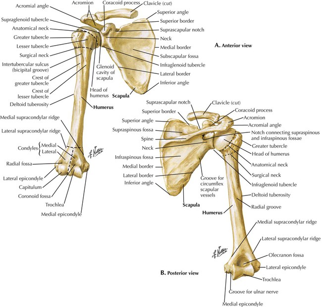
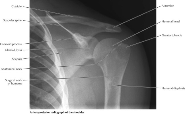
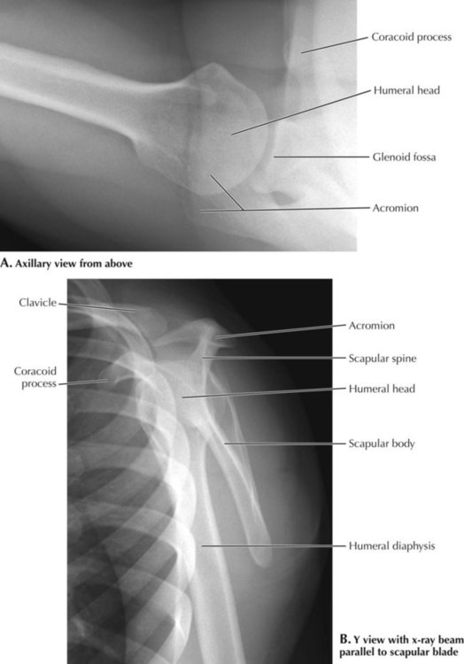
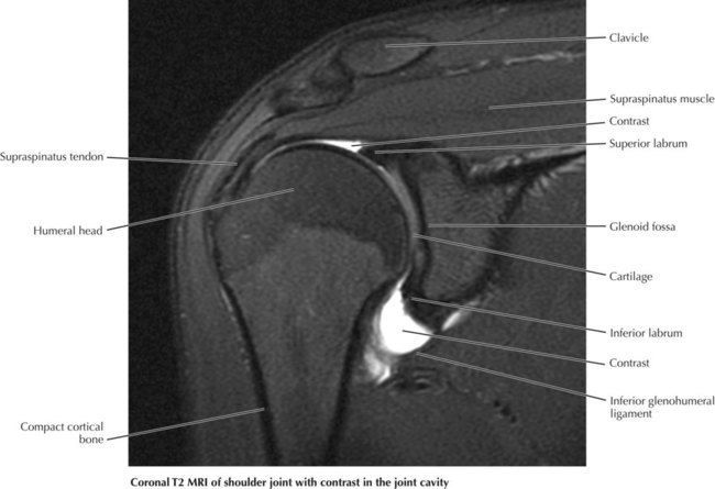
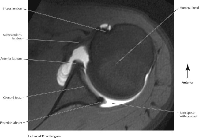
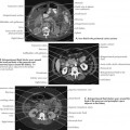
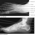
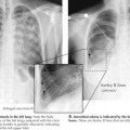
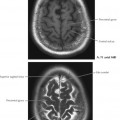
Upper Limbs


Upper Limbs


6.1 Humerus and Scapula


The coracoid (“shaped like a crow’s beak”) process is the most prominent feature on the anterior of the scapula. Three muscles attach to it: pectoralis minor, coracobrachialis, and the short head of the biceps brachii muscle. On the humerus the bicipital groove separates the lesser tubercle of the humerus anteriorly from the greater tubercle laterally. The tendon of the long head of the biceps brachii muscle lies in the groove as it enters the joint cavity to attach to the supraglenoid tubercle of the scapula. The infraglenoid tubercle is the attachment of the long head of the triceps brachii muscle. Posteriorly the scapular spine divides the scapula into supraspinatus and infraspinatus fossae that contain muscles of the same name. The acromion is the termination of the scapular spine; it articulates anteriorly with the clavicle.


image


6.2 Anteroposterior Shoulder X-Ray


Plain films of the shoulder can be good for assessing the bony anatomy for abnormalities that may be a result of such conditions as osteoarthritis or trauma. Three views that are commonly done include the anteroposterior (AP) view, the axillary view, and the Y view. The humeral head should appear as a smooth hemisphere. In the AP view seen here, the medial portion of the humeral head overlaps with the lateral aspect of the glenoid fossa. If a patient has a history of chronic anterior dislocations, there may be a Hill-Sachs deformity, which appears as an indented groove in the upper outer portion of the humeral head. The acromion and acromioclavicular joint should be evaluated for osteophytes (bony projections) that can impinge on the supraspinatus muscle. All the bones should be assessed for fractures, and whatever portion of the lung is visible should be evaluated as well. A radiologist is responsible for everything visualized on a study, and lung cancers have been found incidentally on shoulder films!


image


6.3 Axillary and Y View X-Rays of the Shoulder Joint


In the axillary view of the shoulder shown in A, the arm is abducted 90 degrees, and the beam is projected down through the shoulder. This allows clear visualization of the relation of the glenoid fossa with the humeral head; the fossa should be adjacent to the humeral head. This view is helpful for identifying dislocation of the humeral head and anterior or posterior glenoid rim fractures. On this projection, the acromion is seen overlapping with the humeral head. In the upper right corner of this image is the coracoid process of the scapula. In the Y view x-ray of the scapula (B), the x-ray beam is directed along the scapular spine, and the body of the scapula (stem of the Y) is viewed on edge. The upper limbs of the Y are the coracoid process and scapular spine. This projection aids in evaluation for a shoulder dislocation or a fracture of the scapula.


image


6.5 Coronal T2 MRI of the Shoulder Joint


Most of the clinically significant problems of the four rotator cuff muscles involve the supraspinatus. It may be impinged between the acromion and the greater tubercle or under the acromioclavicular joint if there are downward-pointing osteophytes. It may also be torn. Because MRI provides good soft tissue contrast, it is often the modality of choice for evaluation of the rotator cuff and labrum. This is a T2, fat-saturated, coronal MRI arthrogram of the right shoulder. Note the rim of black surrounding the bones. This is cortex, which is mineral rich and low in water content and thus appears black (absent signal) on both T1- and T2-weighted sequences. The superior and inferior labra also have a low signal and are triangular shaped. Note the low signal of the tendons in this image. Because of their collagenous tissue, tendons are normally uniformly hypointense on all sequences (both T1 and T2). A tendon that is bright on a T2 sequence is often indicative of edema.


image


6.6 Axial T1 Arthrogram of the Shoulder Joint


Note the high signal within the joint space. Normally on T1, synovial fluid should have a low-intermediate signal. The bright signal on this T1 image comes from contrast injected into the joint space using fluoroscopic guidance, which aids in assessment of the labrum. Normal anterior and posterior labra are seen here as triangular shaped, low-signal structures at the periphery of the glenoid fossa. The anterior labrum is typically larger than the posterior labrum and is more commonly involved in tears. A tear or detachment of the labrum would be indicated by the presence of fluid extending between the glenoid and labrum. The long head of the biceps tendon can be seen in the bicipital groove partly surrounded by contrast. It inserts onto the superior labrum, which is above the plane of this section. In athletes who throw a lot, the pull of the biceps tendon onto the superior labrum can cause it to tear in what is referred to as a SLAP lesion (superior labrum anterior to posterior). The superior glenoid is best visualized on an oblique coronal view.


image

Jan 10, 2016 | Posted by in RADIOGRAPHIC ANATOMY | Comments Off on Upper Limbs

Full access? Get Clinical Tree

Get Clinical Tree app for offline access