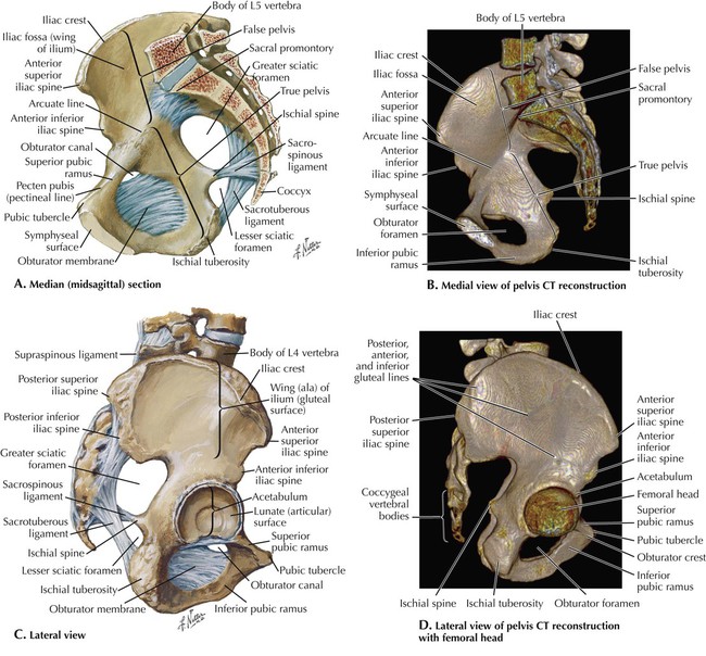
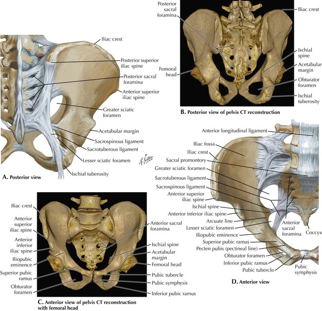
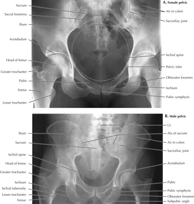
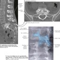
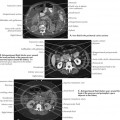
5 Pelvis and Perineum 5.1. BONY FRAMEWORK: MEDIAL AND LATERAL VIEWS 5.2. BONY FRAMEWORK: ANTERIOR AND POSTERIOR VIEWS 5.3. FEMALE AND MALE PELVIC X-RAYS 5.4. FEMALE MIDSAGITTAL SECTION AND CT 5.5. CT VS. MRI IN THE PELVIS 5.6. FEMALE PELVIC CONTENTS 5.7. SEARCH STRATEGY: UPPER FEMALE PELVIS 5.8. SEARCH STRATEGY: LOWER FEMALE PELVIS 5.9. UTERUS, ADNEXA, AND HYSTEROSALPINGOGRAM 5.10. MALE PELVIC CONTENTS 5.11. MALE MIDSAGITTAL SECTION AND CT 5.12. MALE AXIAL CT AND MRI 5.13. CROSS SECTION AT BLADDER-PROSTATE JUNCTION AND CT 5.14. MALE AND FEMALE CORONAL SECTIONS THROUGH THE URINARY BLADDER 5.15. BLADDER RELATIONSHIPS in AXIAL AND CORONAL CT 5.16. CYSTOGRAM 5.1 Bony Framework: Medial and Lateral Views The pelvis consists of the left and right innominate bones and the sacrum. The ilium, pubis, and ischium are fused with one another at the acetabulum of the hip joint to comprise each innominate bone. The pubic bones articulate with each other anteriorly in the midline at the pubic symphysis. We sit on our ischial tuberosity, and the ischial spine separates the greater and lesser sciatic notches posteriorly. The greater or false pelvis consists of the lateral curve of the iliac blades that help support the abdominal viscera. The pubis and ischium make up the lesser or true pelvis that surrounds the birth canal and pelvic viscera. Three-dimensional (3D) computer reconstructions from computed tomography (CT) scans (B and D) are most often used to plan surgical reconstructions in patients with fractures. 5.2 Bony Framework: Anterior and Posterior Views The pelvic brim encircles the pelvic inlet to the birth canal that includes, from front to back, the pubic tubercle, pectin pubis, arcuate line of the ilium, and ala and promontory of the sacrum. The pelvic outlet is bounded by the ischial spines and tip of the coccyx. Laterally are the obturator foramina that are closed off in life by obturator membranes. The sacrospinous and sacrotuberous ligaments convert the sciatic notches into greater and lesser sciatic foramina. 5.3 Female and Male PelviC X-Rays A plain film (x-ray) of the pelvis is a good initial way to look for fractures. CT is sometimes needed to better evaluate fractures or to look for nondisplaced fractures that may be missed on the plain film. Magnetic resonance imaging (MRI) is very sensitive for bone marrow edema and displays bone contusions not seen with plain film or CT. It is also best for tendon, ligament, and other soft tissue injuries. In the x-rays, note the sacroiliac joints, acetabulum of the hip joint, sacral foramina, and pubic symphysis. The obturator foramina appear narrow because of their oblique angle of view. The darker contours over the sacrum and ilium represent air in the colon. A female pelvis has a relatively larger birth canal than a male pelvis. It is wider because of relatively wider ala of the sacrum and relatively wider superior pubic rami and ischiopubic rami. The longer rami result in a wider subpubic angle in the female pelvis compared to that of the male. The sacrum is also angled more posteriorly in the female to enlarge the pelvic outlet. This accounts for the larger greater sciatic notch in the female. 5.4 Female Midsagittal Section and CT Only gold members can continue reading. Log In or Register to continue Share this: Share on X (Opens in new window) X Share on Facebook (Opens in new window) Facebook Related posts: Introduction to Imaging Modalities Back and Spinal Cord Abdomen Lower Limbs Thorax Head and Neck Stay updated, free articles. Join our Telegram channel Join Tags: Netters Introduction to Imaging Jan 10, 2016 | Posted by admin in RADIOGRAPHIC ANATOMY | Comments Off on Pelvis and Perineum Full access? Get Clinical Tree
5 Pelvis and Perineum 5.1. BONY FRAMEWORK: MEDIAL AND LATERAL VIEWS 5.2. BONY FRAMEWORK: ANTERIOR AND POSTERIOR VIEWS 5.3. FEMALE AND MALE PELVIC X-RAYS 5.4. FEMALE MIDSAGITTAL SECTION AND CT 5.5. CT VS. MRI IN THE PELVIS 5.6. FEMALE PELVIC CONTENTS 5.7. SEARCH STRATEGY: UPPER FEMALE PELVIS 5.8. SEARCH STRATEGY: LOWER FEMALE PELVIS 5.9. UTERUS, ADNEXA, AND HYSTEROSALPINGOGRAM 5.10. MALE PELVIC CONTENTS 5.11. MALE MIDSAGITTAL SECTION AND CT 5.12. MALE AXIAL CT AND MRI 5.13. CROSS SECTION AT BLADDER-PROSTATE JUNCTION AND CT 5.14. MALE AND FEMALE CORONAL SECTIONS THROUGH THE URINARY BLADDER 5.15. BLADDER RELATIONSHIPS in AXIAL AND CORONAL CT 5.16. CYSTOGRAM 5.1 Bony Framework: Medial and Lateral Views The pelvis consists of the left and right innominate bones and the sacrum. The ilium, pubis, and ischium are fused with one another at the acetabulum of the hip joint to comprise each innominate bone. The pubic bones articulate with each other anteriorly in the midline at the pubic symphysis. We sit on our ischial tuberosity, and the ischial spine separates the greater and lesser sciatic notches posteriorly. The greater or false pelvis consists of the lateral curve of the iliac blades that help support the abdominal viscera. The pubis and ischium make up the lesser or true pelvis that surrounds the birth canal and pelvic viscera. Three-dimensional (3D) computer reconstructions from computed tomography (CT) scans (B and D) are most often used to plan surgical reconstructions in patients with fractures. 5.2 Bony Framework: Anterior and Posterior Views The pelvic brim encircles the pelvic inlet to the birth canal that includes, from front to back, the pubic tubercle, pectin pubis, arcuate line of the ilium, and ala and promontory of the sacrum. The pelvic outlet is bounded by the ischial spines and tip of the coccyx. Laterally are the obturator foramina that are closed off in life by obturator membranes. The sacrospinous and sacrotuberous ligaments convert the sciatic notches into greater and lesser sciatic foramina. 5.3 Female and Male PelviC X-Rays A plain film (x-ray) of the pelvis is a good initial way to look for fractures. CT is sometimes needed to better evaluate fractures or to look for nondisplaced fractures that may be missed on the plain film. Magnetic resonance imaging (MRI) is very sensitive for bone marrow edema and displays bone contusions not seen with plain film or CT. It is also best for tendon, ligament, and other soft tissue injuries. In the x-rays, note the sacroiliac joints, acetabulum of the hip joint, sacral foramina, and pubic symphysis. The obturator foramina appear narrow because of their oblique angle of view. The darker contours over the sacrum and ilium represent air in the colon. A female pelvis has a relatively larger birth canal than a male pelvis. It is wider because of relatively wider ala of the sacrum and relatively wider superior pubic rami and ischiopubic rami. The longer rami result in a wider subpubic angle in the female pelvis compared to that of the male. The sacrum is also angled more posteriorly in the female to enlarge the pelvic outlet. This accounts for the larger greater sciatic notch in the female. 5.4 Female Midsagittal Section and CT Only gold members can continue reading. Log In or Register to continue Share this: Share on X (Opens in new window) X Share on Facebook (Opens in new window) Facebook Related posts: Introduction to Imaging Modalities Back and Spinal Cord Abdomen Lower Limbs Thorax Head and Neck Stay updated, free articles. Join our Telegram channel Join Tags: Netters Introduction to Imaging Jan 10, 2016 | Posted by admin in RADIOGRAPHIC ANATOMY | Comments Off on Pelvis and Perineum Full access? Get Clinical Tree