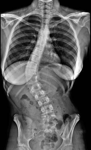
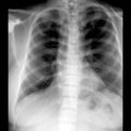
(Left) Anteroposterior radiograph shows idiopathic thoracic dextroscoliosis. Apical vertebra is the most displaced from the midline. Terminal vertebrae show the greatest deviation of the endplates from the horizontal.
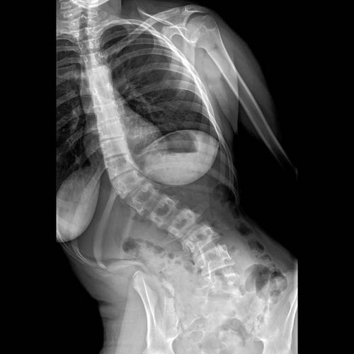
(Right) Lateral radiograph in the same patient shows reversal of usual thoracic kyphosis. Rotational component of the scoliosis can be grossly assessed by the degree of rotation of the ribs .
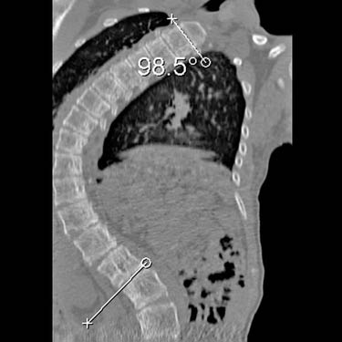
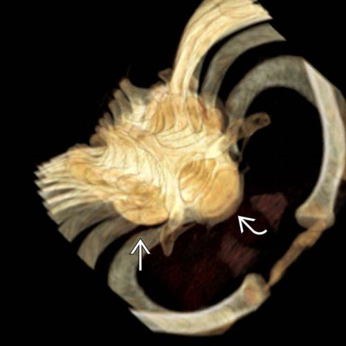
(Left) Axial 3D reformation allows measurement of the rotational component of a scoliosis. This is the angle between the terminal vertebra and the apical vertebra .
(Right) Coronal CT reconstruction shows measurement of a severe neuromuscular scoliosis. The scoliotic curvature is the angle between the vertebrae with the greatest degree of inclination from the horizontal.
(Left) Anteroposterior radiograph shows a single, long curve thoracolumbar scoliosis typical of neuromuscular scoliosis. No vertebral anomalies are present.
(Right) Anteroposterior radiograph in the same patient shows only slight improvement in the curve when the patient bends to the right. Lateral bending films or supine films are used to assess the flexibility of a scoliotic curvature. Rigid curves are less amenable to reduction.
Only gold members can continue reading. Log In or Register to continue
 is the most displaced from the midline. Terminal vertebrae
  show the greatest deviation of the endplates from the horizontal.
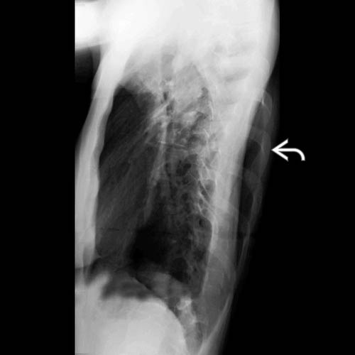
 .
.
 and the apical vertebra
  .
.