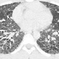
Many people wonder if radiology poses risks to their health. Medical imaging has grown dramatically over the past few decades, and the numbers highlight just how much it has changed. Americans now get over 80 million CT scans yearly, compared to just three million in 1980. These diagnostic tools save countless lives, but they have also significantly increased our overall radiation exposure.
Medical sources now make up 50% of America’s total radiation exposure – a massive jump from 15% in the early 1980s. CT scans alone contribute 24% of all radiation exposure in the United States. These numbers might raise eyebrows, but context helps us understand better. Natural sources expose the average American to about 3 mSv of radiation each year. A chest X-ray gives patients roughly 0.1 mSv – equal to about 10 days of natural background radiation. Modern CT scanners are more efficient and use lower doses than earlier machines, but they still deliver significantly more radiation than standard X-rays. The risk remains small – most scans carry only a slight increase in lifetime cancer risk, though the exact probability depends on the type of scan, radiation dose, and patient factors.
Let me break down what medical radiation really means. We’ll learn about X-ray dangers at high doses and the risks that radiology professionals face. You’ll also discover the best ways to reduce radiation exposure during imaging procedures.
Understanding Radiation Exposure in Radiology
Medical radiation serves as the foundation of modern diagnostic imaging. Healthcare professionals and patients need to understand its nature and effects.
What is Medical Radiation?
Medical radiation specifically means ionizing radiation – high-energy wavelengths or particles that can penetrate body tissues and create detailed internal images. X-rays work differently from light waves. They pass through your body and interact with various tissues based on density. This unique property helps radiologists create detailed images of bones, organs, and soft tissues.
Medical sources now contribute much more to total radiation exposure than they did decades ago. Medical imaging accounts for about 98% of the total radiation dose people receive from all human-made sources. CT scans alone make up 24% of all radiation exposure in the United States.
Radiation Dose Units: mSv, Gy, and Sieverts
Medical contexts use three main units to measure radiation exposure:
Absorbed dose (Gray or Gy) – This measures energy deposited per unit mass (1 Gy = 1 joule/kg). A brain CT scan delivers about 60 milligray (mGy) to the eye’s lens.
Equivalent dose (Sievert or Sv) – This adjusts the absorbed dose based on radiation type. X-rays have a radiation weighting factor of 1.0, which means 1 mGy equals 1 mSv.
Effective dose (Sievert or Sv) – This accounts for radiation type and organ sensitivity. The calculation helps estimate overall risk and lets us compare different types of exposure.
How Radiation Affects the Body at the Cellular Level
Ionizing radiation damages cells in two main ways as it passes through tissue:
DNA strands can break directly. The radiation can also interact with the body’s water molecules to create reactive oxygen species (ROS) and free radicals that damage DNA, proteins, and cell membranes.
Cells respond to damage in three ways:
They repair themselves and return to normal
They die if the damage proves too severe
They repair incorrectly, which might lead to mutations
Cells repair most radiation-induced damage successfully, but repair errors can lead to lasting mutations. This stochastic effect means damage might occur at any radiation level, and higher doses increase the risk.
Health Risks from Radiation Exposure
Medical imaging radiation exposure brings immediate and long-term health risks. The risks change based on dose, frequency, and patient characteristics.
Tissue Reactions vs. Stochastic Effects
Health experts split radiation effects into two distinct categories. Tissue reactions (formerly called deterministic effects) show up only above specific threshold doses. The severity increases with higher exposure. Skin burns, cataracts, and hair loss need high radiation levels to occur. The lens damage threshold sits at 0.5 Gy, while temporary sterility in testes happens at about 0.15 Gy.
Stochastic effects work differently – they have no threshold. The risks increase with dose, but the severity stays the same, whatever the exposure amount. Cancer and hereditary effects fall into this category. Cancer remains the biggest concern in diagnostic imaging.
Why Are X-rays Dangerous in High Doses?
High-dose X-rays damage tissue by disrupting DNA and killing cells. Severe exposure leads to radiation sickness and can be fatal in extreme cases. High-dose procedures like complex interventional fluoroscopy can cause skin to redden and tissue to break down.
Long-Term Cancer Risks from Low-Dose Exposure
Low-dose radiation carries small but real cancer risks. The most extensive longitudinal study of 26 epidemiological studies found clear cancer risk from low-dose exposure. The results showed increased solid cancer risk in 17 of 22 studies. CT scans pose a relatively small lifetime cancer risk – about 1 case per 1,000 people scanned.
Risks of Medical Imaging Radiation in Children
Children face much higher radiation risks for three key reasons. They show more sensitivity to radiation than adults. Their longer life expectancy gives radiation damage more time to turn into cancer. Without proper adjustments, children may receive disproportionately high doses because of their smaller body size.
Children’s risk of developing radiation-related cancer can be several times higher than adults who get similar scans. Head doses of 50-60 mGy link to triple the brain tumor risk. That equals just 2-3 head CT scans with typical settings.
Is Working in Radiology Dangerous?
Healthcare professionals in radiology have a unique role. They must balance diagnostic imaging benefits against occupational hazards. Let’s get into what the actual risks are and how safety measures protect them.
Is Being a Radiology Tech Dangerous?
Radiologic technologists face small but measurable risks. Studies reveal that only 0.53% to 0.87% of radiology technicians will face radiation complications in their lifetime. Notwithstanding that, health workers exposed to ionizing radiation make up over 50% of workers exposed to man-made radiation in France. The original concerns about excessive exposure were valid. Modern technology and strict protocols have dramatically reduced these risks.
Occupational Exposure Limits and Monitoring
Regulatory bodies set clear exposure limits. The Nuclear Regulatory Commission (NRC) has set annual limits of 5,000 mrem (5 rem) for whole-body occupational exposure. These limits are nowhere near as high for pregnant workers – just 500 mrem throughout gestation. Workers who might receive more than 100 millirem yearly must complete radiation safety training.
Protective Equipment and Shielding
Protective equipment is essential for occupational safety. Lead aprons reduce scattered radiation by 90% or more. Thyroid shields with 0.5mm lead equivalence cut exposure by 95%. On top of that, leaded eyeglasses cut radiation exposure to the lens by 90% with consistent use.
Pregnancy and Radiation Safety in Healthcare
Pregnant radiologists can often continue working safely if strict dose limits and protective measures are followed. The International Commission on Radiological Protection states that pregnant workers can work safely if the fetal dose stays below 1 mGy during pregnancy. Research shows that pregnant interventionalists who wear double-lead protection receive about 30 mrem throughout pregnancy. This is significant because it means the exposure is well below the 500 mrem regulatory limit.
Best Practices for Minimizing Radiation Risks
Medical imaging practices today prioritize the reduction of radiation exposure. The “ALARA” principle (As Low As Reasonably Achievable) guides radiation safety protocols and helps avoid unnecessary exposure, even with small doses. Three fundamental protections make ALARA work:
Time: Minimize exposure duration
Distance: Keep away from radiation sources
Shielding: Use protective barriers properly
Clinical judgment plays a key role in selecting the proper imaging test. Healthcare providers can refer to the American College of Radiology Appropriateness Criteria to choose optimal imaging based on clinical scenarios. Non-radiation alternatives like ultrasound or MRI should be the first choice when possible.
Modern dose reduction techniques have improved safety. Pulsed fluoroscopy captures about five images per second, compared to continuous fluoroscopy’s 35 images per second. Automatic exposure control (AEC) adjusts tube current based on the patient’s size and target noise levels.
Clear communication about radiation risks forms the core of patient education. The NIH and AMA recommend writing patient information sheets at a 3rd to 7th-grade reading level. Written consent becomes crucial, especially when imaging pregnant patients or giving contrast to those with previous reactions.
Unnecessary radiation exposure often results from overuse. The United States accounts for a disproportionately high share of global medical radiation use, highlighting concerns about overuse and the need for careful test ordering to avoid potential medical malpractice lawsuits.
Conclusion
Medical radiation from imaging poses real risks, but we need to look at these risks in context. Without doubt, exposure to medical radiation has grown dramatically. It now makes up half of all radiation exposure for Americans, up from just 15% forty years ago. A single chest X-ray gives off only about 0.1 mSv – about the same as 10 days of natural background radiation.
Finding the right balance between diagnostic benefits and potential harm is vital. Cancer risk and other stochastic effects grow with total exposure, though individual procedures carry small risks. Children are substantially more vulnerable because their bodies are more sensitive and they have longer to live. This makes dose adjustment and extra care essential for pediatric imaging.
Radiology professionals face small but measurable risks at work. The core team stays safe through strict exposure limits, proper protective equipment, and regular monitoring. Even pregnant radiologists can work safely with the proper precautions.
Safety comes down to the ALARA principle – keeping radiation “As Low As Reasonably Achievable.” This means choosing the right imaging methods, using dose reduction technology, and avoiding unnecessary scans. Clear communication about risks and benefits helps patients make informed care decisions.
Radiology carries some risks, but when used appropriately, its benefits far outweigh the potential harms. Medical imaging helps save countless lives through early detection and accurate diagnosis. The answer isn’t avoiding these valuable tools – it’s using them wisely with proper safeguards.
Stay updated, free articles. Join our Telegram channel

Full access? Get Clinical Tree


