and Jurgen J. Fütterer2, 3
(1)
Department of Radiological Sciences, Oncology and Pathology, Sapienza University of Rome, Rome, Italy
(2)
Department of Radiology and Nuclear Medicine, Radboudumc, Nijmegen, The Netherlands
(3)
MIRA Institute for Biomedical Technology and Technical Medicine, University of Twente, Enschede, The Netherlands
Leiomyomatous Tumors, Uterus
Uterine leiomyoma (uterine fibroid) is a benign tumor of myometrial (smooth muscle) origin; it is the most common solid benign uterine neoplasm. Its prevalence increases with age and it is more common in black than in white women. It can be solitary or multiple, with different locations within the uterus: intramural (most common), subserosal, and submucosal, some even being pedunculated.
CT: CT imaging is not the investigation of choice for the characterization of pelvic masses. Uterine fibroids are often an incidental finding on CT scans performed for other reasons. The typical finding is an enlarged and irregular uterus or a mass in continuity with the uterus. 4 % of fibroids contain calcifications. Most myomas enhance with contrast similar to normal myometrium.
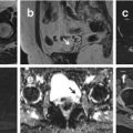
MRI: MRI is the preferred method for accurately characterizing and localizing pelvic masses. Nondegenerated leiomyomas appear isointense to hypointense on T1-weighted images and hypointense on T2-weighted images. The routine use of gadolinium has been shown not to contribute to either fibroid detection or characterization.
Leiomyomatosis
Diffuse leyomiomatosis is a rare condition that consists of diffuse involvement of the myometrium by innumerable small fibroids, which results in symmetrical enlargement of the uterus.
Although it is a completely benign condition, there may be dissemination through the peritoneal cavity or occasionally metastases to distant organs.
Leiomyosarcoma, Uterus
It is a rare malignant tumor, composed entirely of smooth muscle. It accounts for one-third of uterine sarcomas. It may arise in a previously existing benign leiomyoma (sarcomatous transformation) or independently from the smooth muscle cells of the myometrium. Rapid growth and extensive metastasis are frequently encountered with leiomyosarcomas.
Stay updated, free articles. Join our Telegram channel

Full access? Get Clinical Tree