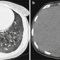
Fig. 12.1
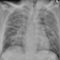
Legionella pneumonia. (a) At day 4 after the onset, chest X-ray demonstrates congestive heart failure. (b) At day 9 after the onset, chest X-ray demonstrates obvious flakes of shadows in the left lung after treatment for congestive heart failure.
Case Study 2
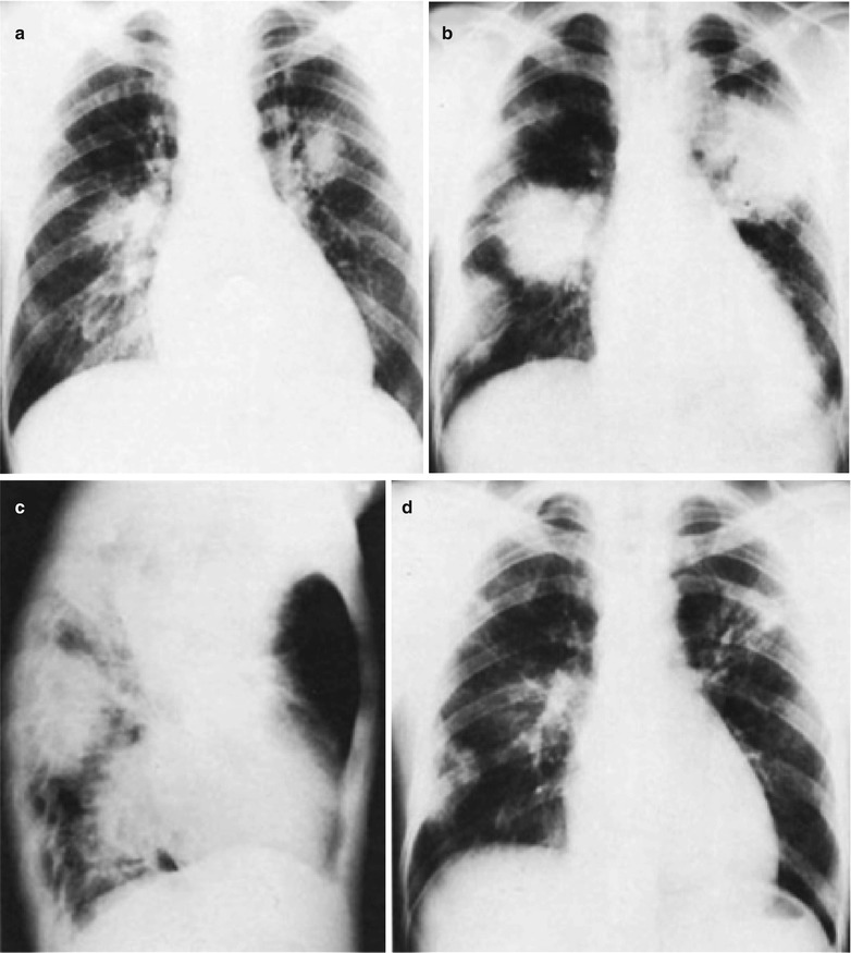
Fig. 12.2
Legionella pneumonia. (a) At the day 4 after the onset, chest X-ray demonstrates blurry shadows around bilateral pulmonary hilum. (b, c) At the day 8 after the onset, chest X-ray demonstrates obviously enlarged lesion range with consolidation shadows. (d) At the convalescence stage, chest X-ray demonstrates flakes of shadows in both lungs
Case Study 3
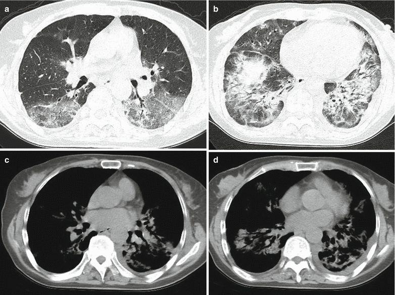
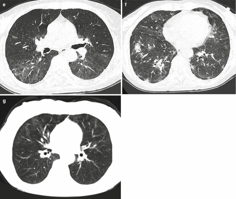
Fig. 12.3
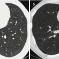
Legionella pneumonia. (a–b) HRCT demonstrates multiple flakes of ground-glass opacities and consolidation shadows in both lungs, with internal air bronchogram sign. (c–d) Mediastinal window demonstrates both lung consolidation and a small quantity of arc-shaped liquid density shadows in both thoracic cavities. (e–f) Reexamination after 2 weeks treatment, HRCT demonstrates obviously absorbed lesions, with scattering patches and consolidation shadows in both lungs. (g) Reexamination after 2 months treatment, CT scanning still demonstrates ground-glass opacities in both lungs
12.7.1.2 CT Scanning
CT scanning demonstrations of Legionella pneumonia are diverse, which is characterized by multiple lobe and segment involvement. Large flakes and patches of shadows are the most common, with accompanying pleura lesions. Cavities can be found, with irregular thick wall or regular thin wall. No fluid level is found, with commonly accompanying large flakes or small patches of shadows. Occasionally, multiple air sacs in different sizes can be found in both lungs, which resemble those seen in Staphylococcus aureus pneumonia. Chest CT scanning demonstrates more lesions and more clearly defined lesions than chest X-ray. Therefore, chest CT scanning provides more reliable evidence for the diagnosis and therapeutic assessment.
Case Study 3
A female patient aged 60 years. She complained of chest distress, shortness of breath, dyspnea, cough without phlegm, and no obvious chest pain for 5 days. There is also fever, with the highest body temperature of around 37.4 °C, with subjective fatigue. Chest X-ray and CT scanning in a local hospital indicated pulmonary inflammation. Pulmonary function test indicated severe restrictive pulmonary ventilation and severe impaired function of pulmonary diffusion. By laboratory test, IgM antibody against LP serotype 1 is positive.
Case Study 4
Legionella pneumonia.
For case detail and figures, please refer to Lei, ZD. et al. Chinese Medical Imaging Technology, 2006, 22 (11): 1668.
12.7.2 Central Nervous System
Legionnaires’ disease is a systemic infectious disease. Currently, the lesions of the central nervous system are believed to be caused by Legionella toxin, with diverse clinical manifestations. In mild cases, flulike symptoms occur, while in severe cases, the symptoms include drowsiness, vomiting, headache, disorientation, limb dyskinesia, disturbance of consciousness, and limb and facial paralysis, with typical pathological reflex and meningeal irritation sign. CT scanning demonstrates low-density shadows in the cerebrum or brainstem and small lesions of hemorrhage. MR imaging demonstrates encephalitis. And FLAIR MRI demonstrates high signal. MR imaging and SPECT demonstrate abnormal brain perfusion. Functional imaging facilitates the diagnosis of Legionella encephalopathy.
Stay updated, free articles. Join our Telegram channel

Full access? Get Clinical Tree


