Location: Type (a): retroperitoneum. Commonly involves kidney, pancreas, and vertebral body for direct extension. Type (b): in the limbs, from piliferous areas. Quite small, less than 2 cm. subcutaneous tumors grow faster and reach a larger size. Type (c): in the lower limbs, more frequently involving the veins, exceptionally the arteries.
Clinical: Type (a): abdominal mass, pain, weight loss, nausea, or vomiting. Type (b): pain, surface changes in the epidermis. Type (c): pain for pressure on nerve close to the affected vessel, edema due to venous compression.
Imaging: On CT hypo- or moderately vascular lesions, type (a): found by CAT scan or angiography that does not show typical aspect. Dislocation of the most important vessels is usual. Type (c): angiographically highly vascularized. On MRI inhomogeneous, iso-/hypointense on T1, with a thick, irregular rim enhancement on contrast T1, and marked inhomogeneity with mixed but mainly high signal intensity on T2.
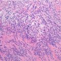
Histopathology: White-gray whorled appearance, more often fleshy masses with foci of necrosis and hemorrhage, and frequent cyst formation. Proliferation of spindle cells with an elongated nucleus with abundant cytoplasm. The nucleus is central and blunt ended or “cigar shaped.” A vacuole close to one end of the nucleus produces a slight indentation so that it becomes concave rather than convex in the contour. Sometimes, cytoplasm may have a “clotted” appearance because of clumping of the myofilaments. Cells are arranged in bundles that form wide waves intersecting with different orientation. A right angle is frequent. Stroma is a delicate mesh between cells. At times, a palisade-like aspect of the nuclei similar to neurinoma is observed. Pleomorphic appearance with highly anaplastic to epithelioid cells is common. Mitotic figures are frequent. Type b is whitish-gray or pinkish, fasciculated aspect, has ill-defined margins by virtue of the intricate blending of tumor fascicles with surrounding collagen and pilar arrector muscle in dermis tumors and well-circumscribed lesions by a pseudocapsule in subcutis tumors. Mitotic figures are frequent. Pure cutaneous (dermal without subcutaneous involvement) leiomyosarcomas have been recently designed as “atypical smooth muscle tumors” because they show a 30 % rate of locale recurrence and no metastasis. Immunohistochemistry is positive for vimentin, pan-muscle actin, smooth muscle actin, desmin, and caldesmon.
Course and Staging: Type (a) highly aggressive, so they may cause death also by local extension. Survival from 0 to 29 % at 5 years. Usually, stage IIB. Type (b): good prognosis, metastases infrequent, local recurrence frequent (50 %), usually stage IA–B. Type (c): poor prognosis, 50 % have metastases; tumors of the small veins seem to have a better prognosis, usually stage I–IIB.
Related posts:


Stay updated, free articles. Join our Telegram channel

Full access? Get Clinical Tree


