WHO-defined regions
Total registered cases by 2010 (1/10,000)
Newly reported cases in 2010 (1/100,000)
Africa
27,111 (0.38)
25,345 (3.53)
America
Southeast Asia
33,953 (0.38)
113,750 (0.64)
37,740 (4.25)
156,254 (8.77)
East Mediterranean
9,046 (0.17)
4,080 (0.67)
West Pacific
8,386 (0.34)
5,055 (0.28)
Total
192,246 (0.34)
228,474 (3.93)
In China, a total of 1,324 cases of leprosy have been newly reported in 2010, with a discovery rate of 0.099/100,000 (Table 13.2). Compared to the year 2009, the newly reported cases decreased by 17.1 % in 2010. By the end of 2010, a total of 6,032 cases of leprosy have been registered in China, with an incidence of 0.45/100,000, including 2,886 cases receiving combined chemotherapy. According to the basic criteria about eradicating leprosy in China with an incidence of lower than 1/100,000, there had been four provinces in China with an incidence of leprosy above that level by the end of 2010, namely, Yunnan, Guizhou, Tibet, and Sichuan. By the end of 2004, there had been 605 hospitals and villages across mainland China with 555 inpatients receiving therapies and totally 18,175 affected cases that had been cured.
Table 13.2
Regional prevalence of leprosy in mainland China in 2010
Epidemic region | Newly reported cases | National percentage (%) | Compared to 2009 (%)a | Compared to 2005 (%) | |
|---|---|---|---|---|---|
Total cases | Incidence (1/100,000) | ||||
North China | 6 | 0.004 | 0.5 | −25.0 | −25.0 |
Northeast | 2 | 0.002 | 0.2 | −33.3 | −66.7 |
East China | 216 | 0.055 | 16.3 | −13.9 | −9.2 |
Middle South | 291 | 0.078 | 22.0 | −23.6 | −26.7 |
Southwest | 766 | 0.389 | 57.8 | −16.6 | −19.8 |
Northwest | 43 | 0.044 | 3.2 | 22.9 | −20.4 |
National | 1,324 | 0.099 | 100.0 | −17.1 | −20.1 |
13.3 Pathogenesis and Pathological Changes
13.3.1 Pathogenesis
The pathogenesis of M. leprae after its invasion into human body is similar to that of mycobacterium tuberculosis. The onset of the disease after invasion of the bacteria, the development of conditions after onset, and the clinical manifestations are all dependent on the immunity of the invaded human body against M. leprae. After its invasion, M. leprae is firstly swallowed by macrophages. After being processed, some antigens are thoroughly expressed on the surface of cell membrane of macrophages, which then collaborate with HLA II type of antigens (DR, DP, and DQ) on the cell membrane of macrophages. And then its recognition by T cells triggers immune responses. In the cases of normal immune responses, T lymphocytes are activated to induce immune responses, with macrophages playing their role to eliminate M. leprae and form the epithelioid cells and Langerhans cells. In the cases of compromised immunity or the cases with changed expressing locus of HLA-DR antigen due to the infection of M. leprae and even expressing impairment, the failed recognition by T cells induces weak immune responses that are unable to eliminate the bacteria. Therefore, the lesions are diffusive and widespread but with slight immune impairments. Leprosy cells containing large quantities of M. leprae form at the site of lesions, which are derived from infected macrophages by large quantities of M. leprae containing lipids. In recent years, scholarly studies about immunological pathogenesis of leprosy indicated that the status of immunity and histocompatibility antigens have direct impacts on the onset of leprosy and its clinical type.
13.3.2 Pathological Changes
13.3.2.1 Tuberculoid Leprosy (TL)
This type commonly occurs in patients with a strong immunity. M. leprae has affinity to nerve tissues, and its invasion causes infiltration of lymphocytes and macrophages around nerve tissues. The infiltrated macrophages may turn into fixed epithelioid cells whose clusters form Langerhans cells cause tuberculoid granuloma. The nerve myelin is often destroyed, with hyperplasia and thickening of neurolemma.
13.3.2.2 Borderline Leprosy (BL)
This type of BL includes three subtypes: borderline tuberculoid leprosy (BTL), mid-borderline leprosy (MBL), and borderline lepromatous leprosy (BLL). The common manifestation of the three subtypes is a narrow subcutaneous non-infiltrative strip. But BTL is characterized by formation of granuloma by intradermal epithelioid cells, rare surrounding lymphocytes, swelling dermic nerves due to infiltration of histiocytes and epithelial cells, as well as a possible small quantity of bacteria in the dermic nerves. MBL is characterized by intradermal extensive granuloma, intradermal histiocyte-based infiltration, slight swelling and cell infiltration of the dermic nerves, and moderate quantities of bacteria at the lesions. BLL is characterized by subdermal macrophagic granulomas, intradermal foam cell-based infiltration, and perineurium in onionskin-like appearance with large quantities of bacteria.
13.3.2.3 Lepromatous Leprosy (LL)
In the cases of LL, the patients lack immunity to fight against M. leprae, and the pathogens spread all over the body along with lymph and blood flows. LL is characterized by extensive atrophy of the skin, thinner epidermis, destructed collagen fibers, and non-infiltrative strip in the subcutaneous papillary layer. Additionally, there is typical leproma in deep dermis, containing histiocytes, macrophages, rare lymphocytes, and rare plasma cells. Macrophages that contain fragmented or granular M. leprae forming a foamy appearance are known as foam cells, namely, leprosy cells, which are a characteristic change of LL. Endothelial cell proliferation in minor vascular vessels containing abundant bacteria commonly develops into necrotic vasculitis in the skin, resulting in ulceration. Characterized by onionskin-like appearance, dermal perineurium has infiltration of histiocytes and plasma cells, with mild swelling of nerve tracts.
13.3.2.4 Other Undifferentiated Leprosy
The cases of this type have no specific manifestations, which occurs commonly in the early stage of leprosy and can be healed by itself or develops into another type of leprosy. The pathological changes include nonspecific inflammation and lymphocytic infiltration around the dermic neurovascular bundles. The detection of M. leprae in the nerve tracts can define the diagnosis.
13.4 Clinical Symptoms and Signs
Leprosy has a chronic onset, with no obvious symptoms in the early stage. The incubation period lasts averagely for 2–5 years, and in some cases, it may be up to above 10 years. The severity of clinical manifestations varies, including self-healed asymptomatic skin rashes in mild cases and progressively destructive conditions in severe cases. The three basic characteristic manifestations of leprosy include loss of skin sensation, edema of peripheral nerves, and the presence of acid-fast bacilli by smear test of tissues at skin lesions. The early symptoms of the disease are nonspecific low-grade fever, general upset, muscular soreness and pain, and strange skin sensation. Advanced lepromatous leprosy commonly involves the eyes, testes, ovaries, bones, liver, spleen, and other tissues and organs.
13.4.1 Main Clinical Manifestations
13.4.1.1 Skin Lesions
Most patients sustain skin lesions of different severity, and the skin lesions are diverse, including macula, papule, node, ulcer, and atrophy. Skin appendages such as hairs, eyebrows, and lanugo may shed off; sweat glands and sebaceous glands may be destroyed causing perspiration stagnation and dry skin. And M. leprae can be detected in skin lesions.
13.4.1.2 Symptoms of Peripheral Nerves
Almost all patients sustain peripheral nerve impairments in different severity. The cases with only neurological symptoms but no skin lesions are defined as simplex neuritis leprosy, which mainly involve terminal nerves and superficial nerve trunks such as ulnar nerves, median nerves, common peroneal nerves, and facial nerves. The nerves are spindle-like, nodular, or evenly thick in shape. In the early stage, superficial sensory dysfunction occurs, followed by sensation loss of consecutively warmth, pain, and touch. The conditions further develop into nerve paralysis or muscle atrophy, resulting in impaired vascular dilation and constriction. Consequently, insufficient blood supply causes malnutrition of limbs, with manifestations of chapped skin, blister, ulcer, cyanosis of fingers and/or toes, swelling, and decreased body temperature.
13.4.2 Clinical Typing
13.4.2.1 Tuberculoid Leprosy (TL)
TL is the most common clinical type, accounting for 60–70 % of leprosy cases. The host of this type has a strong immunity to confine the infection. The main clinical manifestations are skin lesions and peripheral neurological lesions, which are commonly unilateral with a small range and no involvement of organs and mucosa. Therefore, patients of this type have stable conditions. The examinations for M. leprae usually show negative results, but the lepromin test usually shows strongly positive result. This type of leprosy tends to self-heal, with no infectivity and favorable prognosis.
13.4.2.2 Borderline Tuberculoid Leprosy (BTL)
The host of this type has strong immunity that is capable of partly resisting the growth of M. leprae. The skin lesions are in a large quantity and distribute extensively but asymmetrically, with multiple shapes. Many peripheral nerves are involved, with lesions distributing asymmetrically. BTL is usually accompanied by deformities of hands and feet and unilateral numbness of limbs with sole ulcer, burning, or infection of fingers. The examination of skin lesions for bacteria shows positive results. The lepromin test may be weakly positive, suspected positive, or negative. BTL type has a favorable prognosis, which can turn into TL type after standard therapies. Otherwise, due to effects of adverse factors, it may also turn into BL type or LL type.
13.4.2.3 Borderline Leprosy (BL)
BL is characterized by multiple skin lesions in various complex shapes. The nerve lesions are multiple and soft and are moderately swelling. This type of condition can be accompanied by mucosa lesions, lymph node lesions, testis lesions, and organ lesions. The examination of the lesions for M. leprae shows positive result, but the lepromin test is negative. Due to unstable cellular immunity, leprosy reactions and transformation into another type occur.
13.4.2.4 Borderline Lepromatous Leprosy (BLL)
The manifestations of this type are similar to those of lepromatous leprosy, but with a mild severity. In the early stage, the mucosa can be involved, and in the late stage, organs can be involved, with occurrence of saddle nose, ulcer of nasal mucosa, and enlarged lymph nodes. Routine bacteria examination for M. leprae shows strongly positive result, but the lepromin test is negative. The patient has cellular immunity compromised. Due to the unstable immunity of the patients with this clinical type of leprosy, it can develop into TL type or LL type.
13.4.2.5 Lepromatous Leprosy (LL)
This is another immunocompromised extremity of clinical leprosy. The lesions are diffusive and widespread, with slight damages to tissues. The skin lesions are small and multiple, distributing extensively and symmetrically, with light color. The lesions involve thick nerves with no obvious lesions. However, the cases in middle and advanced stages may develop serious muscular atrophy and deformity due to extensive symmetric involvement of nerve trunks (Figs. 13.1 and 13.2). Its occurrence in the face may lead to the formation of typical lion face. The commonly found ulcers in the limbs can hardly be healed. One of the important features of LL is the shedding of eyebrows. The nasal mucosa can be involved early and saddle nose occurs in the advanced stage. The most common ocular lesions are cornea and iris lesions, which may result in permanent vision decrease or blindness. The liver, spleen, testis, and breasts can also be involved. Bone lesions are mainly found at the short bones in the hands and feet, commonly bone destruction and absorption due to involved blood circulation and malnutrition. The above organ lesions are more common in patients in advanced lepromatous leprosy, especially in untreated patients. Generally, immediate death rarely occurs.
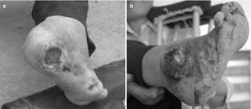

Fig. 13.1


Foot ulceration in a patient with leprosy
Stay updated, free articles. Join our Telegram channel

Full access? Get Clinical Tree


