Fig. 14.1
Leptospirosis with pulmonary bleeding. (a) X-ray at day 1 of hospitalization demonstrates enlarged heart shadow and small patches of shadows in the left upper lung. (b) X-ray at day 2 of hospitalization demonstrates development of the conditions, with patches of blurry shadows in both lungs (Reprint with permission from Wei YF, et al. J Microbiol Immunol Infect, 2012, 45 (3): 251)
Case Study 2
A 46-year-old male vagrant reported a history of sleeping in a forest and eating food bitten by rats 5 days ago. He experienced nausea, diarrhea, myalgia, dizziness, headache, hemoptysis, and high fever with a body temperature of 39 °C. Leptospira was detected in his blood.
For case detail and figures, please refer to Luks AM, et al. Chest, 2003, 123 (2): 639.)
Case Study 3
Two cases of adult males were diagnosed with leptospirosis with pulmonary bleeding.
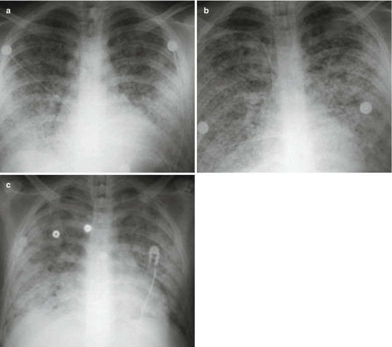
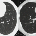
Fig. 14.2
Leptospirosis with pulmonary bleeding. (a) X-ray demonstrates nodular shadows with poorly defined boundaries in the lateral parts of both lower lungs. Some nodular shadows are demonstrated with fusion and the lung apex is comparatively well defined. (b) At the development stage, both lungs are demonstrated with diffuse nodular shadows. (c) Another case of leptospirosis with pulmonary bleeding at the middle or advanced stage. The nodular shadows are demonstrated with fusion in both longs to form diffuse patches of shadows (Reprint with permission from Ketai L, et al. Thorac Imaging, 2006, 21 (4): 265)
14.7.1.2 CT Scanning
CT scanning is superior to X-ray in demonstrating bleeding lesions. By CT scanning, the bleeding lesions are demonstrated as fine spots of shadows due to small volume of bleeding. Along with the increased volume of bleeding, the fine spots of shadows gradually fuse and are enlarged to form small patches, cotton-like, mass-like, and even patches of shadows with extremely blurry boundaries (Figs. 14.3 and 14.4). Meanwhile, in the cases with larger range of bleeding but rare intra-alveolar bleeding, CT scanning demonstrates lesions as ground glass opacities. As bleeding is dynamic and progressive, early demonstrations by radiology may be shadows with uniform morphology. However, in the advanced stage, shadows of various morphologies are demonstrated with mixed existence.
Case Study 4
A male patient aged 19 years experienced headache, neck pain, muscular pain, fever, nausea, vomiting, hemoptysis, and respiratory failure.
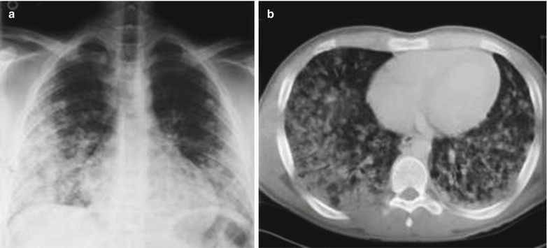
Fig. 14.3
Leptospirosis with pulmonary bleeding. (a) X-ray demonstrates patches of blurry shadows in bilateral middle and lower lung fields. (b) CT scanning demonstrates alveoli and interstitium of both lungs with infiltrative inflammation (Reprint with permission from Kishimoto M, et al. Am J Med Sci, 2004, 328 (2): 116)
Case Study 5
A male patient reported a history of contact to contaminated water by infected rats. He experienced high fever, headache, myalgia, hemoptysis, and jaundice. Bronchoalveolar lavage (BAL) demonstrated pulmonary bleeding. Serological test demonstrated positive.
For case detail and figures, please refer to Marchiori and Müller. J Thorac Imaging, 2002, 17 (2): 151.)
Case Study 6
A male patient reported a history of contact to contaminated water by infected rats. He experienced high fever, headache, myalgia, hemoptysis, and icterus. Autopsy demonstrated the diagnosis of leptospirosis.
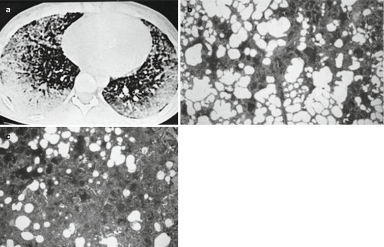
Fig. 14.4
Leptospirosis with pulmonary bleeding. (a) HRCT demonstrates ground-glass and nodular shadows in both lungs and consolidation shadows in the subpleural area. (b) Autopsy under low-power microscope demonstrates extensive pulmonary bleeding (Reprint with permission from Marchiori and Müller, J Thorac Imaging, 2002, 17 (2): 151)
14.7.2 Liver
14.7.2.1 Color Doppler Ultrasound
Color Doppler ultrasound demonstrates mild to moderate hepatomegaly, smooth liver capsule, and weakened and unevenly distributed echoes from the liver, with quite clearly defined vascularization.
14.7.2.2 CT Scanning
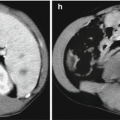
CT scanning demonstrates enlarged liver and multiple low-density lesions in the liver (Fig. 14.5).
Case Study 7
A 61-year-old male farmer experienced fatigue, right upper abdominal pain, and hepatomegaly. Endoscopy demonstrated ulceration at the transverse and ascending colon. Abdominal CT scanning demonstrated multiple low-density lesions in the liver, with slight ring-shaped enhancement. The initial diagnosis was hepatic metastasis of colon carcinoma. However, no malignancies were detected by biopsy of colon and liver tissue. Thereafter, the patient reported a history of close contacts to pigs. The antibody titer of leptospiras was then detected high.
For case detail and figures, please refer to Granito A, et al. World J Gestroenterol, 2004, 10 (16): 2455.
Case Study 8
A male patient aged 62 years experienced fever with a body temperature of 39 °C, icterus, nausea, vomiting, fatigue, and dizziness. By dark field microscopy, leptospiras were observed.
For case detail and figures, please refer to Kaya E, et al. World J Gestroenterol, 2005, 11 (28): 4447.
Case Study 9
A boy aged 10 years complained of headache, fever with a body temperature of 37 °C, abdominal pain, and fatigue for 10 days as well as language impairment for 6 days. Physical examination demonstrated hepatosplenomegaly. He was also detected leptospira positive.
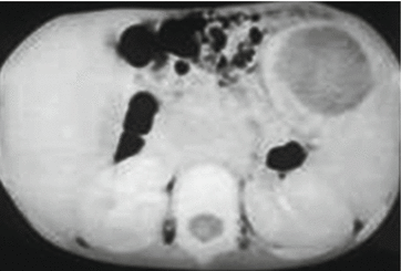
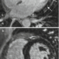
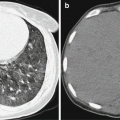
Fig. 14.5
Leptospirosis with hepatic and splenic lesions. CT scanning demonstrates the swelling of spleen, with a size of 6 cm × 4 cm
14.7.3 Brain
Cerebral leptospirosis is a series of clinical symptoms with manifestations of neurological damages caused by leptospiras. The illness course can be divided into the organ lesion stage (the middle stage or complications stage, hereinafter referred to as the cerebral lesions in the complications stage) and the convalescence stage (the late-onset symptoms stage, hereinafter referred to as the cerebral lesions in the late-onset symptoms stage). The lesions of these two stages may coexist, and their clinical manifestations can be hardly distinguished. The relationship between the imaging demonstrations and clinical symptoms is analyzed as the following.
14.7.3.1 Cerebral Lesions at the Complications Stage
The clinical manifestations are characterized by symptoms of encephalitis and meningitis, with severe headache, vomiting, irritation, unconsciousness, neck rigidity, and Kernig sign positive. The imaging demonstration features diffuse cerebral lesions. CT scanning demonstrates normal density, slightly low density, or diffuse cerebral edema. MR imaging demonstrates diffuse multiple spots and flakes of low or equal T1WI signal and high T2WI signal. The signs of demyelination are sometimes demonstrated.
14.7.3.2 Cerebral Lesions in the Late-Onset Symptoms Stage
The symptoms are clinically characterized by reactive meningitis or occlusive cerebral arteries, with manifestations of hemiplegia, aphasia, and multiple repeated transient paralysis. Cerebral angiography demonstrates stenosis of the involved vascular vessels. The imaging demonstrations feature diffuse or focalized lesions. Multiple diffuse lesions are commonly distributed in different areas of unilateral blood vessels, with equal or low T1WI as well as high T2WI signals. Occasionally, the lesions can be found at the interface of cortico-white matters, with typical manifestation of infarction. Local lesions are mostly characterized by signs of cerebral infarction (Fig. 14.6).
Case Study 10
A female patient aged 13 years complained of headache and irritation for 7 days as well as unconsciousness for 3 days. Her body temperature was 36.9 °C. Leptospira was detected positive.
For case detail and figures, please refer to Kurtoğlu MG, et al. Tohoku J Exp Med, 2003, 201 (1): 55.
Case Study 11
Related posts:

Stay updated, free articles. Join our Telegram channel

Full access? Get Clinical Tree


