5
Liver
Pyogenic Abscess
 Spread via:
• Biliary system (obstruction)
 Most common
 For example: cholangiocarcinoma, CBD stone
• Hematogenous spread
 For example: appendicitis, diverticulitis
 Single abscess or multiple abscesses
 More common in the right hepatic lobe
 Bacteria
• Monomicrobial 40%, polymicrobial 40%, culture negative 20%
• Gram-negative organisms most common
• Escherichia coli found in two-thirds of abscesses
• Opportunistic organisms in AIDS patients
 Fungal and mycobacterial
• Blood cultures positive, ∼50% of cases
 Presenting signs and symptoms
• Fever (most common), right upper quadrant abdominal pain/tenderness, jaundice
 Treatment
• Antibiotics and drainage
• Manage etiology of the pyogenic abscess
Amebic Abscess
 Caused by Entamoeba histolytica
 Most common abscess worldwide—infects up to 10% of worldwide population
 Amebic cysts are ingested, passing through stomach and small bowel unharmed → trophozoite in colon → spreads into liver via portal venous system from colon
 Presenting signs and symptoms
• History of recent travel to endemic areas
• Right upper quadrant pain, fever, jaundice, diarrhea
• Labs
 Serologic testing
 Leukocytosis
 Normal bilirubin
 Treatment
• Metronidazole
RADIOLOGY
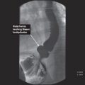
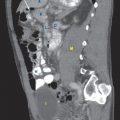
General Liver Abscess (Fig. 5.1)
 Multilocular, rim enhancing mass with focal areas of fluid attenuation representing blood or pus
FIGURE 5.1 A,B
A. Heart
B. Vertebra
C. Kidney
D. Spleen
E. Descending aorta
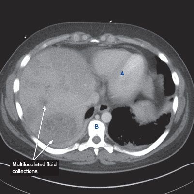
FIGURE 5.1 A
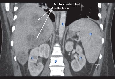
FIGURE 5.1 B
Pyogenic Abscess
 Chest x-ray
• Nonspecific findings—elevated right hemidiaphragm, right-sided pleural effusion, and atelectasis can be seen
 Ultrasound
• Multiloculated fluid collection within the liver, sometimes with posterior acoustic enhancement
 CT findings
• Multilocular fluid collection ± air–fluid levels with areas of peripheral enhancement
Amebic Abscess
 Ultrasound
• Typically, a round hypoechoic, homogeneous lesion with posterior acoustic enhancement
 CT findings
• Round, rim-enhancing hypodense lesions with a peripheral zone of edema
• May contain central septations
Hemangioma
 Most common benign tumor of the liver
 More common in females (3:1)
 If greater than 5 cm then considered a giant hemangioma
 Mostly asymptomatic. Normal LFTs and tumor markers
 If symptomatic: Kasabach–Merritt syndrome (presents with bruising, purpura, thrombocytopenia with consumptive coagulopathy, microangiopathic hemolytic anemia)
 Treatment
• Surgical resection if ruptured, significant change in size, development of Kasabach–Merritt syndrome
RADIOLOGY
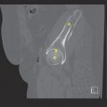
 CT findings (Fig. 5.2)
• Relatively hypodense and well-defined lesion when compared to surrounding liver in precontrast phase
• Early peripheral nodular enhancement, with enhancement equivalent to blood pool
• Centripetal contrast enhancement on more delayed images
 MRI findings
• T1-weighted images may show low signal intensity
• T2-weighted images show high signal intensity
• Peripheral enhancement with equivalent signal intensity to aorta on arterial phase, with centripetal enhancement on more delayed phases
FIGURE 5.2 A,B
A. Stomach
B. Small bowel loops
C. Vertebra
D. Kidney
E. Spleen

FIGURE 5.2 A

Stay updated, free articles. Join our Telegram channel

Full access? Get Clinical Tree


