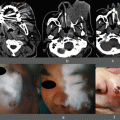
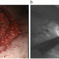
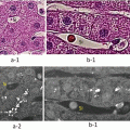
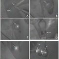
Fig. 33.1
Fluorescence angiography. (a) Intraoperative gross appearance of the right liver graft after the reconstruction of the hepatic artery (arrowhead) and the portal vein (arrow). (b) Following the intravenous injection of ICG, fluorescence imaging delineated continuous blood flow through the anastomotic site (arrowhead). (c) The portal flow (arrow) was subsequently visualized
33.3 Intraoperative Visualization of Veno-occlusive Regions in the Liver Graft
33.3.1 Background
In living-donor liver transplantation using the right liver graft without the middle hepatic vein (MHV), venous occlusion of the MHV tributaries needs to be addressed to avoid adverse events such as necrosis [28], insufficient regeneration [29], and difficulty in postoperative management [30, 31]. The MHV tributaries including V5 draining segment 5 and V8 draining segment 8 were reconstructed or sacrificed based on the institutional criteria. The modality to evaluate success/failure of venous reconstruction and the extent of regions with venous occlusion is limited mainly to intraoperative ultrasound or Doppler ultrasound. Recently, authors’ group evaluated portal uptake function in veno-occlusive regions of the remnant liver or the liver graft using fluorescence imaging following intravenous injection of ICG [21]. This technique was applied to visualize the extent of regions with venous occlusion in the liver graft [22].
33.3.2 Administration of ICG
Following procedure of liver transplantation, the camera head of the florescence imaging system (PDE) was positioned 20 cm above the liver surface, and surgical lights were turned off. The output of the light-emitting diodes (760 nm) was set at 0.21 mW per cm2. ICG (2.5 μg per 1 mL of remnant liver volume calculated based on preoperative computed tomography) was injected intravenously.
33.3.3 Visualization of Regions with Venous Flow After Reconstruction of the Hepatic Venous Tributaries
The FI values on the liver surface increased linearly during first 200 s and then reached a plateau in an average, providing clear demarcation between veno-occlusive regions and non-veno-occlusive regions. [21] Reconstruction of the hepatic veins can be evaluated using ICG fluorescence imaging because FI is lower in veno-occlusive regions than in non-veno-occlusive regions.
Clinical application of this technique is shown in Fig. 33.2. Following placement of a right liver graft and reconstruction of all hepatic vessels, ICG was injected intravenously. FI in regions drained by the right hepatic vein and V8 increased gradually, while FI on the surface of regions drained by V5 was lower than the other regions in the graft. Portal uptake of ICG in the regions drained by V5 was impaired due to sacrifice of the tributaries of V5. By contrast, the drainage area of the right hepatic vein and V8 was not impaired due to reconstruction of those drainage veins and preservation of venous outflow. Doppler ultrasound identifies the flow of the reconstructed vessels and help surgeons to decide success/failure of the reconstruction. This technique is useful to identify the regions with or without venous outflow and assess the level of portal uptake in regions as well. This information complements assessment of Doppler ultrasound and is expected to improve the safety of LT.
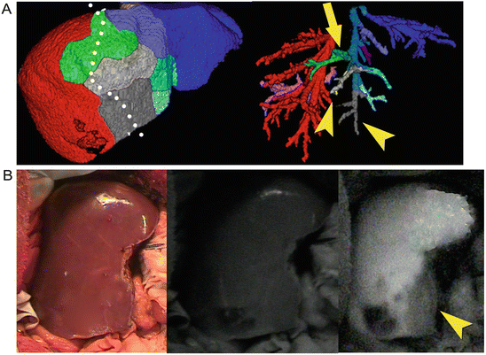

Fig. 33.2
Evaluation of the hepatic vein reconstruction. (a) Procurement of the right liver graft without the middle hepatic vein was scheduled along the border between the left and the right liver (white dotted line). The volume of regions drained by V8 (arrow) was 45 mL (5.3 % of recipient standard liver volume), while the volume of regions drained by two V5s (arrowheads) was 40 mL (4.7 % of recipient standard liver volume). Reconstructions of V8 and the right hepatic veins were scheduled because the drainage areas of V5 in the right liver graft were smaller than those of V8 and the graft volume without venous occlusion (475 mL, 56.3 % of recipient standard liver volume) was sufficient for the recipient (b) Fluorescence imaging revealed a clear demarcation between the veno-occlusive regions (arrowhead) and non-veno-occlusive regions. Reconstruction of the right hepatic vein and V8 was judged to be successful because FI in their drainage areas increased and was higher than veno-occlusive regions in which the corresponding drainage vessels were not reconstructed
These evaluations required approximately 10 min in total. One of advantages of these techniques is that they enable visualization of the degree and the extent of the portal uptake in the regions as a result of hepatic vessel reconstruction. These techniques are expected to more accurately estimate the portal uptake in regions with or without venous occlusion, ensuring the safety of liver resection and liver transplantation.
33.4 Conclusion
ICG fluorescence imaging enables real-time assessment of reconstructed hepatic vessels during LT. Anastomotic sites of the hepatic artery and portal vein were visualized after injection of ICG, and the extent and degree of portal uptake were evaluated referring to fluorescence images on the liver surface. These techniques are expected to complement intraoperative ultrasound which is utilized to assess the hepatic flow after reconstruction, enhancing the safety of LT.
Acknowledgment
This work was supported by grants from the Takeda Science Foundation, the Kanae Foundation for the Promotion of Medical Science, the Pancreas Research Foundation of Japan, the Canon Foundation in Europe, and the Ministry of Education, Culture, Sports, Science and Technology of Japan (No.23689060).
Financial Disclosure
The authors have no relevant affiliations or financial involvement with any organization or entity with a financial interest in or financial conflict with the subject matter or materials discussed in this manuscript. This includes employment, consultancies, honoraria, stock ownership or options, expert testimony, grants or patents received or pending, and royalties. No writing assistance was utilized in the production of this manuscript.
References
1.
Starzl TE, Klintmalm GB, Porter KA, Iwatsuki S, Schroter GP (1981) Liver transplantation with use of cyclosporin a and prednisone. N Engl J Med 305:266–269CrossRefPubMedPubMedCentral
2.
Sugawara Y, Makuuchi M, Kaneko J, Ohkubo T, Imamura H, Kawarasaki H (2002) Correlation between optimal tacrolimus doses and the graft weight in living donor liver transplantation. Clin Transpl 16:102–106CrossRef
3.
Sugawara Y, Makuuchi M, Akamatsu N, Kishi Y, Niiya T, Kaneko J et al (2004) Refinement of venous reconstruction using cryopreserved veins in right liver grafts. Liver Transpl 10:541–547CrossRefPubMed
Stay updated, free articles. Join our Telegram channel

Full access? Get Clinical Tree


