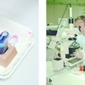
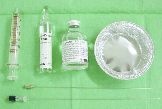
Local Ablative Procedures; Percutaneous Ethanol and Acetic Acid Injection Local ablative injection therapies (e.g., percutaneous ethanol injection) have become established routine procedures for the treatment of hepatocellular carcinoma. They compete with surgical resection and other local ablative procedures such as radiofrequency ablation (RFA)1 and laser-induced thermotherapy (LITT), which are discussed separately in Chapter ▶ 19. Percutaneous ethanol injection (PEI) and percutaneous acetic acid injection (PAI) have yielded very favorable results for (primary) hepatocellular carcinoma but not for metastases from other tumors. This is explained by the cirrhotic transformation of the liver resulting in a predominantly arterial blood supply to the cirrhotic tissue and the frequent encapsulation of hepatocellular carcinoma.2–8 Only a few studies have been published on local ablative injection therapies for metastatic lesions due to the disappointing results.9–12 Note PEI has become an established therapy for hepatocellular carcinoma but not for metastases. Studies to date have not shown a definite survival advantage of radiofrequency thermoablation (RFA or RFTA) over PEI,5 although reports have repeatedly noted a certain trend in that direction. Thus, the local injection of alcohol and other agents should continue to play a key role in the treatment of hepatocellular carcinoma. Remarkably, much more costly local ablative procedures are still being recommended to patients—perhaps uncritically in some cases. The literature comparing RFA and PEI, and the somewhat contradictory results of published findings, will be explored in more detail in Chapter ▶ 19. Most interventionalists have more experience with ethanol than with acetic acid, with the result that PEI is favored at most centers. The differences between the two therapies appear to be marginal.13–15 It has been suggested that PAI requires fewer injections and smaller injection volumes than PEI to achieve the same response, but this is no more than a reported trend.16,17 Both PEI and PAI can be administered in a single- or multiple-session regimen.8,18–20 The advantage of single-session treatment is that in regions and countries with limited health care facilities, definitive treatment can be provided in one visit to the doctor for patients unable to make repeated visits. The advantage of multiple treatment sessions is that they probably yield a better result. It has been found that the injection of large ethanol volumes (100 mL or more) may induce the decompensation of cardiopulmonary diseases such as coronary heart disease or pulmonary hypertension, and therefore these conditions should be excluded before treatment.21 Local ablative procedures can be used as a bridging strategy in patients on a waiting list for a liver transplant or if liver transplantation is contraindicated due to age, comorbidity, or other documentable causes and the risk of surgical treatment (resection) outweighs the potential survival benefit.22–24 Local ablative procedures are appropriate for small, solitary tumors. The prognosis of these lesions is excellent with PEI and is comparable to that following tumor resection. But there are other factors that would weaken the indication for local ablative therapy: a number of tumors are multifocal when first diagnosed in unscreened cases, tumors are often associated with local satellite lesions, and invasion of the portal vein and hepatic veins may occur early in the course of the disease—although data suggest that this invasion does not affect survival after PEI.25 Solitary, well-circumscribed tumors up to 3 cm in size (n = up to 3) are an established indication for local ablation by PEI and PAI. A solitary lesion up to 50 mm in size is an established indication in patients with Child class A or B hepatic cirrhosis. Child class C patients may also benefit in selected cases, depending on the degree of tumor differentiation.26 Tumor invasion of the portal vein or its side branches is not a contraindication to local ablative therapy. It is important to consider the pathogenesis of a hyperregenerative liver nodule, a dysplastic nodule with a low or high grade of dysplasia, well-differentiated hepatocellular carcinoma, and poorly differentiated forms (G1 to G4) since different nodular lesions have different prognostic implications. It has been known for some time that the prognosis of hepatocellular carcinoma depends on the size of the tumor at the time of initial diagnosis,27,28 and this has been confirmed by treatment outcomes in recent large studies.3–6,29 Hepatocellular carcinoma in a Child class C cirrhotic liver appears to have less prognostic importance than in a Child–Pugh class A liver. This is because the tumor itself does not greatly affect the inherently poor prognosis of the advanced liver disease.26 Multifocal hepatocellular carcinomas should be excluded with the highest possible certainty before local ablative therapy. We require histologic confirmation of the tumor before proceeding with local ablative treatment. This confirmation is essential for oncologic reasons in most palliative treatment settings. Other, noninvasive diagnostic criteria have been proposed for the prevention of tumor cell seeding, especially before liver transplantation. According to guidelines, a hypervascular tumor >20 mm is considered to be hepatocellular carcinoma when identified by one contrast-enhanced study (CT, MRI, ultrasound). For tumors 10 to 20 mm in size, two contrast-enhanced studies are required (combined if necessary with elevated AFP levels).29–31 Tumors smaller than 10 mm can be followed at 3 months. Besides general contraindications relating to coagulation status (coagulation disorders and platelet dysfunction) and lack of informed consent, a local ablative procedure may be contraindicated due to comorbidity, poor prognosis, or short life expectancy <3 months. Bleeding time should be determined if the coagulation status is unclear. Another contraindication is ascites, because free fluid increases the complication rate and ascites during diuretic therapy is an unfavorable prognostic factor. Advanced pulmonary hypertension and coronary heart disease are particularly important comorbid conditions, since the injection of large amounts of alcohol may occasionally cause cardiovascular deterioration in these patients. Basic materials Sterile injection needles 0.7 mm in diameter with a long bevel (2 mm), 95% or 96% alcohol, and 10-mL syringes (▶ Fig. 18.1). Fig. 18.1 Tray setup includes sterile 0.7-mm injection needles with a long bevel (2 mm), 95% or 96% alcohol, and 10-mL syringes. Special materials 20-gauge special injection needle, length 20 cm (Peter Pflugbeil), 95% ethanol (Braun Melsungen). What type of needle? Alcohol-resistant material should be used. Various needle types have been used ranging from a simple yellow No. 1 needle to No. 2 needles and needles with multiple side holes (Otto needle) that can provide better dispersal (20-gauge special injection needle, 20 cm, Peter Pflugbeil). There is no hard evidence for the superiority of specific needle types, however. Standard protocols for skin preparation and local anesthesia for percutaneous procedures should be followed (see Chapter ▶ 8). If necessary, local anesthesia may be supplemented by sedation (e.g., with diazepam in patients with liver damage; Chapter ▶ 11). Piritramide is widely preferred over pethidine for analgesia, although there are no study data to support this preference.
18.1 Basic Considerations
18.1.1 What Tumors Are Suitable for Local Ablative Procedures?
18.1.2 Radiofrequency Ablation or Percutaneous Ethanol Injection?
18.1.3 Ethanol or Acetic Acid Injection?
18.1.4 Single or Multiple Sessions?
18.2 Indications
18.2.1 Considerations on Hepatocellular Carcinoma
Factors Determining Prognosis
Histologic Confirmation
18.3 Contraindications
18.4 Practical Aspects
18.4.1 Materials and Equipment
18.4.2 Preparations
Stay updated, free articles. Join our Telegram channel

Full access? Get Clinical Tree


