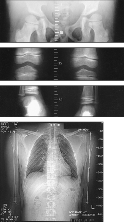
11 The limb to be examined should be positioned as follows: • Adjust and immobilize the limb for an AP projection. • If the two lower limbs are examined simultaneously, separate the ankles 5 to 6 inches (13 to 15 cm) and place the specialized ruler under the pelvis and extended down between the legs. • If the limbs are examined separately, position the patient with a special ruler beneath each limb. • When the knee of the patient’s abnormal side cannot be fully extended, flex the normal knee to the same degree and support each knee on one of a pair of supports of identical size to ensure that the joints are flexed to the same degree and are equidistant from the image receptor (IR). • Localize each joint accurately, and use a skin-marking pencil to indicate the central ray centering point. • Because both sides are examined for comparison and a discrepancy in bone length usually exists, mark the joints of each side after the patient is in the supine position. • With the upper limb, place the marks as follows: for the shoulder joint, over the superior margin of the head of the humerus; for the elbow joint,
LONG BONE MEASUREMENT

Position of Part
Localization of Joints
![]()
Stay updated, free articles. Join our Telegram channel

Full access? Get Clinical Tree


LONG BONE MEASUREMENT


