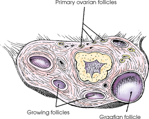
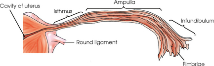
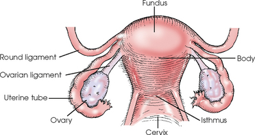
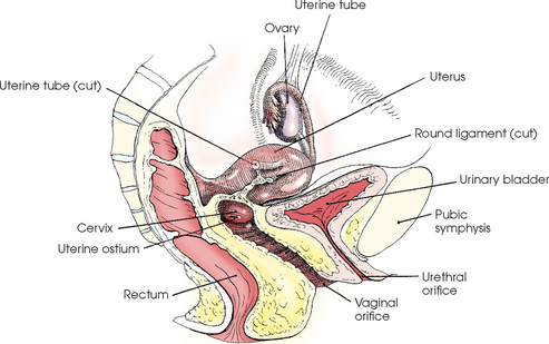
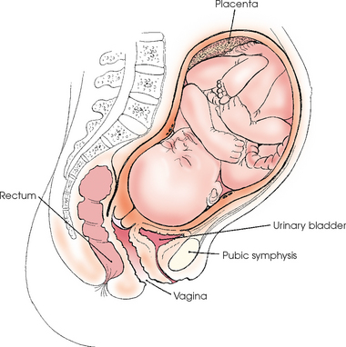
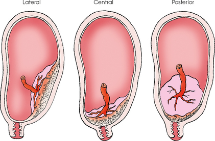
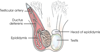
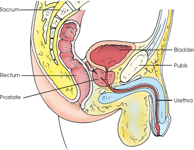
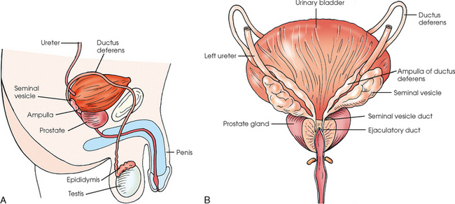
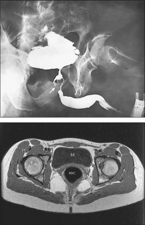
REPRODUCTIVE SYSTEM

19


REPRODUCTIVE SYSTEM


image


SUMMARY OF PROJECTIONS


Female Reproductive System


The female reproductive system consists of an internal and an external group of organs, connected by the vaginal canal. This chapter does not address the anatomy of the external genitalia because those structures do not require radiographic demonstration. The internal genital organs consist of the female gonads, or ovaries, which are two glandular bodies homologous to the male testes, and a system of canals composed of the uterine tubes, uterus, and vagina.


OVARIES


The two ovaries are small, glandular organs with an internal secretion that controls the menstrual cycle and an external secretion containing the ova, or female reproductive cells (Fig. 19-1). Each ovary is shaped approximately like an almond. The ovaries lie one on each side, inferior and posterior to the uterine tube and near the lateral wall of the pelvis. They are attached to the posterior surface of the broad ligament of the uterus by the mesovarium.


The ovary has a core of vascular tissue, the medulla, and an outer portion of glandular tissue termed the cortex. The cortex contains ovarian follicles in all stages of development, and each follicle contains one ovum. A fully developed ovarian follicle is referred to as a graafian follicle. As the minute ovum matures, the size of the follicle and its fluid content increase so that the wall of the follicle’s sac approaches the surface of the ovary and in time ruptures, liberating the ovum and follicular fluid into the peritoneal cavity. Extrusion of an ovum by the rupture of a follicle is called ovulation and usually occurs one time during the menstrual cycle. When the ovum is in the pelvic cavity, it is drawn toward the uterine tube.


UTERINE TUBES


The two uterine tubes, or fallopian tubes, arise from the lateral angle of the uterus, pass laterally above the ovaries, and open into the peritoneal cavity. These tubes collect ova released by the ovaries and convey the cells to the uterine cavity. Each tube is 3 to 5 inches (7.6 to 13 cm) long (Fig. 19-2) and has a small diameter at its uterine end, which opens into the cavity of the uterus by a minute orifice. The tube itself is divided into three parts: the isthmus, the ampulla, and the infundibulum. The isthmus is a short segment near the uterus. The ampulla comprises most of the tube and is wider than the isthmus. The terminal and lateral portion of the tube is the infundibulum and is flared in appearance. The infundibulum ends in a series of irregular prolonged processes called fimbriae. One of the fimbriae is attached either to or near the ovary.


The mucosal lining of the uterine tube contains hairlike projections called cilia. The lining is arranged in folds that increase in number and complexity as they approach the fimbriated extremity of the tube. The cilia draw the ovum into the tube, which then conveys it to the uterine cavity by peristaltic movements. The passage of the ovum through the tube requires several days. Fertilization of the cell occurs in the outer part of the tube, and the fertilized ovum migrates to the uterus for implantation.


UTERUS


The uterus is a pear-shaped, muscular organ (Figs. 19-3 and 19-4). Its primary functions are to receive and retain the fertilized ovum until development of the fetus is complete and, when the fetus is mature, to expel it during birth.


The uterus consists of four parts: the fundus, body, isthmus, and cervix. The fundus is the bluntly rounded, superiormost portion of the uterus. The body narrows from the fundus to the isthmus and is the point of attachment for the ligaments that secure the uterus within the pelvis. The isthmus (superior part of the cervix), a constricted area between the body and the cervix, is approximately ½ inch (1.3 cm) long. The cervix is the cylindric vaginal end of the uterus and is approximately 1 inch (2.5 cm) long. The vagina is attached around the circumference of the cervix.


The nulliparous uterus (i.e., the uterus of a woman who has not given birth) is approximately 3 inches (7.6 cm) in length, almost half of which represents the length of the cervix. The cervix is approximately ¾ inch (1.9 cm) in diameter. During pregnancy, the body of the uterus gradually expands into the abdominal cavity, reaching the epigastric region in the 8th month. After parturition, the organ shrinks to almost its original size but undergoes characteristic changes in shape.


The uterus is situated in the central part of the pelvic cavity, where it lies posterior and superior to the urinary bladder and anterior to the rectal ampulla. The long axis, which is slightly concave anteriorly, is directed inferiorly and posteriorly at a near right angle to the axis of the vaginal canal into which the lower end of the cervix projects.


The cavity of the body of the uterus, or the uterine cavity proper, is triangular in shape when viewed in the frontal plane. The canal of the cervix is dilated in the center and constricted at each extremity. The proximal end of the canal is continuous with the canal of the isthmus. The distal orifice is called the uterine ostium.


The mucosal lining of the uterine cavity is called the endometrium. This lining undergoes cyclic changes, called the menstrual cycle, at about 4-week intervals from puberty to menopause. During each premenstrual period, the endometrium is prepared for the implantation and nutrition of the fertilized ovum. If fertilization has not occurred, the menstrual flow of blood and necrosed particles of uterine mucosa ensues.


FETAL DEVELOPMENT


During the implantation process, the fertilized ovum, called a zygote, is passed from the uterine tube into the uterine cavity, where it adheres to and becomes embedded in the uterine lining. About 2 weeks after fertilization of the ovum, the embryo begins to appear. The embryo becomes a fetus 9 weeks after fertilization and assumes a human appearance (Fig. 19-5).


During the first 2 weeks of embryonic development, the growing fertilized ovum is primarily concerned with the establishment of its nutritive and protective covering, the chorion and the amnion. As the chorion develops, it forms (1) the outer layer of the protective membranes enclosing the embryo and (2) the embryonic portion of the placenta, by which the umbilical cord is attached to the mother’s uterus and through which food is supplied to and waste is removed from the fetus. The amnion, often referred to as the “bag of water” by the laity, forms the inner layer of the fetal membranes and contains amniotic fluid in which the fetus floats. After the birth, the uterine lining is expelled with the fetal membranes and the placenta, constituting the afterbirth. A new endometrium is then regenerated.


The fertilized ovum usually becomes embedded near the fundus of the uterine cavity, most frequently on the anterior or posterior wall. Implantation occasionally occurs so low, however, that the fully developed placenta encroaches on or obstructs the cervical canal. This condition results in premature separation of the placenta, termed placenta previa (Fig. 19-6).


Male Reproductive System


The male genital system consists of a pair of male gonads, the testes, which produce spermatozoa; two excretory channels, the ductus deferens, or vas deferens; the prostate; the ejaculatory ducts; the seminal vesicles; and a pair of bulbourethral glands, which produce secretions that are added to the secretions of the testes and ductal mucosa to constitute the final product of seminal fluid. The penis, the scrotum, and the structures enclosed by the scrotal sac (testes, epididymides, spermatic cords, and part of the ductus deferens) are the external genital organs.


TESTES


The testes are ovoid bodies averaging 1½ inches (3.8 cm) in length and about 1 inch (2.5 cm) in width and depth (Fig. 19-7). Each testis is divided into 200 to 300 partial compartments that constitute the glandular substance of the testis. Each compartment houses one or more convoluted, germ cell–producing tubules. These tubules converge and unite to form 15 to 20 ductules that emerge from the testis to enter the head of the epididymis.


The epididymis is an oblong structure that is attached to the superior and lateroposterior aspects of the testis. The ductules leading out of the testis enter the head of the epididymis to become continuous with the coiled and convoluted ductules that comprise this structure. As the ductules pass inferiorly, they progressively unite to form the main duct, which is continuous with the ductus deferens.


SEMINAL VESICLES


The two seminal vesicles are sacculated structures about 2 inches (5 cm) long (Fig. 19-9). They are situated obliquely on the lateroposterior surface of the bladder, where, from the level of the ureterocystic junction, each slants inferiorly and medially to the base of the prostate. Each ampulla of the ductus deferens lies along the medial border of the seminal vesicle to form the ejaculatory duct.


PROSTATE


The prostate, an accessory genital organ, is a cone-shaped organ and averages 1¼ inches (3.2 cm) in length. The prostate encircles the proximal portion of the male urethra and, extending from the bladder neck to the pelvic floor, lies in front of the rectal ampulla approximately 1 inch (2.5 cm) posterior to the lower two thirds of the pubic symphysis (see Fig. 19-9). The prostate comprises muscular and glandular tissue. The ducts of the prostate open into the prostatic portion of the urethra.


Because of advances in diagnostic ultrasound imaging, radiographic examinations of the male reproductive system are performed less often than in the past. The prostate can be ultrasonically imaged through the urine-filled bladder or using a special rectal transducer. The seminal ducts can be imaged when the rectum is filled with an ultrasound gel and a special rectal transducer is used. Testicular ultrasonic scans are performed to evaluate a palpable mass or an enlarged testis and to check for metastasis. Most testicular scans are performed because of a palpable mass or an enlarged testis.


Mar 3, 2016 | Posted by in GENERAL RADIOLOGY | Comments Off on REPRODUCTIVE SYSTEM

Full access? Get Clinical Tree

Get Clinical Tree app for offline access