Chapter 14 Lumbar Zygapophysial Joint Nerve (Medial Branch) Injection, Oblique Approach
Note: Please see page ii for a list of anatomical terms/abbreviations used in this book.
Lumbar medial branch blocks are diagnostic procedures that are indicated to determine whether a patient’s axial pain is caused by the zygapophysial joints under study. Medial branch blocks are used to determine if a patient is a candidate for radiofrequency neurotomy.1
The lumbar facet joints were first suggested as a source of low back and lower extremity pain by Goldthwait in 1911.2 Provocation maneuvers have been comprehensively studied in the lumbar spine, and they have widely varying sensitivity, specificity, and interrater variability.3 Schwarzer concluded that, although the lumbar zygapophysial joint may be a source of pain in a substantial minority of patients, a “facet syndrome” was unable to be demonstrated clinically. The prevalence of chronic lumbar zygapophysial joint pain is as high as 15% among younger patients4 and as high as 40% among older patients.5
The radiographic approach to the performance of diagnostic blocks of the lumbar zygapophysial joint innervation was confirmed to be target specific. It was further refined by Dreyfuss, with defined target points and needle trajectories required to provide specific anesthetization of the zygapophysial joint.6 The volume of local anesthetic injected should be limited to 0.4 mL to 0.5 mL to maintain the diagnostic specificity of the injection.
Reporting of Diagnostic Results
Diagnostic accuracy is dependent on the reliable evaluation of the patient’s response to the block. The “double-block” protocol (as described in the International Spine Intervention Society Guidelines1) is not universal, and it has been challenged on the basis of both its cost effectiveness7 and the consideration that the radiofrequency neurotomy procedure carries no greater risk of complications as compared with the diagnostic block.9 The percentage of postblock improvement above which a patient may be considered to be a candidate for radiofrequency denervation (i.e., 50% versus 80%) has been studied, and it has been concluded that the optimal threshold cannot be determined.9
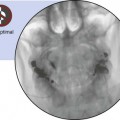
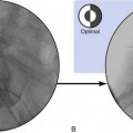
 Trajectory View
 Confirm the level (with the anteroposterior view).


Related posts:
 Atlantoaxial Joint Intraarticular Injection
 Lumbar Zygapophysial Joint Nerve (Medial Branch) Radiofrequency Neurotomy, Posterior Approach
Stay updated, free articles. Join our Telegram channel

Full access? Get Clinical Tree


