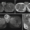
Fig. 29.1
Cerebral malaria. (a, b) Transverse T2WI and FLAIR demonstrate symmetrical high signal in bilateral thalamus. (c, d) DWI and ADC demonstrate no limited diffusion of the lesions (Reprinted with permission from Yadav et al. 2008, 49(5):566)
Case Study 4
A male patient aged 30 years complained of headache, fatigue, and consequent coma. By GCS, he was scored 6 points. By laboratory tests, a large quantity of P. falciparum was detected by blood smear.
(For case detail and figures, please refer to Cordoliani YS et al. AJNR, 1998, 9 (5): 871.)
Case Study 5
A female patient aged 13 years was admitted to the hospital due to frequent convulsions. She suffered from malaria 1 month ago, with detected P. falciparum by blood smear. By neurological examination, she was normal. EEG demonstrated θ wave in the right temporal frontal lobe.
(For case detail and figures, please refer to Cordoliani YS et al. AJNR, 1998, 9 (5): 871.)
Case Study 6
A male patient aged 23 years was diagnosed with recurrent cerebral malaria. His GCS was scored 5.8 points. By blood smear, P. falciparum was detected.
(For case detail and figures, please refer to Cordoliani YS et al. AJNR, 1998, 9 (5): 871.)
Case Study 7
A female patient aged 31 years complained of periodic fever, chills, headache, nausea, and vomiting for 1 week. By blood smear, P. falciparum was detected.
(For case detail and figures, please refer to Nickerson JP et al. AJNR, 2009, 30 (6): e85.)
29.7.1.3 PET
The metabolic rate of glucose in the cerebral cortex diffusively decreases in experimental monkeys, but the changes in the basal ganglia are not obvious.
29.7.2 Pulmonary Malaria
29.7.2.1 X-Ray
According to different severities of the conditions, pulmonary X-ray demonstrations have great variance, which can be divided into the following five types.
Bronchitis Type
The demonstrations are commonly thickened and increased pulmonary markings in both lungs. These findings are mainly distributed in the middle and inner areas of both lower lung fields, and the findings in the outer area are usually along with interstitial changes. These demonstrations are nonspecific.
Interstitial Pneumonia Type
Concurrent with more, thickened and blurry pulmonary markings, X-ray demonstrates reticular shadow and small spots of changes in the interlobular septa. These findings are mainly distributed in the middle and outer areas. In some cases, these findings are distributed in the whole lungs (Fig. 29.2).
Bronchial Pneumonia Type
X-ray demonstrates the type with patches of blurry shadow in the thickened lung markings of both lower lung fields.
Lobar Pneumonia Type
X-ray demonstrates large flakes of cloudy high-density shadows that are distributed in pulmonary lobes or segments. The consolidation of pulmonary segments is characterized by lesions at the fundus (Fig. 29.3).
Pulmonary Edema Type
For the demonstrations, see Fig. 29.4. In addition, pleural thickening and pleural effusion can also be observable.
Case Study 8
A male patient aged 37 years complained of fever and cough for 10 days. By blood smear, plasmodia were detected positive.
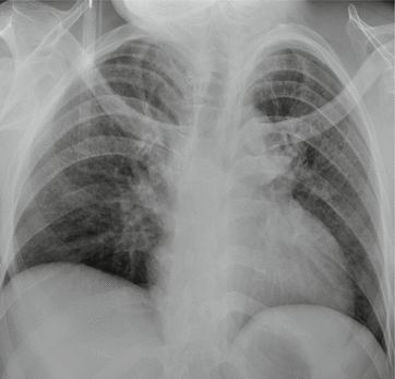

Fig. 29.2
Pulmonary malaria (interstitial pneumonia type). X-ray demonstrates more thickened and blurry lung markings in both lungs as well as reticular and small spots of shadows with increased density
Case Study 9
A boy aged 3.6 years complained of high fever and shortness of breath for 6 days. By blood smear, plasmodia were detected positive.
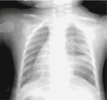

Fig. 29.3
Pulmonary malaria (lobar pneumonia type). X-ray demonstrates wedge-shaped dense shadow in the right middle lung lobe, with quite straight boundary
Case Study 10
A male patient aged 40 years complained of fever and paroxysmal dyspnea for 1 day. By blood smear, plasmodia were detected positive.
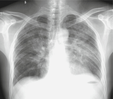

Fig. 29.4
Pulmonary malaria, with acute pulmonary edema and pleural effusion. X-ray demonstrates increased lung markings in both lungs, butterfly wing-shaped patches of shadows in the inner and middle areas of both lungs, and blunt bilateral costophrenic angles
29.7.2.2 CT Scanning
Pulmonary Edema
Symmetrical large flakes of effusive shadows in both lungs can be observed (Fig. 29.5).
Bronchitis
The bronchovascular bundles in both lungs are demonstrated to be thickened, increased, and deranged, which are especially obvious in the inferior lobes of both lungs (Fig. 29.6).
Pneumonia
The bronchovascular bundles are demonstrated to be thickened, increased, and deranged. There are patches, large flakes, and even segmental or lobar consolidation shadows with the pulmonary hilum as the center (Fig. 29.6).
Pleural Effusion
Case Study 11
A female patient aged 33 years complained of fever, cough, and chest pain for half a month. By blood smear, plasmodia were detected positive.
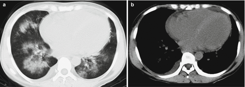

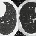
Fig. 29.5
Pulmonary malaria with pulmonary edema, pericardial effusion, and pleural effusion. (a, b) CT scanning demonstrates pulmonary edema in both lungs, enlarged heart, and a small quantity of liquid density shadows in the pericardial cavity, with right arch-shaped liquid density shadow
Case Study 12
A female patient aged 43 years complained of fever, cough, and chest pain for 7 days. By blood smear, plasmodia were detected positive.
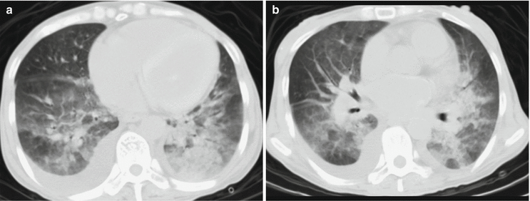

Fig. 29.6
Pulmonary malaria with pulmonary edema, pericardial effusion, and pleural effusion. (a, b) CT scanning demonstrates thickened, increased, and deranged bronchovascular bundles in both lungs, patches of consolidation shadows with the pulmonary hilum as the center, and small quantities of pleural effusion in both lungs
Case Study 13
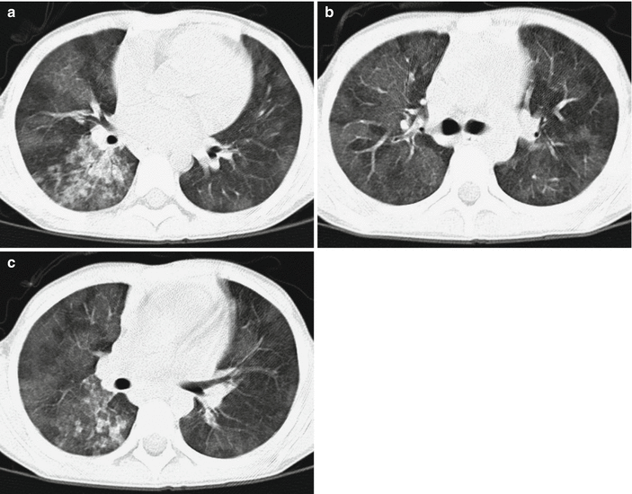
Fig. 29.7
Pulmonary malaria with alveolar edema. (a–c) CT scanning demonstrates diffusive ground-glass opacities in both lungs
29.7.2.3 PET
The metabolic rate of glucose in both lungs is demonstrated with diffusive decrease.
29.7.3 Abdominal Lesions
Abdominal malaria most commonly involves the liver and spleen, followed by the kidneys and gastrointestinal tract. The manifestations include hepatomegaly, liver dysfunction, gallbladder wall edema, splenomegaly or megalosplenia, spleen infarction, spleen rupture and hemorrhage, portal hypertension, gastrointestinal wall swelling, ascites, and mild edema of both kidneys.
29.7.3.1 Ultrasound
Hepatosplenomegaly is commonly demonstrated. There are also thickened echoes from the liver parenchyma, dilated hepatic portal vein and splenic vein as well as ascites. In the cases with spontaneous spleen rupture, the area under the splenic capsule on the diaphragmatic surface is demonstrated with crescent-shaped low echo. Ultrasonography and color Doppler ultrasound can provide valuable information for the diagnosis of spleen infarction, which is demonstrated with enlarged spleen and singular or multiple wedge-shaped or irregular low echo area in the spleen parenchyma. In the cases with complicating hemorrhage, the hemorrhage lesions are demonstrated with high echo. Ultrasonography demonstrates irregular or wedge-shaped filling defects in the spleen. Gallbladder wall edema is demonstrated with thickened gallbladder wall and decreased echo. Kidney edema is demonstrated with poorly defined corticomedullary interface and decreased echo.
29.7.3.2 CT Scanning
Hepatosplenomegaly is commonly demonstrated, and in some cases even megalosplenia, with enlarged volume. By plain scanning, the density of the enlarged spleen and the enlarged liver is demonstrated to be decreased, while by contrast scanning, the density is progressively enhanced, with stronger enhancement of the liver than that of the spleen (Fig. 29.8). Occasionally, the liver is enlarged with increased density, which is speculated to be the result of hemosiderosis in the liver due to extensive rupture of erythrocytes (Fig. 29.9). By plain scanning, spleen infarction is demonstrated as multiple wedge-shaped, bar-shaped, or map-like low-density lesions at the margin of the spleen, whose pathogenesis may be related to hyperplasia of reticuloendothelial system due to hyperfunctional removal of the spleen. By contrast scanning, infarction of the spleen is demonstrated with no enhancement of the lesions. And the wedge-shaped lesions are typical demonstration of splenic infarction. Radiological studies of animal models have demonstrated that bar-shaped low-density lesions are organized thrombi formed by infected erythrocytes in the dilated splenic vein. Kim et al. reported that the lesions in the spleen are reversible. By follow-up reexaminations, the bar-shaped low-density lesions in the spleen can disappear, with simultaneous normal size of the spleen. The liver dysfunction is demonstrated with intrahepatic lymphatic stasis (Fig. 29.10). Megalosplenia and multiple splenic infarctions are common in the cases of severe P. falciparum malaria (Fig. 29.11). In addition, there are also edema surrounding the gallbladder wall and the portal vein, ascites, spontaneous rupture of the spleen, and subcapsular hemorrhage, which are common in the patients with P. vivax malaria.
Stay updated, free articles. Join our Telegram channel

Full access? Get Clinical Tree


