
* For Peritoneal Cavity–Pelvis, see Chapter 3 ( pp. 166-205 ).
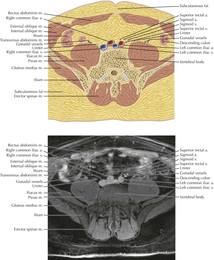
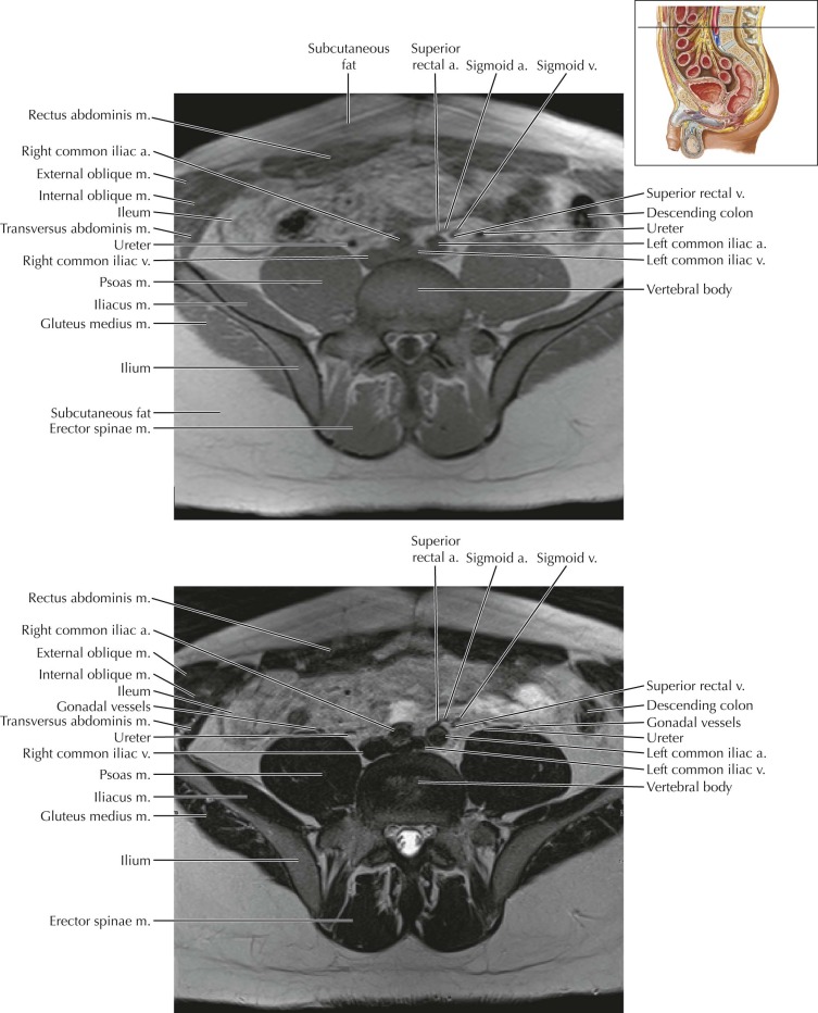
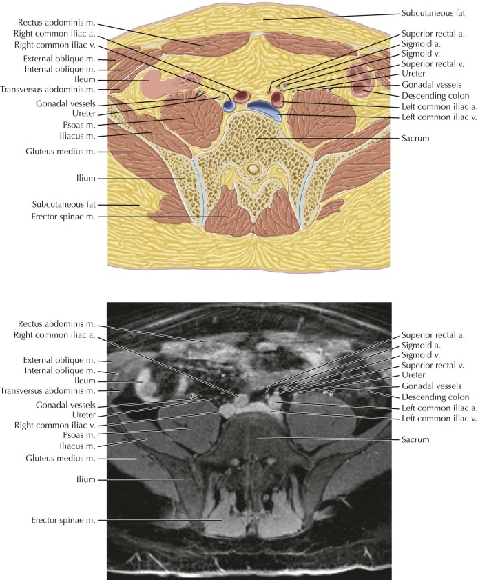
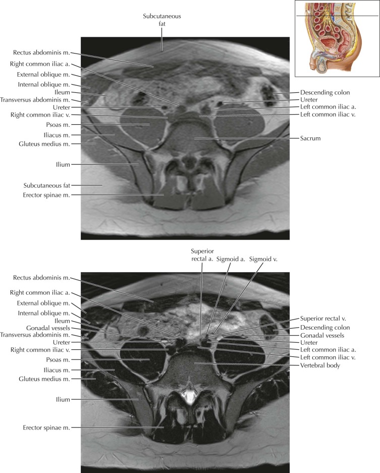
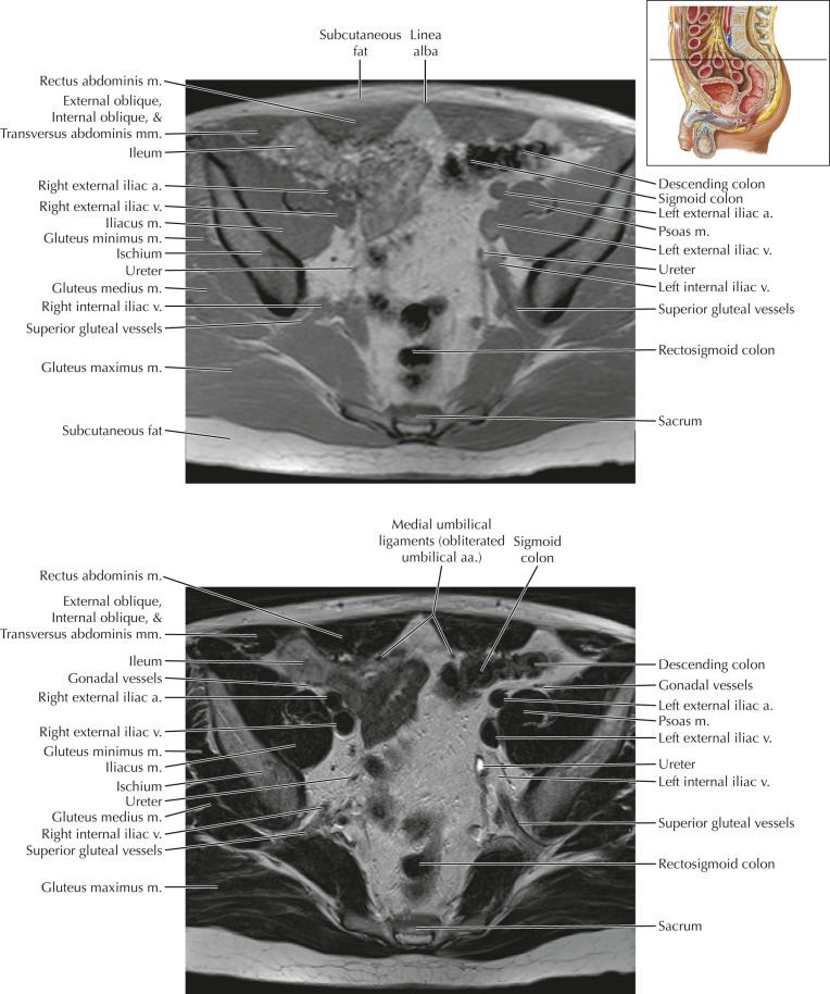
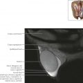
Male Pelvis Axial 1
Normal Anatomy
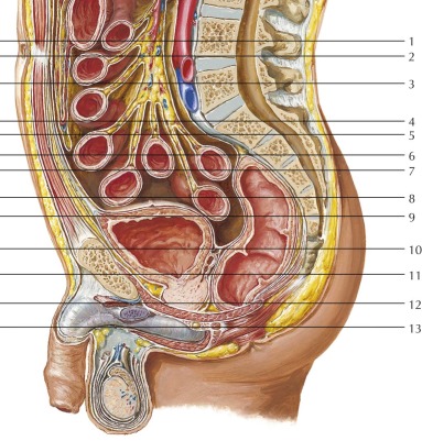
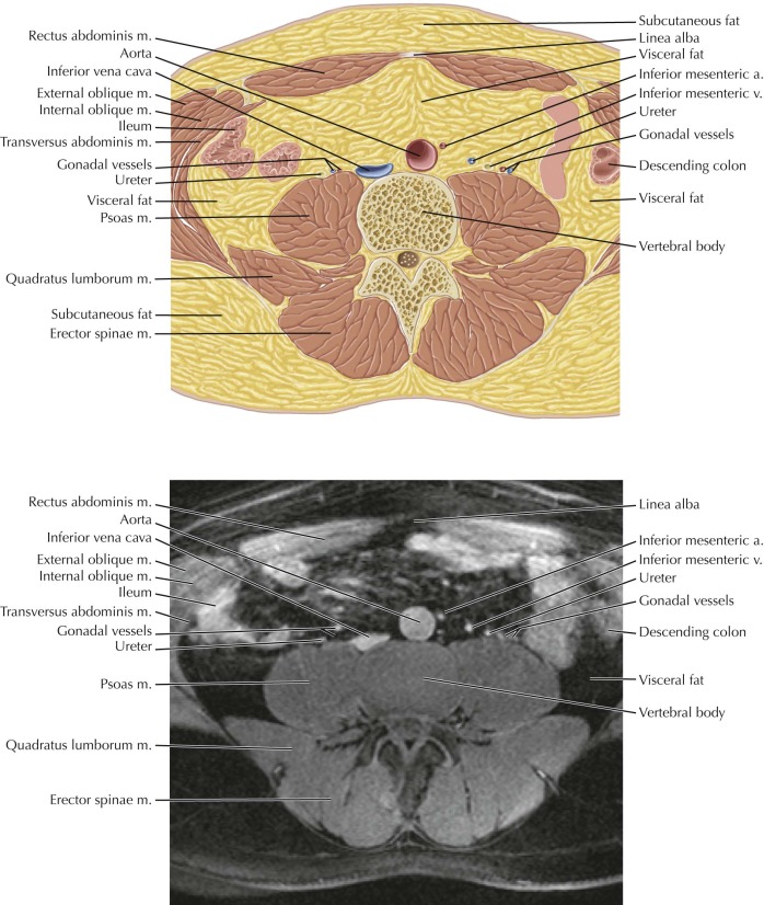
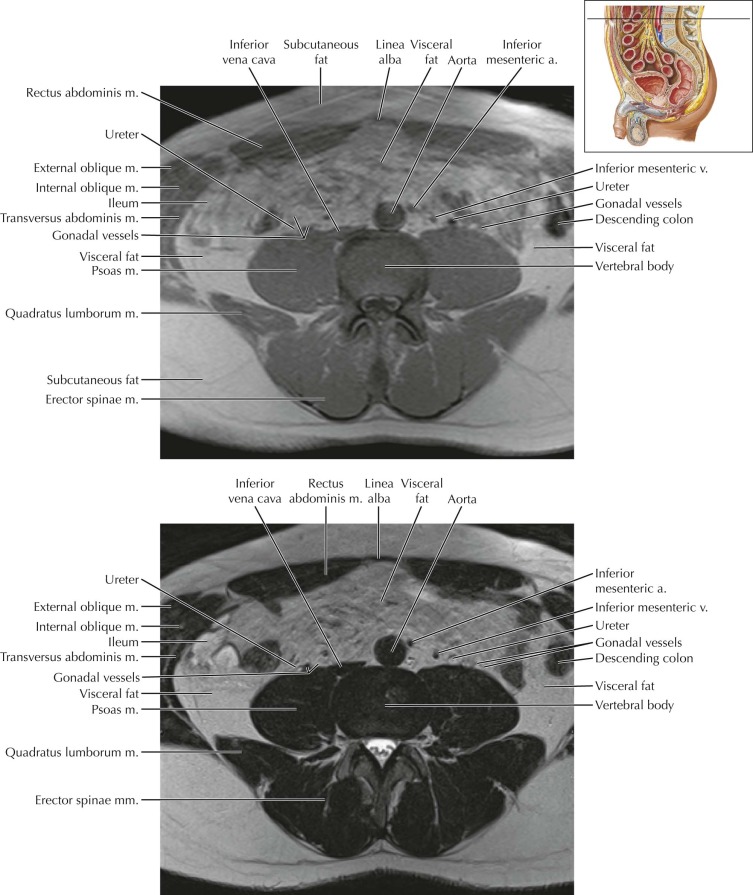
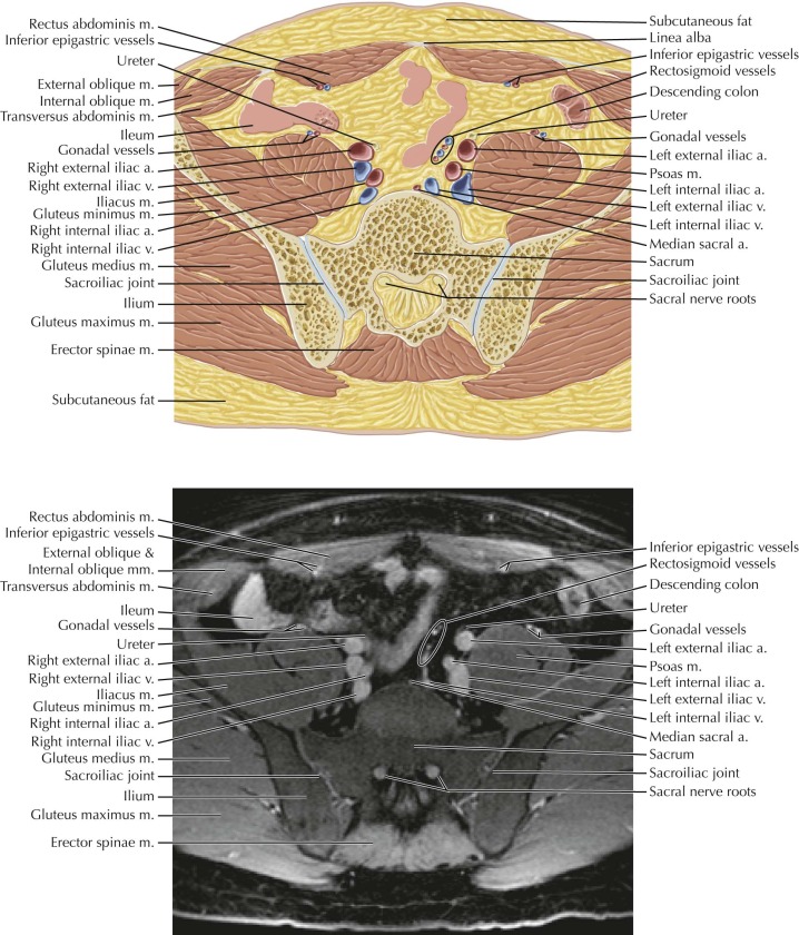
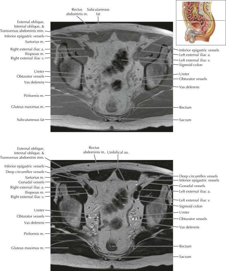
The gonadal vessels refer to the testicular artery and testicular vein in males and the ovarian artery and ovarian vein in females. The gonadal arteries arise directly from the aorta and carry blood to the gonads (testicles or ovaries). The right gonadal vein drains into the inferior vena cava (IVC), and the left gonadal vein drains into the left renal vein. On this image in the lower abdomen, the gonadal vessels are seen coursing anterior to the psoas muscles, adjacent to the ureters.

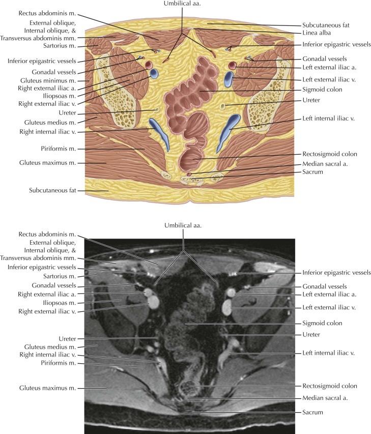
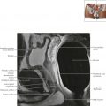
Male Pelvis Axial 6
Normal Anatomy
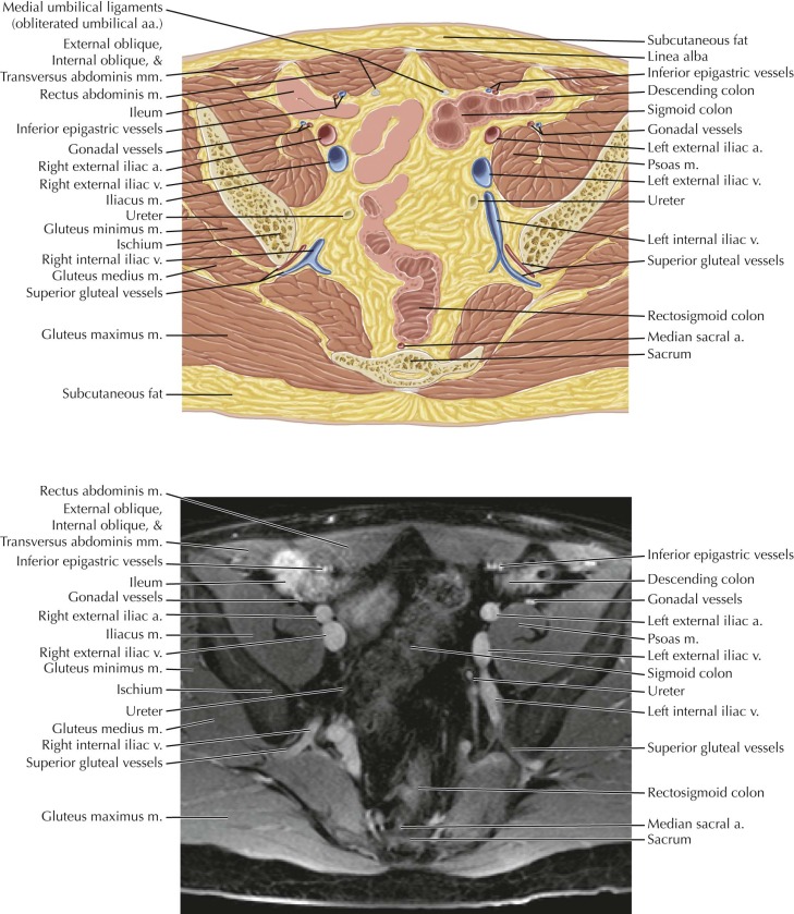
The superior gluteal artery is the largest branch of the internal iliac artery and appears as the continuation of the posterior division of the internal iliac artery. On this image, the superior gluteal vessels are seen traveling through the greater sciatic foramen just above the piriformis muscle. Other branches of the posterior division of the internal iliac artery include the iliolumbar and lateral sacral arteries.
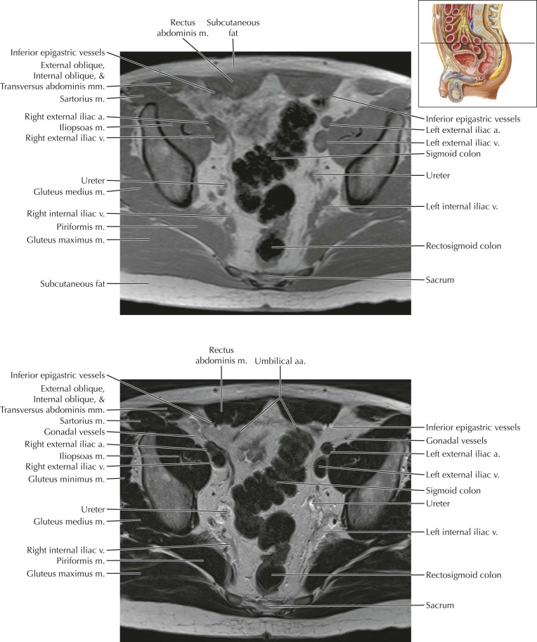
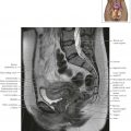
Male Pelvis Axial 8
Normal Anatomy
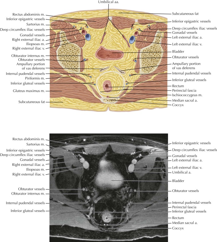
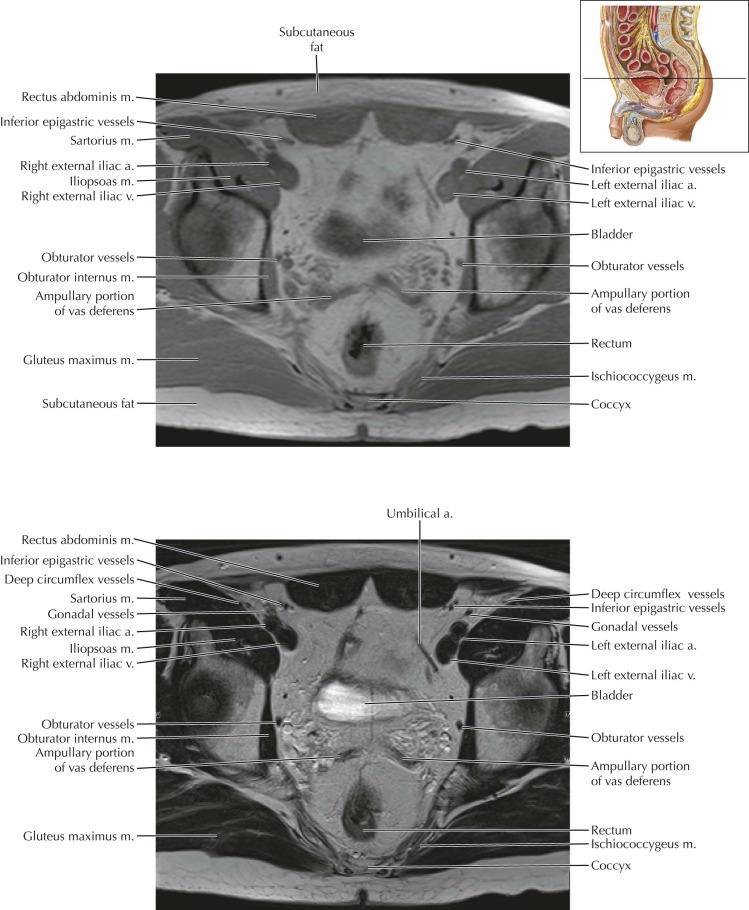
The paired vasa deferentia are continuations of the epididymal tails (cauda epididymidis), which ascend from the scrotum into the pelvis through the inguinal canals, course along the lateral pelvic walls, cross over (anterior and superior to) the ureters, and then curve along the superomedial surface of the seminal vesicles, where they dilate to form the ampullary portions of the vasa deferentia.
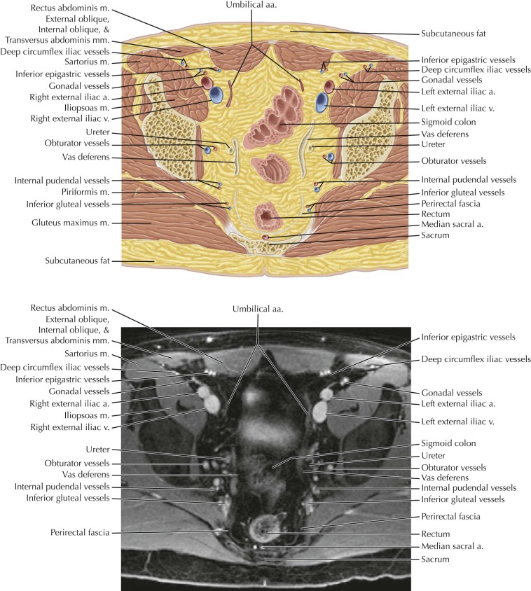
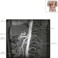
Male Pelvis Axial 9
Normal Anatomy
Several of the main branches of the anterior division of the internal iliac artery can be seen on this image, including the umbilical artery, which usually gives off the superior vesical artery; the obturator artery, which exits the pelvis through the obturator canal; and the inferior gluteal and internal pudendal arteries, which exit through the greater sciatic foramen, with the internal pudendal giving off the inferior rectal artery. Other branches from the anterior division of the internal iliac artery include the inferior vesical artery (whereas the middle vesical artery usually arises from the superior vesical artery), the middle rectal artery, the deferential artery in men (which more often arises from the superior vesical artery), and the uterine and vaginal arteries in women.
Related posts:
Stay updated, free articles. Join our Telegram channel

Full access? Get Clinical Tree


