Chapter 28
Masseter Muscle Venous Malformation
Epidemiology
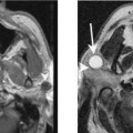
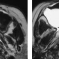
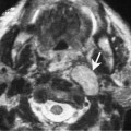
It is now believed that cavernous hemangiomas represent venous malformations. They usually present at birth but may present in late childhood or early adulthood. These lesions grow slowly in proportion to body growth and they do not involute. They may also undergo spontaneous growth spurts in response to changes in hormonal environment such as puberty and pregnancy. Venous malformations are most commonly detected in the tongue, buccal space, and neck. They are usually superficially located but deep intramuscular involvement may also be seen.
Clinical Findings
Venous malformations are soft, compressible, and nontender lesions. If they are located superficially, they appear bluish. Masseter muscle venous malformations may mimic a parotid mass or enlargement of the masseter muscle. They are not associated with bruit or thrills.
Pathology
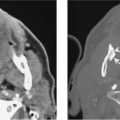
Venous malformations are characterized by cystic dilatation of venous lumen, and thrombosis may be seen in some large vessels. These vessel walls are thin and lined with matured endothelium. Large malformations may be associated with thrombocytopenia or intravascular coagulopathy. Small lesions are well circumscribed but some larger lesions may show an infiltrative pattern. Phleboliths are commonly detected in adults.
Treatment