Abnormalities of the medial ligaments and posterior tibial tendon can occur because of acute injury or chronic instability or malalignment. Medial ankle injuries may occur because of pronation or supination–external rotation injuries. Deltoid ligament injuries have a significant impact on lateral ankle instability but can be overlooked in patients with lateral ligament injuries. Posterior tibial tendon dysfunction is usually associated with spring ligament or flexor retinaculum injury. Tarsal tunnel syndrome, accessory flexor muscles, and subtalar coalition should be considered as well as ligament and tendon tears in differential diagnosis of chronic medial ankle pain.
Key points
- •
Abnormalities of the medial ligaments and posterior tibial tendon can occur because of acute injury or chronic instability or malalignment.
- •
Medial ankle injuries may occur because of pronation or supination–external rotation injuries.
- •
Deltoid ligament injuries have a significant impact on lateral ankle instability but can be overlooked in patients with lateral ligament injuries.
- •
Posterior tibial tendon dysfunction is usually associated with spring ligament or flexor retinaculum injury.
- •
Tarsal tunnel syndrome, accessory flexor muscles, and subtalar coalition should be considered as well as ligament and tendon tears in differential diagnosis of chronic medial ankle pain.
Introduction
The medial soft tissue anatomy of the ankle is complex; the ligaments and posterior tibial tendon are closely interrelated both anatomically and functionally. The medial soft tissues may be acutely injured, or may undergo degeneration caused by hindfoot instability or malalignment. Abnormalities may be limited to the medial side of the foot, or occur in conjunction with lateral hindfoot abnormalities. Clinically, medial ankle abnormalities are often underestimated, or overshadowed by lateral injuries, and magnetic resonance (MR) imaging is useful in showing the full extent of injury and guiding surgical management.
A systematic analysis of medial soft tissue structures on MR imaging should begin with the deltoid ligament, followed by the flexor retinaculum, the spring ligament, and the posterior tibial tendon. The tarsal tunnel is examined next, and the intrinsic muscles of the foot. In addition, a search for tarsal coalition should be made because this diagnosis is often overlooked, especially in subtle cases. The medial findings should be analyzed in conjunction with lateral abnormalities, which may have precipitated the medial findings, or may have resulted from them. Integrating the entire MR imaging picture usually enables radiologists to diagnose the type of injury and to guide treatment.
Imaging protocols
Images obtained through the hindfoot should be aligned with the axis of the talus. The axial plane is the long axis of the talus, and the coronal plane is perpendicular to it. At least 1 T1-weighted sequence should be obtained to evaluate for bone marrow. Fluid-sensitive sequences should be acquired in all 3 planes. A second short echo time plane, either T1 or proton density, is recommended to improve identification of normal anatomy and variants. The field of view is generally 12 to 14 cm, and a dedicated ankle coil is preferred.
Anatomy
The medial ankle ligaments are closely associated anatomically and functionally. A robust understanding of their interconnections is necessary to evaluate medial ankle pain.
Deltoid Ligament Anatomy Overview
The constituent parts of deltoid ligament anatomy have been debated by numerous anatomists, perhaps because cadaveric studies tend to be performed in an elderly population that has a high likelihood of prior ankle injury. The appearance of the ligament on MR imaging is fairly constant ( Figs. 1–3 ).
Deep Deltoid Ligament Anatomy
The deep deltoid ligament prevents lateral talar shift and external rotation of the talus. It has 2 bands. The posterior band of the deep ligament (often called simply the deep deltoid) is the larger of the bands. It is a short, cone-shaped ligament that arises from the posterior margin of the anterior colliculus and from the posterior colliculus. It courses inferiorly, posteriorly, and laterally from the medial malleolus to insert on the fovea at the medial margin of the talar body. Because of its obliquity, it is not visualized in its entirety on a single axial image, and the talar attachment is seen on slices inferior to the tip of the medial malleolus. The ligament has a striated appearance similar to that of the anterior cruciate ligament of the knee, and it is normal to see a small amount of high signal intensity between the fibers. The individual fibers should always appear taut and sharply demarcated. The small anterior band of the deep ligament inserts on the medial talus at the junction of the talar neck and body, and is not always visible on MR imaging.
Superficial Deltoid Ligament Anatomy
The superficial deltoid ligament helps maintain rotational stability of the ankle. It has multiple bands that originate from the superficial surface of the medial malleolus and together make the fanlike shape that gives the ligament its name. The tibiocalcaneal band is the strongest band and inserts on the medial margin of the sustentaculum tali. Although cadaveric studies indicate that it may be absent, it is always visible on coronal MR imaging in patients who have not had a deltoid ligament injury. The tibiospring band merges with the superomedial band of the spring ligament, and is also best evaluated on coronal images. The tibionavicular band is deep to the posterior tibial tendon, and also merges with the superomedial band of the spring ligament. The anterior tibiotalar band is variably present, seen on axial or coronal images. The posterior tibiotalar band inserts on the body of the talus, posterior to the medial malleolus, and is best seen on axial images.
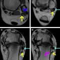
At their origin from the superficial margin of the medial malleolus, the superficial deltoid fibers merge with the periosteum of the malleolus, which in turn merges with the flexor retinaculum (see Fig. 2 ). This continuous sheet formed by the superficial deltoid, periosteum, and flexor retinaculum has been termed the medial malleolar fascial sleeve. The fascial sleeve can be detached completely in twisting injuries, explaining the frequent association of superficial deltoid ligament injuries with injuries of the flexor retinaculum. This relationship should always be scrutinized on fluid-sensitive axial images.
Flexor Retinaculum Anatomy
The flexor retinaculum originates from the superficial margin of the medial malleolus, and extends both laterally and inferiorly. Horizontally oriented fibers insert on the deep flexor fascia (see Fig. 2 ). They maintain the position of the flexor tendons posterior to the medial malleolus. Longitudinally oriented fibers extend inferiorly from the medial malleolus to form the roof to the tarsal tunnel, merging with the fascia of the abductor hallucis muscle and inserting on the calcaneus (see Fig. 1 A). On MR imaging, the normal flexor retinaculum forms a thin, taut line that is low signal intensity on all sequences. Axial images are key to evaluate the insertion of the flexor retinaculum onto a pointed promontory at the posteromedial corner of the tibia.
Spring Ligament Anatomy
The spring ligament helps maintain the medial arch of the foot, although its importance has been debated. The spring ligament is composed of 3 bands. The superomedial spring ligament forms a broad sling, arising from the surface of the sustentaculum tali and inserting on the superomedial surface of the navicular, dorsal to the insertion of the tibialis posterior tendon. It merges with the tibiospring band of the superficial deltoid ligament (see Fig. 3 A). There are 2 deep plantar bands of the spring ligament (see Fig. 3 B). The medioplantar oblique ligament is a narrow straplike ligament that arises immediately anterior to the sustentaculum tali, and has a diagonal course, inserting on the median eminence of the navicular. The inferoplantar ligament is a short, broad ligament that arises from the calcaneus just lateral to the medioplantar oblique ligament and inserts on the lateral beak of the navicular. A joint recess of the talonavicular joint lies between the 2 plantar bands. The plantar fascicles of the spring ligament are usually best seen on sequential axial images. The superomedial band is best seen on coronal images.
Tibiocalcaneonavicular Ligament
It is evident from the earlier descriptions that the tibionavicular, tibiospring, and superomedial spring ligament are closely linked, and they coalesce on MR imaging. In practice, they can be described together as the tibiocalcaneonavicular ligament or the spring ligament complex.
Posterior Tibial Tendon Anatomy
The posterior tibial muscle is the most important dynamic stabilizer of the medial longitudinal arch of the foot. It originates in the calf, and becomes tendinous in the distal one-third of the leg. At the ankle, the posterior tibial tendon is the most medial of the extrinsic flexor tendons and is held in place in a shallow groove behind the medial malleolus by the flexor retinaculum (see Fig. 2 ). It continues into the hindfoot within the tarsal tunnel, superficial to the deltoid ligament (see Figs. 1 A and 3 A), and has its primary insertion on the plantar aspect of the median eminence of the navicular. Small tendon slips continue anterior to the navicular to insert on the second and third cuneiforms, the bases of the second to fourth metatarsals, and the cuboid. A recurrent slip inserts on the sustentaculum tali.
On MR imaging, the normal tendon is twice the size of the adjacent flexor digitorum longus tendon. A small amount of fluid is often seen in its tendon sheath, and does not indicate tenosynovitis. The tendon normally shows slightly increased signal intensity as it inserts on the navicular. The anterior tendon slips to the cuneiforms, metatarsals, and cuboid are well seen on MR imaging and are rarely abnormal. A small recurrent tendon slip inserts on the anterior margin of the sustentaculum tali, and may be injured but is hard to distinguish from the spring ligament on imaging.
Accessory Navicular Bone
The accessory navicular bone (also called the os tibiale externum) is a normal variant that can be associated with posterior tibial tendon dysfunction. A type 1 accessory navicular is a sesamoid in the distal tendon, and is not associated with tendon abnormalities. A type 2 accessory navicular is an accessory center of ossification, connected to the main portion of the navicular by a synchondrosis. The posterior tibial tendon attaches at least partly to the ossicle. This construct is vulnerable, and increases likelihood of acute or chronic posterior tibial tendon injury.
Tarsal Tunnel Anatomy
Students first learning the anatomy of the ankle are sometimes confused by 3 similar-sounding structures: the tarsal tunnel, the tarsal sinus (sinus tarsi), and the tarsal canal. It becomes easy to remember the location of the tarsal tunnel if you remember that it is analogous to the carpal tunnel: a space with a fibrous roof and bony floor, transited by nerves, vessels and tendons, within which the nerve is vulnerable to compression. In the case of the tarsal tunnel, the vulnerable nerves are the tibial nerve and its branches. The tarsal sinus is a lateral space between the calcaneus and talus, and the tarsal canal is the posteromedial continuation of the sinus.
The tibial nerve and its branches are most easily evaluated within the tarsal tunnel on fluid-sensitive coronal or axial MR imaging. The nerve is lower in signal intensity than the veins, but higher in signal intensity than the tendons (see Fig. 1 A). Fascicles should be visible within it. The nerve branches in the tarsal tunnel into a medial calcaneal nerve, and medial and lateral plantar nerves. The branching pattern may vary.
Deltoid ligament injuries
Acute Deltoid Injuries
Deltoid ligament injuries were at one time thought to be uncommon, but are now known to be fairly common. They are significantly less common than medial malleolar fractures. Injuries may occur either because of pronation or because of supination–external rotation injury.
Pronation injuries apply a direct abduction force to the deep deltoid ligament and may result in medial malleolar fracture or deltoid ligament tear ( Fig. 4 ). The deltoid can be injured in isolation, but, in more severe injuries, there is a fibular fracture or a tear of the tibiofibular syndesmosis.
Both ankle sprain (injury to the lateral collateral ligament complex) and Weber B fractures are caused by supination–external rotation injuries. A supination–external rotation injury occurs when the foot is planted and inverted; the tibia rotates internally on the planted talus. By convention, ankle injuries are described by the force applied on the talus, hence the term supination–external rotation.
The twisting component of the supination–external injury mechanism may also result in deltoid ligament injury. The risk of deltoid ligament injury increases with the severity of the lateral collateral ligament injury. A clinical focus on the lateral side of the ankle can lead physicians to overlook associated medial ligamentous injuries, delaying diagnosis of medial injury. Physical examination for the presence of medial injuries is not reliable in the setting of acute lateral ankle injury and so stress radiographs are often used to diagnose deep deltoid injuries. However, one study of stress radiographs and MR imaging in lateral malleolar fractures found that stress radiographs may be falsely positive in patients who have only a partial rupture of the deep deltoid by MR imaging criteria, and who do not need operative treatment.
MR imaging is uncommonly performed for acute ankle sprain, but should be considered in severe cases ( Fig. 5 ) because the extent of injuries may not be apparent clinically. In reading MR imaging of cases with multiple injuries, radiologists are in danger of experiencing satisfaction of search. One area where injuries are often overlooked is the proximal origin of the superficial deltoid. This area should be carefully scrutinized on axial images (see Fig. 5 A).


