Following a brief description of the normal anatomy and biomechanics of the midfoot, this article focuses on MR imaging features of common osseous, tendon, and ligament abnormalities that affect the midfoot. Discussion of the anatomy and pathology affecting the Chopart and Lisfranc joint complexes, both of which play important roles in linking the midfoot to the hindfoot and the forefoot respectively, is also included.
Key points
- •
The navicular, cuboid, and 3 cuneiform bones form the midfoot, the anatomic region located between the Chopart and Lisfranc joints.
- •
Midfoot pathology, involving the osseous and soft tissue structures at the midfoot and at the junction of the midfoot with the hindfoot (Chopart joint complex) and forefoot (Lisfranc joint complex), is a common, albeit elusive cause for pain.
- •
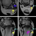
Navicular, cuboid and cuneiform fractures represent radiographically occult causes of foot pain that often require evaluation with MR imaging.
- •
MR imaging is the modality of choice for detection of several tendon and ligamentous pathology about the midfoot.
Related posts:

Stay updated, free articles. Join our Telegram channel

Full access? Get Clinical Tree


